Ms. Kumkum Agarwal
Sarojini Naidu Govt. Girls P.G. College, Bhopal [M.P.] Email: [email protected]
Address for correspondence
Access this article online www.japer.in
Antioxidant activity and Phytochemical analysis of
Hyptis
suaveolens (L.) Poit
INTRODUCTION
Reactive oxygen species (ROS) is a term that
encompasses all highly reactive, oxygen containing
molecules including free radicals, which includes the
hydroxyl radical, the superoxide anion radical,
hydrogen peroxide, singlet oxygen, nitric oxide
radical, hypochlorite radical and various lipid
peroxides. All these are capable of reacting with
membrane lipids, nucleic acids, proteins and enzymes
and other small molecules resulting in cellular
damage. To protect the cells and organ system of the
body against ROS, humans have evolved a highly
sophisticated and complex antioxidant protection
system. It involves a variety of components both
endogenous and exogenous in origin that function
interactively and synergistically to neutralize free
radicals and include nutrient derived antioxidants
(Vitamin C, E , beta carotene and polyphenols),
antioxidant enzymes, metal binding proteins and
numerous other plant derived phytonutrients. The
human body has multifaceted structure of natural
enzymatic and non-enzymatic antioxidant resistance,
which counteracts the detrimental effects of free
radicals and other oxidants. Substantial data indicate
that food rich in antioxidants may be of chief
significance in disease prevention. [1]
Oxidative stress has been implicated in the
pathogenesis of many human diseases like
Alzheimer's , Parkinson's, neurodegenerative diseases,
the pathologies caused by diabetes, rheumatoid
arthritis, and cancer. It also mediates a wide range of
renal impairments, from acute renal failure,
obstructive nephropathy, hyperlipidemia, and
glomerular damage to chronic renal failure. [2]
Studies show that oxidative stress is increased along
with increased lipid peroxidation and decrease in
antioxidative defense in acute renal failure patients
undergoing hemodialysis and urolithiasis. The
positive role of Vitamin E was found to be synergistic
with ascorbic acid to ameliorate oxidative stress. [3-4]
In a study by Felix Grases et.al it was concluded that
the antioxidant activity of herbal extracts could have
an important role in avoidance of calcium oxalate
monohydrate papillary calculi formation. [5-6]
Several laboratories reported that oxalate causes
renal tubular injury by increasing generation of free
radicals. In a study by Sivagnanum et.al showed that Research
Research
Research ArticleArticleArticleArticle
Hyptis suaveolens (L) Poit. commonly known as Vilayati tulsi,belongs to the family Lamiaceae. Production of reactive oxygen species (ROS) causes various diseases and cellular anomalies in human beings. Antioxidants inhibit generation of reactive species, or scavenge them, or raise the levels of endogenous antioxidant defenses. In this study the antioxidant activity as well as phytochemical analysis of Hyptis suaveolens L. was undertaken. Results revealed that Hyptis suaveolens (L) Poit. has potent antioxidant ability of 69.46% at 100 µg/ml concentration and IC50 value at 40.91 µg/ml concentration and a
good correlation was found to exist between concentration of extract and % inhibition with r2=0.995. Phytochemical analysis revealed the presence of alkaloids, carbohydrates, reducing sugars, flavonoids, glycoside, tannin, phenolic compounds, protein, amino acids, triterpenoids and steroids as well as 0.88 mg/gm of photosynthetic pigments, 0.0004275 mg/g of ascorbic acid while 0.105 mg/g of foliar phenol content was found to be present.
Keywords: Tulsi, antioxidant, oxidative stress, diseases, phytochemical. ABSTRACT
ABSTRACT ABSTRACT ABSTRACT Kumkum Agarwal*
RanjanaVarma1
Department of Botany, Sarojini Naidu Govt. Girls P.G. College, Shivaji Nagar, Bhopal-462016, Madhya Pradesh, India.
1Department of Botany, Sarojini
Naidu Govt. Girls P.G. College, Shivaji Nagar,Bhopal-462016, Madhya Pradesh, India.
vitamin E which is considered to be a potent
antioxidant, completely prevented calcium oxalate
deposition, by preventing peroxidative injury and
restoring renal tissue antioxidants and glutathione
redox balance. [7] Antioxidant refers to a compound
that can delay or inhibit the oxidation of lipids or
other molecules by inhibiting the initiation or
propagation of oxidative chain reactions and which
can thus prevent or repair damage done to the body’s
cells by oxygen.[8]
Thus the use of antioxidants in pharmacology is
intensively studied, particularly as treatments for
neurodegenerative diseases and renal diseases etc. [9]
Therefore, it is important to assess antioxidant
activity of the plants used in the herbal medicine
either to elucidate the mechanism of their
pharmacological action or to provide information on
antioxidant activity of these herbal plants. All the
natural anti-oxidants though safer but show lower
antioxidant activity than the synthetic anti-oxidants
like BHA (butylated hydroxyanisole) and BHT
(butylated hydroxytoulene), but as these are
considered to be promoters of carcinogenesis so need
exists for safer, economic natural anti-oxidants with
high anti-oxidant activity. The use of DPPH as a
reagent for screening the antioxidant activity of small
molecules and pure compounds or plant extracts has
been reported. [10] The phytochemicals from plants,
particularly flavonoids and other polyphenols, have
been reported to inhibit the propagation of free
radical reactions, to protect the human body from
disease, and to retard lipid oxidative rancidity.[8] They
are primary or secondary metabolites having a range
of biochemical and physiological effects. Because
many phytochemicals are not well characterized and
their mode of action is not well established so
research on these physiologically active plant
components is currently an area of intense effort.
Hyptis suaveolens (L) Poit. commonly known as
Vilayati tulsi belongs to the family Lamiaceae or the
Mint family. It is a shrubby, scented plant with blue or
purple small flowers. The plant has been considered
as a weed, distributed throughout the tropics and
subtropics. Common in open uncultivated areas, rocky
dry substratum, on roadsides and waste grounds.
Flowering and fruiting occurs during October till
January.
Almost all parts of this plant are being used in
traditional medicine as well as it has immense
ethnomedicinal importance for treating various
diseases. And the lot of research work done on this
plant has also revealed various other activities in this
plant.
Its leaf methanolic extract has shown various
activities like antimicrobial and [11] antibacterial
activity [12] it was found effective against
methicillin-resistant Staphylococcus aureus [13] Similarly its
toxicity and hypoglycemic activity was studied. [14] It
was also screened for antifungal activity [15] while leaf
and seed methanolic extracts was screened for
insecticidal activity. [16] Various researchers have
studied its antioxidant activity. Pradeep et.al (2011)
[17] found that aqueous extract of Hyptis suaveolens
L.showed a protective effect on the antioxidant status
of the animals which was evident from the low lipid
peroxidation levels, similarly the alcoholic extract [18]
and petroleum ether leaf extract [19] showed
antioxidant activity.
The antioxidant activity of the essential oil was
determined by using two complementary methods:
DPPH (1,1-diphenyl-2-picrylhydrazyl ) and ABTS
(2,2'-azinobis-(3-ethylbenzothiazoline-6-sulfonic
acid) free radical decolorization assay. Hyptis
suaveolens L. showed IC50 values of 3721 μg/ml. [8]
similarly essential oil was screened by DPPH assay
and was found to show an IC50 value of 3.72mg/ml
whereas the TEAC value determined by ABTS assay
was 65.02µM/mg. [20] The aqueous extract of Hyptis
suaveolens L. leaf showed 98.06 % inhibition in DPPH
radical. [21] The methanolic extract of leaves of Hyptis
suaveolens exhibited potent antioxidant activity in
DPPH assay with IC50 value of 14.04µg/ml. [22] Novel
phenolic compound,from Hyptis suaveolens L.was
damage and related degenerative diseases involving
metabolic stress and genotoxicity. [23]
The polyphenol extracts of Hyptis leaf (0.4–1.6 μg/ml)
caused a dose-dependent significant decrease in the
MDA (malondialdehyde) contents in the brain. [24] The
plant has been reported to be rich in plant chemicals.
Phytochemical screening of methanol fraction of
whole plant of Hyptis showed the presence of
alkaloids, flavones, flavonols, terpenoids, tannins,
aldehydes and ketones. The methanol soluble fraction
of the plant showed the least growth inhibitory effect
on fungi- Candida albicans and Aspergillus niger. [25]
Similarly methanolic leaf extract showed the presence
of alkaloids, tannins, along with steroids, triterpinoids,
reducing sugars, phenolic compounds, flavonoids,
saponins, anthraquinones and amino acids. [26] Thus
the present study was undertaken to re-investigate
the antioxidant activity and quantitative and
qualitative phytochemical screening of alcoholic
extract of Hyptis suaveolens (L) Poit. growing in
Bhopal district.
MATERIALS AND METHOD
Plant collection, identification and preparation of
extract
The plant was collected from Kolar road, Bhopal,
Madhya Pradesh, during the month of January 2013
and plant was identified with the help of regional
floras [27] and taxonomists and finally confirmed with
the herbarium of Botanical Survey of India (BSI),
Allahabad, voucher specimen No. 1234-126.01-605.
Fresh plant, after collection was shade dried at room
temperature. Plant material was then grinded in a
Crompton & Greaves mixer and grinder, and then the
powdered plant material 100 gm was extracted with
250ml alcohol by Soxhlet apparatus for 72 hours.
Then the extract was concentrated in vacuo to dryness
at 30-40°c temperature, obtaining dried extract. The
dried extract was stored in refrigerator until used for
further analysis.
Measurement of antioxidant activity
DPPH radical scavenging assay-
The procedure for estimating the antioxidant activity
involved UV-Spectrophotometric determination.[28]
Three solutions i.e. standard, test and control were
prepared.
Different solutions (25 - 100μg/ml) of ascorbic acid
were prepared in methanol. 1.5 ml of each solution of
ascorbic acid were mixed with 1.5 ml of 200μM DPPH
solution and incubated for 30 min at room
temperature in dark. Absorbance of each solution was
taken against blank at 517 nm. Different solutions of
the given sample and control were prepared to give
concentrations (50 - 200μg/ml). 1.5 ml of each
solution of given sample was mixed with 200 μM
DPPH solution and incubated at room temperature in
dark. Absorbance of each solution of given sample was
taken against blank at 517 nm.
Percentage antioxidant activity of plant extract and
Ascorbic acid was calculated by using
formula:
I % = Ac – (At- Ab) X 100 At
where
I% = percentage inhibition
Ac = absorbance of control (methanol and 200 μM
DPPH solution)
At = absorbance of ascorbic acid / given sample with
DPPH solution.
Ab =absorbance of ascorbic acid / given sample
without DPPH solution.
Phytochemical analysis
Phytochemical testing was performed to assess the
various phytoconstituents present in alcoholic extract
of Hyptis suaveolens L. Quanlitative analysis [29] of its
extract was performed to determine the presence or
absence of carbohydrates, proteins and amino acids,
glycosides, alkaloids, flavonoids, saponin,
triterpenoids and steroids, tannin and phenolic
compounds. While quantitative analysis was
performed to determine the amount of chlorophyll,
ascorbic acid as well as foliar phenols present in fresh
Qualitative analysis:
Tests for carbohydrates and reducing sugars:
Molish test:
2 ml of aqueous extract was treated with 2 drops of
alcoholic α-naphthol solution in a test tube and then 1
ml of concentrated sulphuric acid was added carefully
along the sides of the test tube. Formation of violet
ring at the junction indicate the presence of
carbohydrates.
Barfoed’s test:
1 ml of extract and Barfoed’s reagent were mixed in a
test tube and heated on water bath for 2 minutes. Red
colour due to formation of cupric oxide indicates the
presence of monosaccharide.
Fehling’s test:
To 1 ml of aqueous extract, 1 ml of Fehling’s A and 1
ml of Fehling’s B solutions were added in a test tube
and heated in the water bath for 10 minutes.
Formation of red precipitate indicates the presence of
reducing sugar.
Benedict’s test:
Equal volume of Benedict’s reagent and extract were
mixed in a test tube and heated in the water bath for
5-10 minutes. Solution appears green, yellow or red
depending on the amount of reducing sugar present in
the test solution which indicated the presence of
reducing sugar.
Tests for protein and amino acids:
Ninhydrin test:
3 ml of the test solution was heated with 3 drops of
5% Ninhydrin solution in a water bath for 10 minutes.
Formation of blue colour indicates the presence of
amino acids.
Tests for glycosides:
Borntrager’s test:
To 3 ml of test solution, dilute sulphuric acid was
added, boiled for 5 minutes and filtered. To the cold
filtrate, equal volume of benzene or chloroform was
added and shake it welled. The organic solvent layer
was separated and ammonia was added to it.
Formation of pink to red color in ammonical layer
indicates presence of anthraquinone glycosides.
Legal’s test:
1 ml of test solution was dissolved in pyridine. 1 ml of
sodium nitropruside solution was added and made
alkaline using 10% sodium hydroxide solution.
Formation of pink to blood red color indicates the
presence of Cardiac glycosides.
Keller-killiani test:
To 2 ml of test solution, 3 ml of glacial acetic acid and
1 drop of 5% ferric chloride were added in a test tube.
Add carefully 0.5 ml of concentrated sulphuric acid by
the side of the test tube. Formation of blue color in the
acetic acid layer indicates the presence of Cardiac
glycosides.
Tests for alkaloids:
To the extract, dilute hydrochloric acid was added,
shake it well and filtered. With the filtrate, the
following tests were performed.
Mayer’s test:
To 2-3 ml of filtrate, few drops of Mayer’s reagent
were added along sides of tube. Formation of white or
creamy precipitate indicates the presence of alkaloids.
Hager’s test:
To 1-2 ml of filtrate, few drops of Hager’s reagent
were added in a test tube. Formation of yellow color
precipitate indicates the presence of alkaloids.
Wagner’s test:
To 1-2 ml of filtrate, few drops of Wagner’s reagent
were added in a test tube. Formation of reddish brown
precipitate indicates the presence of alkaloids.
Tests for flavonoids:
Lead acetate test:
The extract was treated with few drops of lead acetate
solution. Formation of yellow precipitate may indicate
the presence of flavonoids.
Alkaline reagent test:
The extract was treated with few drops of sodium
hydroxide separately in a test tube. Formation of
intense yellow color, which becomes color less on
addition of few drops of dilute acid, indicate presence
Shinoda test:
To the extract, 5 ml (95%) of ethanol was added. The
mixture was treated with few fragments of
magnesium turning, followed by drop wise addition of
concentrated hydrochloric acid. Formation of pink
color indicate presence of flavonoids.
Test for saponin:
Foam test:
The extract was diluted with distilled water and
shaken in graduated cylinder for 15 minutes. The
formation of layer of foam indicates the presence of
saponins.
Tests for triterpenoids and steroids:
Salkowski’s test:
The extract was treated with chloroform and filtered.
The filtrate was added with few drops of concentrated
sulphuric acid, shaken and allowed to stand. If the
lower layers turns red, sterol are present. Presence of
golden yellow layer at bottom indicates the presence
of triterpenes.
Libermann-burchard’s test:
The extract was treated with chloroform. To this
solution few drops of acetic anhydride were added,
boiled and cooled. Concentrated sulphuric acid was
added through the sides of the test tube. Formation of
brown ring at the junction of two layers, if upper layer
turned green, indicate presence of steroids and
formation of deep red color indicate presence of
triterpenoids.
Tests for tannin and phenolic compounds:
Ferric chloride test:
Some amount of extract was dissolved in distilled
water. To this solution 2 ml of 5% ferric chloride
solution was added. Formation of blue, green or violet
color indicates presence of phenolic compounds.
Lead acetate test:
Some amount of extract was dissolved in distilled
water. To this solution few drops of lead acetate
solution was added. Formation of white precipitate
indicates presence of phenolic compounds.
Dilute iodine solution test:
To 2-3 ml of extract, few drops of dilute iodine
solution were added. Formation of transient red color
indicates presence of phenolic compounds.
Quantitative analysis:
Photosynthetic pigments content:-
Photosynthetic pigments in O.G. leaf was analyzed by
the method proposed by Machlachalan & Zalik (1963)
and Duxbury & Yentsch (1956). Chlorophyll-a,
Chlorophyll-b and carotenoids were determined by
extracting the pigments from fresh leaf samples.
Leaves were washed with distilled water and were cut
into small pieces, 100 mg of these leaf pieces were put
in 5 ml of 80% acetone and were crushed in a mortar
pestle with little acid washed sand. The resultant
extract was centrifuged at 5000 rpm for 5 minutes.
Supernatant was collected and sediment was washed
with 1 ml of 80% acetone and centrifuged again. The
supernatant thus obtained is added to previous one
and the total final volume of the supernatant was
made upto 10 ml by adding 80% acetone. Optical
density of the supernatant so obtained was recorded
at 480, 510, 645 and 663 nm wavelengths, using
Systronics Digital spectrophotometer Model-166,
against blank carried out throughout the process.
The amounts of Chlorophyll-a, Chlorophyll-b and
carotenoids were estimated in terms of mg/gm fresh
weight of the leaf was calculated by using the
following formulae (Machlachalan & Zalik, 1963; and
Duxbury & Yentsch, 1956):
Chlorophyll-a = 12.3 D663 – 0.86 D645 X V (mg/gm fresh wt.) d x 1000 x w
Chlorophyll-b = 19.3 D645 – 3.6 D645 X V (mg/gm fresh wt.) d x 1000 x w
Total Chlorophyll = Chlorophyll-a + Chlorophyll-b (mg/gm fresh wt.)
Carotenoides = 7.6 D480 – 1.49 D510 X V mg/gm fresh wt.) d x 1000 x w
Where V, is the volume of chlorophyll solution in
acetone, d, is the light path, and w, is the fresh weight
of plant part in gms.
Ascorbic acid content:-
Ascorbic acid content was estimated by using method
proposed by Schaffert and Kingsley (1955). Reagents
(Trichloro acetic acid), 2 % 2-4 DNPH ( 2-4 Dinitro
phenyl hydrazine), 85 % Sulphuric acid, 10 %
Thiourea
Extraction of ascorbic acid:
2 gms of plant material was crushed with 100 ml of 4
% TCA and then the contents were centrifuged at
5000 rpm. 20 ml of supernatant was taken and was
mixed with ½ teaspoon of activated charcoal. This was
shaken well and then filtered. From the filtrate, 4 ml
filtrate was used for further analysis. To 4 ml extract 1
drop of thiourea and 1 ml of 2-4 DNPH were added,
the test tube containing sample was placed in a boiling
water bath exactly for 10 minutes. After which the
tubes were placed in a beaker containing crushed ice
and 5 ml of 85% sulphuric acid was added to the
sample slowly drop by drop and mixed by rotating the
test tube placed in crushed ice. The sample was kept
as such for 10 minutes and then optical density of this
sample was read by using Systronics digital
Spectrophotometer, Model- 166 at 515 nm against
blank carried throughout the process. The µg ascorbic
acid content at a given optical density was determined
with the help of a standard curve prepared by taking
known amounts of ascorbic acid.
Standardization:
0.1 % solution of ascorbic acid was prepared by
adding 100 mg of ascorbic acid in 100 ml of 4 % TCA.
This served as the stock solution. From this stock
solution, working ascorbic acid solution of 0.002 %
concentration was prepared by mixing 0.02 ml of
stock solution with 98 ml of 4% TCA. Aliquots of this
working ascorbic acid solution were prepared as
follows in separate test tubes. 0.1 ml, 0.15 ml, 0.2 ml,
0.25 ml, 0.3 ml, upto 1.0 ml. Each aliquot was diluted
upto 4 ml by 4 % TCA. 1 drop of thiourea and 1 ml of
2-4 DNPH was added to each sample and the test
tubes were placed in boiling waterbath for 10
minutes. Optical density of the samples was read at
515nm. From these optical densities and solutions of
known concentrations, standard curve was plotted.
With the help of standard curve ascorbic acid contents
in samples were obtained. Total amount of ascorbic
acid present in the sample was calculated by using the
following formula:
Total ascorbic acid = µg ascorbic acid X V W X 1000
Where, µg ascorbic acid = Concentration obtained
from standard curve
V = Total volume of the sample
W= Weight of the plant sample.
Foliar phenol content:
Foliar phenols were estimated by the method
proposed by Bray & Thorpe (1954). 200 mg of fresh
plant material was homogenized with 10ml of 70%
ethanol and centrifuged at 6000 rpm for 10 minutes.
Residue was subjected to repeated washings with 70
% ethanol followed by centrifugation each time.
Supernatant so obtained was concentrated by
allowing evaporation overnight and by using a
separating funnel the concentrate was partitioned
through light petroleum to remove chlorophyll. To 1
ml extract in a test tube, 2 ml of 20% sodium
carbonate solution and 1 ml 1N Folin Phenol reagent
were added. The reaction mixture was immediately
kept in boiling water bath exactly for 1 minute and
cooled to room temperature before the optical density
of this blue colored solution was measured at 650nm
by using Systronics digital spectrophotometer,
Model-166.
The µg phenol content at a given optical density was
determined with the help of a standard curve
prepared with known amount of quinine.
Total amount of phenol in the sample was obtained by
using the following formula:
Phenol content = µg phenol X V (mg/gm of fresh wt.) W X 1000
Where, µg phenol = concentration obtained from the
standard curve
V = total volume of the mixture
W = weight of the sample.
Results
In the present work, DPPH assay was utilized for
evaluating the free radical scavenging action of Hyptis
standard compound and its IC50 value was determined
to be 4.69 µg/ml (Figure1). Results show that,
methanolic extract of leaf of Hyptis suaveolens L. has
potent antioxidant ability with an IC50 value at
40.91µg/ml concentration and minimum % inhibition
of 45.30% at 25 µg/ml and highest % inhibition was
found to be 69.46% at 100 µg/ml concentration
(Figure 2) which is comparable with that of ascorbic
acid 82.05% at100 µg/ml. It was also found that a
good correlation exists between the concentration of
extract and % inhibition, with r2 = 0.995 and the
regression equation shows that the % inhibition is the
dependent variable while concentration is
independent and the % inhibition of DPPH free
radical, increased with the increase in the
concentration of extract.
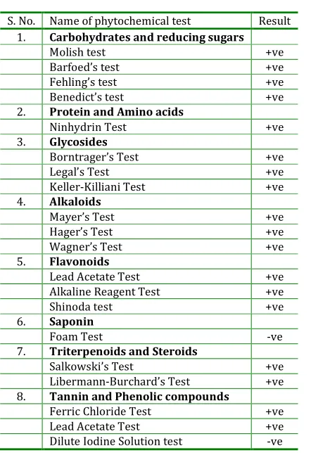
The results of qualitative phytochemical
analysis of Hyptis suaveolens L.methanolic extract
revealed the presence of alkaloids, carbohydrates,
reducing sugars, flavonoids, glycoside, tannin,
phenolic compounds, protein, amino acids,
triterpenoids and steroids (Table1). While
quantitative phytochemical analysis showed the
presence of 0.88 mg/gm fresh weight of
photosynthetic pigments, 0.0004275 mg/g fresh
weight of ascorbic acid while 0.105 mg/g fresh weight
of foliar phenol content.
DISCUSSION
The DPPH assay is a very simple method for screening
small anti-oxidant molecules, because the reaction can
be observed visually and intensity can be analyzed
using common spectrophotometric assay. The stable
radical DPPH has been used widely for the
determination of primary anti-oxidant activity. The
DPPH anti-oxidant assay is based on the ability of
DPPH a stable free radical, to decolorize in the
presence of anti-oxidants. Qualitative and quantitative
phytochemical analysis showed that the plant is rich
in flavonoids and phenols that are considered to be
potent anti-oxidants, so this activity may be
attributable to the presence of these chemicals in the
extract. As the earlier studies by Felix G et.al (2006)
[5] and [6] Bharathi B.K (2013) have shown that the
antioxidant activity of herbal extracts have an
important role in avoidance of calcium oxalate
monohydrate papillary calculi formation, thus this extract will be further screened for its antiurolithiatic
potential in our near future studies. And these in vitro
results should be confirmed in vivo so as to develop
potent antioxidant from this plant, as this property of
the extract will be advantageous in preventing
oxidative stress and in preventing the various other
diseases caused by it. Further the extract could be
fractioned and fractions could be analyzed for
extracting the particular active principle responsible
for this activity.
CONCLUSION
In the present work the inhibition of DPPH free
radicals and phytochemical screening of Hyptis
suaveolens Poit. methanolic extract was studied. The
extract showed potent radical scavenging ability and
the percentage inhibition was found directly
proportional to the increase in concentration or
percentage of the plant extract. The extract may
contain phytochemicals that cause inhibition mainly
phenols and flavonoids. This property of plant may be
important in preventing oxidative stress related
diseases. These in vitro results should be confirmed in
vivo.Literature review has shown that very few
studies on antioxidant potential of Hyptis suaveolens
Poit. methanolic extract has been undertaken and to
the best of our knowledge till now, no such study on
antioxidant activity of Hyptis by DPPH assay has been
done in Bhopal district. The mechanism by which it
exerts its effects remains unknown, so the mechanism
as well as chemicals responsible could be isolated and
studied in future.
ACKNOWLEDGEMENT
The author would like to acknowledge the Principal of
Sarojini Naidu Govt. Girls. P.G. College, Bhopal as well
Department of Botany, Teaching and nonteaching staff
of the college for their cooperation. Sincere gratitude
is due to Dr. Madhuri Modak Madam, Proffesor,
Motilal Vigyan Mahavidyalaya, Bhopal for arranging
plant identification. The authors are thankful to the
Director, PBRI, Bhopal (M.P., India) for providing the
necessary facilities to carry out this research work. No
funding agency is involved in this research work.
Fig. 1: Graph represent regression curve of Ascorbic acid by DPPH assay
Table1: Qualitative phytochemical screening of Hyptis suaveolens L
S. No. Name of phytochemical test Result
1. Carbohydrates and reducing sugars
Molish test +ve
Barfoed’s test +ve
Fehling’s test +ve
Benedict’s test +ve
2. Protein and Amino acids
Ninhydrin Test +ve
3. Glycosides
Borntrager’s Test +ve
Legal’s Test +ve
Keller-Killiani Test +ve
4. Alkaloids
Mayer’s Test +ve
Hager’s Test +ve
Wagner’s Test +ve
5. Flavonoids
Lead Acetate Test +ve
Alkaline Reagent Test +ve
Shinoda test +ve
6. Saponin
Foam Test -ve
7. Triterpenoids and Steroids
Salkowski’s Test +ve
Libermann-Burchard’s Test +ve
8. Tannin and Phenolic compounds
Ferric Chloride Test +ve
Lead Acetate Test +ve
Dilute Iodine Solution test -ve
Fig. 2: Graph represent regression curve of Hyptis suaveolens leaf methanolic extract (HSLME) by DPPH
assay method
REFERENCES
1. Rajiv S, Santosh S, Sugandha S.. Antioxidants
Accelerate Cellular Health; International Journal Of Green Pharmacy. 2010; 4(3):212-212.
2. Devinder S, Rajnendrapal K, Vikas C, and Kanwaljit C.
Antioxidants in the Prevention of Renal
Disease.Journal of Medicinal Food. 2006; 9 : 443–450. 3. Shelgikar P.J., Deshpande K.H, Sardeshmukh A.S,
Katkam R.V, Suryakarl A.N. (2005).Role of oxidants and antioxidants in ARF patients undergoing
hemodialysis. Indian J Nephrol. 2005; 15: 73-76.
4. Vasavidevi. V., Kishor. H., Adinath. N.,Rajesh. D. and Raghavendra. V. Depleted Nitrite And Enhanced
Oxidative Stress In Urolithiasis. Indian Journal of Clinical biochemistry. 2006; 21: 177-180.
5. Felix G, Rafael M., Isabel G, Pilar S, Antonia C.
Phytotherapy and renal stones: the role of
antioxidants. A pilot study in Wistar rats; Urol
Res.2009; 37:35–40.
6. Bharathi B.,Shivakumar H, Soumya N. Depleted
Antioxidant Vitamins And Enhanced Oxidative Stress
In Urolithiasis. International Journal Of Pharmacy And Biological Sciences.2013; 3: 71-75.
7. Sivagnanam T and Mani M. (2005).Vitamin E therapy
prevents hyperoxaluria-induced calcium oxalate
crystal deposition in the kidney by improving renal
tissue antioxidant status. B. J. U.International. 2005;
9(6):11 7 – 1 2 6.
8. Suganya T and Sombat C. Comparison of Antioxidant
and Antimicrobial Activities of Essential Oils from
Hyptis suaveolens and Alpinia galangal Growing in Northern Thailand; CMU. J. Nat. Sci. 2007; 6: 32-42. 9. Siddharthan S, Yi-Zhong C, Harold C, Mei S.
anti-oxidants from 133 Indian Medicinal plants. Food Chemistry. 2007; 102: 928-953.
10. Agrolo A., Sant A. Antioxidant activity of leaf extracts
from Bauhinia monandra. Bioresource Technology. 2004; 95: 229-233.
11. Satish V, Padmaa M, Usha G, Ravichandrian V.
Antimicrobial on the extract of Cocculus hirsutus
Linn. and Hytis suaveolens Poit. Indian Journal of Natural Products and Resources. 2010; 1:49-52.
12. Kavitha H.and Satish S. Antibacterial potential of crude extract of some medicinal plants on plant and
human pathogenic bacteria. Journal of Pharmacy Research. 2010; 3:2964-2967.
13. Daniyan S, Galadima M, IJah U, Odama L, Yusuf A and Abbas Y. Antibacterial Activity of some Nigerian
plants against methicillin-resistant Staphylococcus
aureus (MRSA) and methicillin-sensitive
Staphylococcus aureus (MSSA) ;Advanced Scholars in Medicine. 2011;1:08-14.
14. Danmalam, U. , Abdullahi, L., Agunu, A. And Musa, K. Acute Toxicity Studies And Hypoglycemic Activity Of
The Methanol Extract Of The Leaves Of Hyptis Suaveolens Poit. (Lamiaceae); Nigerian Journal Of Pharmaceutical Sciences.2009; 8: 87-92.
15. Varaprasad B, Prasanth K, Chandrasekhar N and
Somasekhar P. Antifungal activity of selected plant
extracts against phytopathogenic fungi Aspergillus
niger F2723.Indian Journal of Science and Technology.2009; 2: 87-90.
16. Musa A, Dike M and Onu.I Evaluation of Nitta (Hyptis suaveolens Poit.) Seed and Leaf Extracts and Seed Powder for the Control of Trogoderma granarium
Everts (Coleoptera: Dermestidae) in Stored
Groundnut. American-Eurasian Journal of
Agronomy.2009; 2: 176-179.
17. Pradeep, V., Nivethetha M., Radhika. J., Jothi G. Andbrindha P. Role of Hyptis suaveolens (l.) Poit in maintaining the Antioxidant status in carbon
tetrachloride induced Hepatotoxicity in albino rats;
Journal of Cell and Tissue Research.2011; 11: 2563 – 2566.
18. Bhagwat D., Umathe S. The Immunomodulatory
Activity Of Hyptis suaveolens (L.) Poit., Family- Lamiaceae; Indian Journal Of Pharmacology. 2003;
35: 128-136.
19. Iwalokun B,Okoh H, Ajibaye O, Akindele S, Egbuna K.
Photo-Aerative alterations of Antioxidant,
Antibacterial and Antiplasmodial Activity of Hyptis
suaveolens Petroleum ether, Leaf Extract.;
International Journal of Malaria and Tropical diseases.2009; 5:148-154.
20. Witayapan N, Sombat C, Siriporn O. Antioxidant and
antimicrobial activities of Hyptis suaveolens essential oil. Sci. Pharm.2007; 75:35-46.
21. Nithya N and Balakrishnan K. Evaluation of some
Medicinal Plants for their Antioxidant Properties.
International Journal of PharmTech Research. 2011;
3: 381-385.
22. Gavani U. and Paarakh P. Antioxidant Activity Of
Hyptis suaveolens Poit., International Journal Of Pharmacology.2008; 4: 227-229.
23. Hadi G., Behrouz J, Sampath K and Prakash H
(2011).Antioxidant Properties of Hyptis suaveolens
(L.) Poit. and Characterization Of A Novel
Antioxidant Compound. World Congress On
Biotechnology. 2011; 524.
24. Ganiyu Oboh. Polyphenol Extracts from Hyptis suaveolens Leaves Inhibit Fe2+-induced Lipid Peroxidation in Brain. International Journal of Biomedical and Pharmaceutical Sciences. 2008; 1: 41-46.
25. Mbatchou V, Abdullatif S. and Glover R.
Phytochemical Screening of Solvent Extracts from
Hyptis suaveolens LAM for Fungal Growth Inhibition.
Pakistan Journal of Nutrition. 2010; 9: 358-361. 26. Britto A., Gracelin D. Phytochemical screening and
antibacterial activity of a few Medicial plants against
Xanthomonas campestris; Pharmacologyonline. 2011;
2: 271-277.
27. Oommachan M. The Flora of Bhopal (Angiosperms); Chapter 8: Systematic enumeration of species;
Ed.1.Bhopal; J.K Jain Bros. 1976; 311-312.
28. Ramya Premanath And N. Lakshmidevi. ‘Studies On
Anti-Oxidant Activity of Tinospora cardifolia (Miers).
Journal Of American Science. 2010; 6:736-743. 29. Kokate C, Purohit A. and Gokhale S (2006)
Pharmacognosy; 23 ed., Nirali prakashan: 493-497.
How to cite this article: Kumkum Agarwal* Ranjana
Varma1; Antioxidant activity and Phytochemical analysis of Hyptis suaveolens (L.) Poit; J. Adv. Pharm. Edu. & Res. 2013: 3(4): 541-549.