Dysfunction in Transfusion-Dependent Thalassemia Patients
Amirataollah Hiradfar MD 1,2,*, Ahmad Jamei Khosroshahi MD 1,3, Aziz Hasanzadeh MD 1,2, Maryam Banihosseinian MD 4
1. Pediatric Health Research Center, Tabriz University of Medical Sciences, Iran 2. Pediatric Department, Tabriz Children Hospital, Iran
3. Pediatric Department, Tabriz Shahid Madani Hospital, Iran 4. Thalassemia Clinic, Tabriz Children Hospital, Iran
*Corresponding author: Amirataollah Hiradfar , MD, Pediatric Health Research Center, Tabriz University of Medical Sciences, Iran. E-mail: [email protected]
Received: 13 July 2017 Accepted: 25 August 2018 Abstract
Background: The purpose of this study was to investigate efficacy of high dose vitamin D in improving left ventricular ejection fraction (LVEF) in thalassemia patients with heart failure and vitamin D deficiency. Materials and Methods: This clinical trial study was conducted on 16 chronically transfused thalassemia patients and ventricular dysfunction with vitamin D deficiency between December and Jun 2018 in Thalassemia clinic, Tabriz Children Hospital. Mean age of the patients was 11.15 ± 3.61 years ranged from 8 to 18 years old. A serum 25-hydroxy vitamin D3 (25-OHD3) level less than 30ng/dl was considered vitamin D deficiency in this study. LVEF less than 55% was indicated as poor pump function. The patients received 50,000 IU of vitamin D3 weekly for 8 weeks. Data on LVEF and serum 25-OHD3 were compared before and after completing the treatment. Moreover, adverse effects were recorded during the study.
Results: Means of serum 25-OHD3 levels, before and after the study, were 13.10±5.91ng/ml and 51.03±4.31ng/ml, respectively (p=0.01). Means of LVEF were13.10±5.91% and 50.27±11.93% before and after the study, respectively (p=0.03). Means of serum ferritin levels were 3913±2229 ng/ml (ranged from 1246 to11000ng/ml). Mean of cardiac magnetic resonance imaging (MRI) T2* of the patients was 11.51±5.34ms. Serum parathyroid hormone (PTH) levels of the patients decreased from the beginning of the study to the end of the eighth week (94.28 ± 18.35 vs 43.66 ± 17.31ng/ml) (p=0.03). There was a positive correlation between mean of serum 25-OHD3 level and cardiac MRI T2* parameter at the beginning of the study (r=0.001). There was a positive correlation between in the increase of mean serum 25-OHD3 and LVEF percent at the end of study (r=0.001).
Conclusion: Results showed that vitamin D3 was effective and safe in improving LVEF and cardiac dysfunction in transfusion-dependent thalassemia patients with vitamin D deficiency.
Keywords: Cardiac dysfunction, Thalassemia, Ventricular Dysfunction, Vitamin D
Introduction
Despite the increasing awareness about the use of dairy products enriched with vitamin D, vitamin D deficiency and its potential complications remain a health issue in today's world (1, 2). In transfusion-dependent thalassemia patients, metabolic needs during the disease and the rise in iron levels following frequent transfusions, along with the high global prevalence of vitamin D deficiency, further increase the prevalence of vitamin D deficiency complications (2-9).
Vitamin D receptors are found in all body tissues and have a major role in the regulation of total calcium hemostasis (8, 9). In thalassemia patients, clinical symptoms of vitamin D deficiency are often mistaken for the symptoms of chronic anemia or complications of iron-overload treatments (10). Reduced back and joint pains, increased muscle strength, and improved osteopenia after treatment of vitamin D deficiency in transfusion-dependent thalassemia patients have been reported in several studies (8-12).
Hiradfar et al
Iran J Ped Hematol Oncol. 2018, Vol 8. No 4, 228-236 229 Significant relationships have been
established between low serum 25-OHD3 levels and cardiac dysfunction, muscle weakness, refractory heart failure, and insulin-secretion dysfunction (13-16). Frequent transfusions and hemolysis over time lead to iron overload and iron deposition in the tissues of transfusion-dependent thalassemia patients (3, 10, 13). Deposition of non-transferrin bound iron (NTBI) in the myocardial tissue and the resultant cardiac hemosiderosis are the main causes of mortality in thalassemia patients (3, 10, 13, and 17-21). Accumulation of iron also disrupts vitamin D synthesis by disrupting hydroxylation processes (3, 10, 13).
Increased parathyroid hormone (PTH) levels secondary to vitamin D deficiency and the role of L-type voltage-dependent calcium channels (LVDCC) in promoting NTBI entry into myocardial cells according to recent studies have assumed a more significant part in the onset and progress of heart failure in thalassemia patients (13, 14, 22).
The present clinical trial study was conducted in collaboration with the Iranian Thalassemia Society (East Azerbaijan Provincial Office) and with support from the Health Research Center of Tabriz University of Medical Sciences to assess the importance of vitamin D deficiency treatment in improving the cardiac function and LVEF in transfusion-dependent thalassemia patients.
Materials and Methods
The present clinical trial study was conducted over 6 months (from December to Jun 2018) on eligible transfusion-dependent thalassemia patients. Following the approval of the project proposal by the Research Deputyship of Tabriz Medical science University (Code of
ethics:IR.TBZ.MED.REC.1396.1287) and
registration at the Iranian Clinical Trials site (Code:IRCT20150618022805N2), a total of 153 transfusion-dependent thalassemia patients based on the
Thalassemia Society Records were screened according to our defined inclusion criteria.
The data were recorded by a skilled collaborator from the Thalassemia Society in all the stages of the present study. The study objectives were fully explained to all the participating patients or parents, and the consent was expressed to cooperate in the study.
Systematically, all transfusion-dependent thalassemia patients undergo liver, kidney, and serum ferritin tests once every 3 months. Additionally, the cardiac function is monitored annually using echocardiography and cardiac MRI T2* (23-28). At the request of the cardiologist; however, the interval between these cardiac assessments may be shorter in some patients with cardiac hemosiderosis. In the current study, the resting left ventricular ejection fraction (LVEF) was considered as an indicator of left ventricular function. The LVEF is currently accepted and recommended by
the American College of
Cardiology/American Heart Association (ACC/AHA) as the preferred method for the noninvasive assessment of the cardiac function (23).
All the patients underwent
echocardiography at study
commencement, and an LVEF equal to or greater than 55% was considered normal (23). Based on the current standards and via cardiac MRI T2*, the patients were categorized into 3 risk groups: severe hemosiderosis (T2* values <10 ms), moderate hemosiderosis (T2* values = 10– 14 ms), and mild hemosiderosis (T2* values = 15–20 ms) (25). Up-to-date cardiac MRI T2* data were extracted from the patients' clinical records.
Serum 25-OHD3 level formed the basis of vitamin D assessment in the present study (29, 30). In most transfusion-dependent thalassemia patients, serum 25-OHD3 is measured annually. In the present study, a serum 25-OHD3 level below 20 ng/mL
was considered deficient and between 20 and 29 ng/mL inadequate (31).
Inclusion Criteria
1. Transfusion-dependent β-thalassemia major or intermediate patients with regular monthly transfusions
2. Aged ≤ 18 year old
3. Minimum of 1 year since the initiation of oral or intravenous iron chelating agents
4. No active liver or kidney disease 5. LVEF < 55%
6. Symptomatic heart failure and receiving medication therapy (Stage C) based on the ACC/AHA guidelines
7. Serum 25-OHD3 < 30 ng/mL 8. No history of vitamin D injection
in the preceding 6 months
Exclusion criteria
The presence of congenital heart disease, diagnosis of malignancy, and pericardial effusion
All the patients were given 50,000 IU of vitamin D3 soft gelatin capsule (D-VIGEL® Daana pharmaceutical Company) once a week for 8 weeks. The transfusion and heart failure treatment protocols were not changed during vitamin D therapy. The serum levels of calcium, phosphorus, magnesium, alkaline phosphatase, and 25-OHD3 were monitored at study commencement and subsequently at end of the fourth and eighth weeks of high-dose vitamin D therapy. Serum 25-OHD3 was
measured using
electrochemiluminescence. The serum levels of calcium, phosphorus, magnesium, alkaline phosphatase were checked with spectrophotomeric method by SELETRA E autoanalyser during the study. The serum PTH level was assessed at the start of the study and at the end of the eighth week. The cardiac function was monitored and recorded through the calculation of the LVEF in electrocardiography by a cardiologist at study commencement and thereafter at the end of the fourth and
eighth weeks of the study. The serum 25-OHD3 level and the LVEF were reassessed at the end of the sixth month.
In the present study, a serum 25-OHD3 level exceeding 100ng/ml was considered toxic. All the patients were examined for the clinical evidence of vitamin D poisoning, including hypercalcemia, nausea, vomiting, low appetite, epigastric pain, constipation or diarrhea, and renal failure.
By the end of the eighth week, the patients with a minimum serum 25-OHD3 level of 30 ng/mL received 50,000 IU of vitamin D3 soft gelatin capsule (D-VIGEL® Daana pharmaceutical Company) maintenance therapy once a month. At the end of the sixth month, serum 25-OHD3 level and the LVEF were reassessed.
The data were analyzed using SPSS, version 20, and a P value less than 0.05 was considered statistically significant. In time-trend analysis, the paired t-test was performed to evaluate the effects of high-dose vitamin D supplementation on the serum levels of 25-OHD3 and the LVEF. Kolmogorov smirnov (K-S) test and repeated measures Anova were used to evaluate quantitative variables for normality of the distribution. The Pearson correlation test was applied to show the correlation between the LVEF and the other study variables.
Results
Out of the 153 transfusion-dependent thalassemia patients, 16 (7 women and 9 men) at a mean age of 11.15 ± 3.61 years (ranged from 8-18years) were entered in the final study according to our defined inclusion criteria. No significant difference was observed between the female and male patients in terms of serum calcium, phosphorus, alkaline phosphatase, magnesium, PTH, or 25-OHD3 (Table I). All the participants regularly received 15 ml/kg transfusion every 3 to 4 weeks. The mean age at transfusion initiation was 4.17 ± 3.88 months (ranged from 2 to12 months) (Table II). All the patients
Hiradfar et al
Iran J Ped Hematol Oncol. 2018, Vol 8. No 4, 228-236 231 received chelating therapy simultaneously.
A variety of conventional chelating agents were used by the patients (Table 2). The mean age at heart failure therapy initiation was 11.75 ± 5.89 years (ranged from 7 to 17 years) in the present study. Calcium-channel blockers were also taken by the patients for the treatment of their heart failure. In the present study, the mean serum ferritin level in the 6-month period prior to study commencement was 3913 ± 2229 ng/ml (ranged from 1246 to11000 ng/ml). The mean of serum 25-OHD3 level was 13.10±5.91 ng/ml (ranged from 3 to 27 ng/ml) at the start of the study. In 84% of the patients, the serum 25-OHD3 level was < 20 ng/ml. Based on cardiac MRI T2* parameters, 7 patients (3 girls and 4 boys) had severe cardiac hemosiderosis. The serum ferritin level in the patients with severe cardiac hemosiderosis was higher than that in the other patients (4345 ± 1877 and 3085 ± 2467 ng/ml, respectively) (P = 0.04). There was a positive correlation between mean of serum 25-OHD3 level and cardiac MRI T2* parameter at the beginning of the study (r=0.001). The serum 25-OHD3 level was lower in the patients with severe cardiac hemosiderosis than in the other patients (8.77 ± 1.28 ng/ml, respectively) (P = 0.04). The serum 25-OHD3 level exhibited a significant rise in both boys and girls at the end of the eighth week of high-dose vitamin D therapy (51.12 ± 11.00 and 49.23 ± 13.37 ng/ml , correspondingly) (P = 0.01). The clinical symptoms of vitamin D poisoning were not observed in any patient, and the
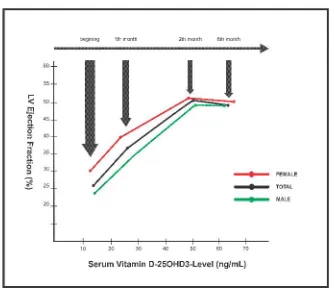
highest serum 25-OHD3 level was 93ng/ml in the course of the present study. The mean increase in the LVEF before and 8 weeks after study commencement was 26.89 ± 11.68 and 51.03±4.31%, correspondingly (P = 0.03), with no significant difference between the male and female patients (P = 0.5). The mean serum PTH decreased from the beginning of the study to the end of the eighth week (94.28±18.35 and 43.66±17.31ng/ml, respectively) (p=0.03). There was a positive correlation between increasing in mean serum 25-OHD3 and LVEF percent at the end of study (r=0.001). The LVEF became normal following the normalization of the serum 25-OHD3 level (≥ 30 ng/ml) in 6 (37.50%) patients. The LVEF increased threefold its initial value and reached 62.50% in 10 patients following the rise in serum 25-OHD3 (≥ 30 ng/ml) (Table 3). The positive trend of a concurrent rise in the serum 25-OHD3 level and the LVEF continued in all the patients until the end of the study (Figure1). The LVEF had a marked improvement in the patients with severe cardiac hemosiderosis (7/16 patients) compared with the other patients (P = 0.03). No significant difference was observed in the serum 25-OHD3 level and the LVEF at the end of the sixth month by comparison with the end of the second month (table 3) (P = 0.4). In the present study, the increasing trend of the serum 25-OHD3 level and the LVEF was positive in both sexes, with the difference between them failing to constitute statistical significance (P = 0.2) (Figure1).
Efficacy of Vitamin D in Improving Ventricular Dysfunction in Transfusion-Dependent Thalassemia Patients
232 Iran J Ped Hematol Oncol. 2018, Vol 8. No 4, 228-236 TableI. Mean ± SD of the parameters at the beginning of the study
Gender (number) Age (years) Serum Ferritin (ng/ml) Serum 25-OHD3 (ng/ml) Serum Calcium (mg/dl) Serum Phosphorous (mg/dl) Serum Magnesium (mg/ml) Serum Alkaline Phosphatase (U/L) Serum PTH (ng/ml) LVEF (%) T2* score (ms) Male (9) 12.15 ± 4.72 3859 ± 1975 13.68 ± 6.51 ٨٫٩٨ ± ٠٫٧٤ ٢٫٨٣ ± ٠٫٢٦ ١٫٨٢ ± ٠٫٤٠ 440.00 ± 160.50 99.00 ± 17.94 24.06 ± 10.20 10.94 ± 1.30 Female (7) 10.82 ± 3.65 3980 ± 2590 12.38 ± 5.25 ٩٫٤١ ± ٠٫٧٨ ٢٫٦٠ ± ٠٫٣٦ ١٫٧٥ ± ٠٫٤٢ 541.84 ± 352.15 88.30 ± 18.24 30.38 ± 12.82 12.23 ± 5.66 Total (16) 11.15 ± 3.61 ٣٩١٣ ± ٢٢٢٩ ١٣٫١٠ ± ٥٫٩١ ٩٫١٧ ± ٠٫٧٤ ٢٫٧٢ ± ٠٫٣٢ ١٫٧٩ ± ٠٫٤١ ٤٨٥٫٦٥ ± ٢٦٣ ٩٤٫٢٨ ± ١٨٫٣٥ ٢٦٫٨٩ ± ١١٫٦٨ ١١٫٥١ ± ٥٫٣٤
Table II. Blood transfusion and iron chelating protocols
Gender (number) Start of blood transfusion (months) Start of heart failure (years)
Iron Chelating Protocols
DEFERROXAMINE DEFERRIZIROX
DEFERROXAMINE + DEFERRIPIRONE DEFERRIPIRONE Male (9) 5.45 ± 2.34 9.6٠ ± ٫ ٠ 99
4 1 4 1
Female (7)
3 .٣ 5 ± ٫ ١ 67
١5 .٠ 7 ± 2.0١
1 2 2 1
Total (16) ٫ ٤ 17±3.88 (2-14) 12.75±5.89 (7-17) 5 (31%) 3 (18.75%) 6 (37.5%) 2 (12.5%)
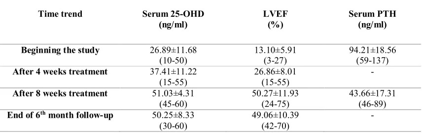
Table III. Serum 25-OHD3 levels, LVEF and serum PTH during the study
Time trend Serum 25-OHD
(ng/ml)
LVEF (%)
Serum PTH (ng/ml) Beginning the study 26.89±11.68
(10-50)
13.10±5.91 (3-27)
94.21±18.56 (59-137) After 4 weeks treatment 37.41±11.22
(15-55)
26.86±8.01 (15-55)
-
After 8 weeks treatment 51.03±4.31 (45-60)
50.27±11.93 (24-75)
43.66±17.31 (46-89) End of 6th month follow-up 50.25±8.33
(30-60)
49.06±10.39 (42-70)
-
Iran J Ped Hematol Oncol. 2018, Vol 8. No 4, 228-236 233 Figure 1. Improving of LVEF with increasing
in serum 25-OHD3 levels during the study
Discussion
The present study reassertd the high prevalence of vitamin D deficiency across the society at large and in transfusion-dependent thalassemia patients in particular. All the transfusion-dependent thalassemia patients with symptomatic heart failure participating in this study suffered from vitamin D deficiency. This study revealed that the patients with severe cardiac hemosiderosis also had a serious concomitant problem of low serum 25-OHD3 levels and high serum ferritin levels.
A positive and ascending trend of the LVEF in the present study population occurred concurrently with the administration of a high dose of vitamin D for 8 consecutive weeks. The 6 months follow-up after the successful treatment of vitamin D deficiency showed the sustained ascending trend of the LVEF and the relative improvement in the cardiac function in comparison with the baseline values in the whole study population. However, the LVEF became normal (≥55%) in less than half of the patients. There was no difference between male and female patients in terms of the rise in the LVEF and the serum 25-OHD3 level in this study. Vitamin D supplementation at conventional maintenance doses (400–800
IU/daily) in transfusion-dependent thalassemia patients is inadequate for maintaining the appropriate supply of vitamin D, and using higher doses of vitamin D is deemed a favorable and harmless method for reducing the complications of vitamin D deficiency (32, 33). Lummila et al., (1998) were first to propose the role of vitamin D deficiency in cardiac dysfunction in patients with chronic kidney failure (34). In their study, treatment with vitamin D improved the patients' cardiac function (34).
A study conducted by Wood et al., showed the association between vitamin D deficiency and increased PTH levels on the one hand and the role of LVDCC along with iron deposition in the myocardial tissue on the other in increasing the risk of cardiac dysfunction in transfusion-dependent thalassemia patients (13). Serum PTH levels (secondary hyperparathyroidism) increase in response to vitamin D deficiency to maintain calcium-cycling hemostasis (35).
The rise in PTH and iron overload in transfusion-dependent thalassemia patients with vitamin D deficiency leads to increased heart rates, intracellular myocardial calcium contents, and increased secretions of natriuretic peptide—eventually resulting in cardiac hypertrophy (22, 36, 37).
Treatment of vitamin D deficiency reduces the secretion of brain natriuretic peptide (BNP) and stops the process of cardiac hypertrophy in transfusion-dependent thalassemia patients (38, 39). Murine data show that LVDCC has a key role in the stimulation of NTBI transfer into the myocardium (22). The increase in PTH secondary to vitamin D deficiency stimulates LVDCC and causes cardiac hemosiderosis in thalassemia patients (13, 14, 22). Recent evidence suggests that increased TNF-α and reduced interleukin-10 during vitamin D deficiency are
Efficacy of Vitamin D in Improving Ventricular Dysfunction in Transfusion-Dependent Thalassemia Patients
234 Iran J Ped Hematol Oncol. 2018, Vol 8. No 4, 228-236
accompanied by a severe risk of atherosclerosis (40-42).
Schleithoff et al., found a significant reduction in PTH and TNF-α level in patients with chronic heart failure following vitamin D supplementation by comparison with a placebo group (40). Reduced cardiac function following increased PTH levels alone in the absence of rising circulating NTBI has also been explained in calcium-cycling dysfunction in the myocardium (13). In their study, Dejkhamron et al., found no significant relationship between vitamin D deficiency and cardiac iron or its function in transfusion-dependent thalassemia patients (35).
Conclusion
The high prevalence of vitamin D deficiency and its serious complications in transfusion-dependent thalassemia patients shows the need for regular biannual monitoring of serum 25-OHD3 levels with a view to diagnosing vitamin D deficiency early and thus reducing its complications. The present study showed that high-dose vitamin D supplementation for the treatment of vitamin D deficiency is an effective and healthy method for increasing the LVEF and improving the quality of life of transfusion-dependent thalassemia patients with symptomatic heart failure.
Conflict of interest
There is no conflict of interest to declare.
References
1. Holick MF. Vitamin D deficiency. N Engl J Med 2007; 357:266–281.
2. Tangpricha V, Pearce EN, Chen TC, Holick MF. Vitamin D insufficiency among free-living healthy young adults. Am J Med 2002; 112:659–662.
3. Napoli N, Carmina E, Bucchieri S, Sferrazza C, Rini GB, Di Fede G. Low serum levels of 25-hydroxy vitamin D in adults affected by thalassemia major or intermedia. Bone 2006; 38:888–892.
4. Vogiatzi MG, Macklin EA, Trachtenberg FL, Fung EB, Cheung AM, Vichinsky E, et al. Differences in the prevalence of growth, endocrine and vitamin D abnormalities among the various thalassaemia syndromes in North America. Br J Haematol 2009; 146 :546-556.
5. Vogiatzi MG,Macklin EA,Fung EB, Cheung AM, Vichinsky E, Olivieri N, et al. Bone disease in thalassemia: A frequent and still unresolved problem. J Bone Miner Res 2009; 24: 543–557. 6. Fung EB, Aguilar C, Micaily I, Haines D, Lal A. Treatment of vitamin D deficiency in transfusion-dependent thalassemia. Am J Hematol 2011; 86:871– 873.
7. Shamshirsaz AA, Bekheirnia MR, Kamgar M, Pourzahedgilani N, Bouzari N, Habibzadeh M, et al. Metabolic and endocrinologic complications in beta-thalassemia major: a multicenter study in Tehran. BMC Endocr Disord2003; 12; 3(1):4-10.
8. Singh K, Kumar R, Shukla A, Phadke SR, Agarwal S. Status of 25-hydroxyvitamin D deficiency and effect of vitamin Dreceptor gene polymorphisms on bone mineral density in thalassemia patients of North India. Hematology 2012; 17:291296-291299.
9. Nakavachara P, Viprakasit V. Children with hemoglobin E/ßthalassemia have a high risk of being vitamin D deficient even if they get abundant sun exposure: A study from thailand. Pediatr Blood Cancer 2013;; 60(10):1683-1688. 10. Soliman A, De Sanctis V, Yassin M. Vitamin D status in thalassemia major: An update. Mediterr J Hematol Infect Dis 2013; e2013057-e2013064.
11. Soliman A, Adel A, Wagdy M, Al Ali M, ElMulla N. Calcium homeostasis in 40 adolescents with beta-thalassemia major: a case-control study of the effects of intramuscular injection of a megadose of cholecalciferol. Pediatr Endocrinol Rev 2008;6 ( 1):149-154.
Iran J Ped Hematol Oncol. 2018, Vol 8. No 4, 228-236 235 12. Soliman AT, Adel A, Wagdy M,
Alali M, Aziz Bedair EM. Manifestations of severe vitamin D deficiency in adolescents: effects of intramuscular injection of a megadose of cholecalciferol. J Trop Ped 2011; 57:303-306.
13. Wood JC, Claster S, Carson S, Menteer JD, Hofstra T, Khanna R, et al. Vitamin D deficiency, cardiac iron and cardiac function in thalassemia major. Br J Haematol 2008; 141:891-894.
14. Lowry F. Vitamin D, heart dysfunction tied in thalassemia. Fam Pract News2008; 1:1-4.
15. Pfeifer M, Begerow B, Minne HW. Vitamin D and muscle function. Osteoporos Int 2002; 13:187–194.
16. Chiu KC, Chu A, Go VL, Saad MF. Hypovitaminosis D is associated with insulin resistance and beta cell dysfunction. Am J Clin Nutr2004; 79:820–825.
17. Wood JC, Enriquez C, Ghugre N, Otto-Duessel M, Aguilar M, Nelson MD, et al. Physiology and pathophysiology of iron cardiomyopathy in thalassemia. Ann N Y Acard Sci 2005; 1054:386-395. 18. Cogliandro T, Derchi G, Mancuso L, Mayer MC, Pannone B, Pepe A, et al. . Giudeline recomendations for heart complications in thalassemia major. J Cardiovasc Med 2008; 9:515-525.
19. Pennell DJ, Porter JB, Cappellini MD, El-Beshlawy A, Chan LL, Aydinok Y, et al. Efficasy of deferasirox in reducing and preventing cardiac iron overload in beta-thalassemia. Blood 2013; 128:281-230.
20. Kirk P, Roughton M, Porter JB, Walker JM, Tanner MA, Patel J, et al. Thalassaemia major and the heart, a toxic cardiomyopathytamed? Heart J 2013; 99:827-834.
21. Claster S, Wood JC, Noetzli L, Carson SM, Hofstra TC, Khanna R, et al. Nutritional deficiencies in iron overload patients with hemoglobinopathies. Am J Hematol 2009; 84:344-348.
22. Oudit GY, Sun H, Trivieri MG, Koch SE, Dawood F, Ackerley C, et al. L-type Ca(2+) channels provide a major pathway for iron entry into cardiomyocytes in iron-overload cardiomyopathy. Nat Med2003; 9:1187– 1194.
23. Gibbons RJ, Abrams J, Chatterjee K, Daley J, Deedwania PC, Douglas JS, et al. ACC/AHA 2002 guideline update for the management of patients with chronic stable angina J Am Coll Cardiol 2003;41(1):159-68.
24. Anderson LJ, Holden S, DavisB, Prescott E, Charrier CC, Bunce NH, et al. Cardiovascular T2-star (T2*) magnetic resonance for the early diagnosis of myocardial iron overload. Eur Heart J 2002; 22:2171-2179.
25. Carpenter JP, He T, Kirk P, Roughton M, Anderson LJ, de Noronha SV, et al. On T2* magnetic resonance and cardiac iron. Citculation 2011; 123:1519-1528.
26. Kirk P, Roughton M, Porter JB, Walker JM, Tanner MA, Patel J, et al. Cardiac T2* magnetic resonance for prediction of cardiac complications in thalassemia major. Circulation 2009; 120:1961-1968.
27. Maggio A, Vitrano A, Calvaruso G, Barone R, Rigano P, Mancuso L, et al. Serial echocardiographic left ventricular ejection fraction measurements: a tool for detecting thalassemia major patients at risk of cardiac death. Blood Cells Mol Dis 2013; 50:241-246.
28. Vogel M, Anderson LJ, Holden S, Deanfield JE, Pennell DJ, Walker JM. Tissue Doppler echocardiography in patients with thalassemia detects early myocardial dysfunction related to myocardial iron overload. Eur Heart J 2013; 24:113-119.
29. Condamine L, Vztovsnik F, Friedlander G, Menaa C, Garabedian M. Local action of phosphate depletion and insulinlike growth factor–I on in vitro
Efficacy of Vitamin D in Improving Ventricular Dysfunction in Transfusion-Dependent Thalassemia Patients
236 Iran J Ped Hematol Oncol. 2018, Vol 8. No 4, 228-236
production of 1,25dihydroxyvitamin D3 by cultured mammalian kidney cells. J Clin Invest 1994;94:1673-1679.
30. Wright NM, Papadea N, Wentz B, Hollis B, Willi S, Bell NH. Increased serum 1,25 dihydroxyvitamin D after growth hormone administration is not parathyroid hormone–mediated. Calcif Tissue Int 1997; 61:101-103.
31. Dawson-Hughes B, Heaney RP, Holick MF, Lips P, Meunier PJ, Vieth R. Estimates of optimal vitamin D status. Osteoporos Int 2005; 16:713–716.
32. Soliman A, Adel A, Wagdy M, Al Ali M, ElMulla N. Calcium homeostasis in 40 adolescents with beta-thalassemia major: A case-control study of the effects of intramuscular injection of a megadose of cholecalciferol. Pediatr Endocrinol Rev 2008; 6 :149–154.
33. Fung EB, Aguilar C, Micaily I, Haines D, Lal A. Treatment of vitamin D deficiency in transfusion-dependent thalassemia. Am J Hematol 2011; 86(10):871-873.
34. Lemmila S, Saha H, Virtanen V, Ala-Houhala I, Pasternack A. Effect of intravenous calcitriol on cardiac systolic and diastolic function in patients on hemodialysis. Am J Nephrol1998; 18:404– 410.
35. Dejkhamron P, Wejaphikul K, Mahatumarat T, Silvilairat S, Charoenkwan P, Saekho S, Unachak K. Vitamin D deficiency and its relationship with cardiac iron and function in patients with transfusion-dependent thalassemia at
Chiang Mai University Hospital. Pediatr Hematol Oncol 2018; 35(1):52-59.
36. Pilz S, Tomaschitz A, Drechsler C, Dekker JM, März W. Vitamin D deficiency and myocardial disease. Mol Nutr Food Res 2010; 54:1103–1113. 37. Pilz S, Tomaschitz A, März W, Drechsler C, Ritz E, Zittermann A, et al. Vitamin D, cardiovascular disease and mortality. Clin Endocrinol 2011; 75:575– 584.
38. Tamez H, Zoccali C, Packham D, Wenger J, Bhan I, Appelbaum E, et al. Vitamin D reduces left atrial volume in patients with left ventricular hypertrophy and chronic kidney disease. Am Heart J 2012; 164:902–909.
39. Ambarwati L, Endah
Rahayuningsih S, Setiabudiawan B. Association between vitamin D levels and left ventricular function and NT-proBNP levels among thalassemia major children with iron overload. Ann Pediatr Cardiol. 2016; 9(2): 126-131.
40. Schleithoff S, Zittermann A, Tenderich G, Berthold HK, Stehle P, Koerfer R. Vitamin D supplementation improves cytokine profile in patients with congestive heart failure: A double blind, randomized, placebo-controlled trial. Am J Clin Nutr 2006; 83:754–759.
41. Camil F, Rogal K, Sypniewska G. Vitamin D and its role in cardiovascular disease. JLM 2010; 46:75–79.
42. Norman PE, Powell JT. Vitamin D and cardiovascular disease. Circ Res 2014; 114:379–393.