Different Methods for Isolation and Preliminary Identification of
Azotobacter
Z. Mazinani1 ; M. Aminafshar2* ; A. Asgharzadeh3 ; M. Chamani4
1: Department of Biotechnology, Science and Research Branch, Islamic Azad University, Tehran, Iran
2: Department of Genetics & Animal Breeding, Faculty of Agriculture and Natural Resources, Science and Research Branch, Islamic Azad University, Tehran, Iran
3: Department of Soil Biology, Soil and Water Researches Institute, Karaj, Iran
4: Department of Animal Science, Faculty of Agriculture and Natural Resources, Science and Research Branch, Islamic Azad University, Tehran, Iran
Received: December, 2, 2012 Accepted: May, 1, 2013
ABSTRACT
Plant growth promoting rhizobacteria (PGPR) are known to influence plant growth by various direct or indirect mechanisms. Thirty–two strains were isolated from 15 soils sampled in central Iran, by using and comparing three different methods. The screening of soil samples by means of soil paste– plate method combined with isolation on mannitol agar proved to be the best strategy in terms of reliability and selectivity. These test isolates were biochemically characterized. These isolates were screened in vitro and identified by using BIBI(Bioinformatics Bacterial Identification Tool). BIBI was designed to automate DNA sequence analysis for bacterial identification in the different fields. BIBI relies on the use of BLAST and CLUSTAL W programs applied to different subsets of sequences extracted from GenBank. These sequences are filtered and stored in a new database, which is adapted to bacterial identification.
INTROBUCTION
Plant growth promoting rhizobacteria (PGPR) are a heterogeneous group of bacteria that can be found in the rhizosphere, at root surfaces and in association with roots, which can improve the extent or quality of plant growth directly and or indirectly. In last few decades a large array of bacteria including species of Pseudomonas, Azospirillum, Azotobacter,
Klebsiella, Enterobacter, Alcaligens,
Arthobacter, Burkholderia, Bacillus and Serratia have reported to enhance plant growth
(Kloepper et al., 1989; Okon & Labandera-
Gonzalez, 1994; Glick, 1995). The direct promotion by PGPR entails either providing the plant with a plant growth promoting substances that is synthesized by the bacterium or facilitating the uptake of certain plant nutrients from the environment. The indirect promotion of plant growth occurs when PGPR lessen or prevent the deleterious effect of one or more phytopathogenic microorganisms. The exact mechanisms by which PGPR promote plant growth are not fully understood, but are thought to include (i) the ability to produce or change the concentration of plant growth regulators like indoleacetic acid, gibberellic acid, cytokinins and ethylene (Arshad & Frankenberger, 1993; Glick, 1995), (ii) asymbiotic N2 fixation (Boddey & Dobereiner, 1995), (iii) antagonism against phytopathogenic microorganisms by production of siderophores (Scher & Baker,
1982), antibiotics (Shanahan et al., 1992) and
cyanide (Flaishman et al., 1996), (iv) solubilization of mineral phosphates and other nutrients (De Freitas et al., 1997; Gaur, 1990). Most popular bacteria studied and exploited as biocontrol agent includes the species of fluorescent Pseudomonas and Bacillus. Some PGPR may promote plant growth indirectly by affecting symbiotic N2 fixation, nodulation or nodule occupancy (Fuhrmann & Wollum, 1989). However, role of cyanide production is contradictory as it may be associated with deleterious as well as beneficial rhizobacteria (Bakker & Schippers, 1987; Alstrom & Burns, 1989).
In addition to these traits, plant growth promoting bacterial strains must be rhizospheric competent, able to survive and
colonize in the rhizospheric soil (Cattelan et
al., 1999). Unfortunately, the interaction between associative PGPR and plants can be unstable. The good results obtained in vitro cannot always be dependably reproduced under field conditions (Chanway & Holl, 1993;
Zhender et al., 1999). The variability in the
performance of PGPR may be due to various environmental factors that may affect their growth and exert their effect on the plant. The environmental factors include climate, weather conditions, soil characteristics or the composition or activity of the indigenous microbial flora of the soil. To achieve the maximum growth promoting interaction between PGPR and nursery seedlings it is important to discover how the rhizobacteria exerting their effects on plant and whether the effects are altered by various environmental factors, including the presence of other microorganisms (Bent et al., 2001).
MATERIALS AND METHODS
SOIL SAMPLING
Soil samples were collected during Spring in different regions of central Iran from cultivated soils. Samples were withdrawn at a depth of 10–15 cm below the surface, collected into sterile vials as described by Kole & Altosaar (1988), sieved through a 4–mm–mesh sieve, and stored at 4°C.
ISOLATION
Three different isolation methods were used: (a) streaking of serial soil dilutions on plates containing Ashby medium containing (per 1l):
20 g mannitol, 0.2 g K2HPO4, 0.2 g MgSO4–
7H2O, 0.2 g NaCl, 0.1 g K2SO4, 5 g CaCO3,
single soil grains (Pochon, 1954) methods realised as follows: about 30–50 g of each soil sample were accurately mixed with 20% (v/w) of sterile water with 0.5–1.0 g of mannitol, 0.5
g of CaCO3, 0.12 ml of 10% aqueous K2HPO4
solution, 0.12 ml of 10% aqueous MgSO4
solution. The soil paste, prepared in a porcelain mortar, was transferred and pressed inside a petri dish with a sterile spatula to obtain a smooth and levelled surface. After 3–7 days incubation at 27–30°C, the soil paste–plates presenting growth of Azotobacter were revealed by the appearance of slimy, glistening colonies, turning brown with aging if produced by the species A. chroococcum. Subsequently, in order to carry out isolation, soil samples resulted positive for the presence of these free– living nitrogen–fixing bacteria were subjected to sowing of single grains on the Mannitol-agar medium proposed by Pochon (1954), containing (per 1l): 10 g mannitol, 0.5 g
K2HPO4, 0.2 g MgSO4 .7H2O, 0.1 g NaCl, 1.0
g yeast extract, 3.0 g CaCO3, 20 g agar
(Becking, 1981).
All the isolates were purified by streaking on NA plates. Long–term storage of the purified isolates was at –80°C in the LG broth medium with 50% (w/v) glycerol.
SCREENING OF ISOLATES
The bacterial isolates were characterized by their cultural conditions, morphological and biochemical characteristics (utilization of glucose, fructose, maltose, raffinose, trehalose, growth at diffrerent temperatures, catalase, oxidase, Gram-stain reaction) using standard methods (Cappuccino & Sherman, 1992).
MOLECULAR ANALYSIS
For molecular analysis, one isolete was grown for 2–3 days on LG. Crude template DNA was extracted using alkaline lysis method(Rademaker & de Bruijin, 1997). The 16S rRNA gene was amplified by means of universal primers 27f and 1495r (Weisburg et al., 1991). The PCR reaction was run for 35 cycles as follows: denaturation at 94°C for 1 min, annealing at 55 °C for 1 min, elongation at 72 °C for 2 min. An initial denaturation step at 95 °C for 4 min and a final extension step at
72 °C for 15 min was also performed and the PCR product was sequenced by biobasic company(Canada).
BIOINFORMATICS ANALYSIS
For bioinformatic analysis, we used a specific bioinformatics tool dedicated to bacterial identification (BIBI, for Bioinformatics Bacterial Identification) in order to simplify sequences analysis within a bacterial identification framework. BIBI fully automates and speeds up different operations for the treatment of sequences. BIBI, which can be
accessed at http://pbil.univ –lyon1.fr/bibi/,
enables the identification of a microorganism from a gene fragment sequence of previously described cultured bacteria.
The program implements a chaining of two well-known tools: BLAST (Altschul et al., 1997) and CLUSTAL W (Thompson et al., 1994). CLUSTAL W runs are accelerated by the use of prealigned BLAST results. BIBI is written in standard ANSIC language, and the interface is implemented in HTML–PHP. Analysis of an unknown sequence proceeds in four phases: search for matching sequences, sequence extraction and parsing, sequence alignment, and display of results.
RESULTS AND DISCUSSION
SOIL SAMOLING
The sampling strategy described in this work was chosen taking into account the different parameters influencing the presence of azotobacteria in soil. Since distribution of Azotobacter in the rhizosphere is not dependent on the type of plant (Kole & Altosaar, 1988) soil samples were indifferently collected from the rhizosphere of gramineum.
ISOLATION METHODS
The three methods utilised in the present work were described by different authors as feasible for Azotobacter isolation. Method ‘a’ allowed the direct isolation of Azotobacter like colonies on selective Ashby medium from 18 out of 15 soil samples utilised. All members of genus
Azotobacter produced slimy, glistening,
Method ‘b’ was tested on 15 soil samples. As expected, growth of Azotobacter strains on Winogradsky solution was revealed by an increase of turbidity and the appearance of a thin pellicle on the liquid surface. Moreover, growth of most Azotobacter was accompanied by the production of diffusible pigments. Although, the results observed with the strains in Winogradsky solution were unambiguous, those obtained after inoculation with soil sample dilutions were not so easily understandable. Indeed, growth of different microorganisms (e.g. aerobic and microaerophilic species in the nearby of the liquid surface) led to the production of a milky and creamy pellicle, browning with aging, and to a significant increase in turbidity which rendered the observation of diffusible pigments not possible. Consequently, due to the impossibility to individuate the positive tubes for Azotobacter, pellicles coming from all the tubes showing growth were streaked onto Ashby medium. In this way, two objectives were contemporary pursued: the individuation of Azotobacter like colonies and the achievement of pure cultures. As a result, 14 Azotobacter like colonies were isolated from 15 soil samples screened.
According to method ‘c’, soil samples to be employed in the isolation step were selected by
means of the soil paste–plate technique, thanks to the appearing of slimy and glistening colonies. upon the smoothed soil paste surface. 21 out of 15 soil samples screened were therefore selected and utilised for Azotobacter isolation onto mannitol medium, through the direct sow of single soil grains. The utilisation of this combined method led to the isolation of 15 Azobacter–like cultures.
SCREENING OF THE ISOLATES
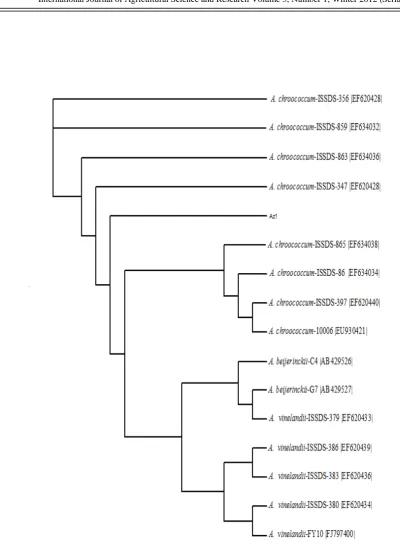
On the basis of cultural, morphological and biochemical characteristics a total of 32 soil isolates were grouped into Azotobacter (Table 1) as described in Bergey’s Manual of Determinative Bacteriology (Holt et al., 1994). Also, Results obtained of sequencing indicated
that strain Az1 belonging to genus
Azotobacter. Phylogenetic tree are displayed by Java applet: Jalview (version 1.7) [http://www2.ebi.ac.uk/_michele/jalview/])
(Fig. 1). The tree revealed that Az1 is similar
with strains A. chroococcum ISSD- 356, A.
chroococcum ISSD- 859, A. chroococcum
ISSD- 863, A. chroococcum ISSD- 347, A.
chroococcum ISSD- 865, A. chroococcum
ISSD- 86, A. chroococcum ISSD- 397, A.
Table 1: Biochemical and morphologic characterization of the test isolates
Azotobacter Biochemical and morphologic characters
32 Number of isolates
-
Gram reaction
+
Catalase
+
Oxidase
Carbohydrate utilization:
+
Glucose
+
Fructose
+
Maltose
+
Raffinose
+
Trehalose
+
CONCLUSION
This research work firstly aimed to compare three different methods reported in literature for the isolation of free N-fixing bacteria from soil samples, in order to individuate the most effective one. In second instance, it aimed to verify whether LG medium, described up to now as a selective substrate for the isolation of Azotobacter can be successfully employed to screen soil isolates for a presumptive recognition of microorganisms belonging to the genus of interest.
In conclusion, our results showed that the most reliable strategy for the isolation and preliminary identification of Azotobacter is given by the combination of paste–plate and soil grains sowing methods followed by screening on LG.
ACKNOWLEDGMENTS
This research was supported by the Islamic Azad University, Science and Research Branch. We would like to express our gratitude to the expert of the Biotechnology Laboratory, Yashar Madadkar.
REFERENCES
1. Altschul, S. F., Madden, T. L., Schaffer, A. A., Zhang, J., & Lipman, D. J. (1997). Gapped BLAST and PSI-BLAST: a new generation of protein database search programs. Nucleic Acids Res, 25(3), 3389–3402.
2. Alstrom, S., & Burns, R.G. (1989). Cyanide production by rhizobacteria as a possible mechanism of plant growth inhibition. Biol Fertil Soil, 7(1), 232–238.
3. Arshad, M., & Frankenberger, J. r.(1993). Microbial production of plant growth regulators. Soil Microbial Ecology. New York: Marcel and Dekker.
4. Augier, J.(1956). A propos de la numeration des Azotobacter en milieu liquide. Annales de l’Institut Pasteur, 66(44), 759–765.
5. Bakker, A.W., & Schippers, B. (1987). Microbial cyanide production in the rhizosphere in relation to potato yield
reduction and Pseudomanas sp. mediated plant
growth stimulation. Soil Biol Biochem, 19(3),
451–457.
6. Becking, J.H. (1981). The family Azotobacteraceae. Berlin: Springer, Heidelberg.
7. Bent, E., Tuzun, S., Chanway, C.P., & Enebak, S. ( 2001). Alterations in plant growth and in root hormone levels of lodgepole pines inoculated with rhizobacteria. Can. J. Microbiol, 47(2), 793–800.
8. Boddey, R.M., Dobereiner, J., 1995. Nitrogen fixation associated with grasses and cereals: recent progress and perspectives for the future. Fert. Res. 42, 241–250.
9. Brown, M.E., Burlingham, S.K., & Jackson, R.M. (1962). Studies on Azotobacter species in soil. I. Comparison of media and techniques for counting Azotobacter in soil. Plant and Soil, 17(4), 309–319.
10. Cappuccino, J.C., & Sherman, N. (1992). In: Microbiology: A Laboratory Manual, New York: Benjamin/Cummings
11.Cattelan, A.J., Hartel, P.G. & Fuhrmann, J.J. (1999). Screening of plant growth_promoting rhizobacteria to promote early soybean growth. Soil Sci, 63(3), 1670– 1680.
12. Chanway, C.P. & Holl, F.B. (1993). First year yield performance of spruce seedlings inoculated with plant growth promoting rhizobacteria. J. Microbiol, 39(4), 1084–1088.
13. De Freitas, J.R., Banerjee, M.R., & Germida, J.J. (1997). Phosphate solubilizing rhizobacteria enhance the growth and yield but not phosphorus uptake of canola. Biol. Fertil. Soil, 24(5), 358–364.
producing strains of Pseudomonas putida. Plant Microbe Interact, 9(4), 642–645.
15. Fuhrmann, J.J., & Wollum II, A.G. (1989).
Nodulation competition among Bradyrhizobium japonicum strains as influenced by rhizosphere bacteria and iron availability. Biol. Fertil. Soil, 7(4), 108–112.
16. Gaur, A.C. (1990). Physiological functions of phosphate solubilizing microorganisms. Phosphate Solubilizing Microorganisms as
Biofertilizers. Omega Scientific Publishers,
5(1), 16–72.
17. Glick, B.R. (1995). The enhancement of plant growth by free living bacteria. Can. J. Microbiol, 41(2), 109–114.
18. Holt, J.G., Krieg, N.R., Sneath, P.H.A., Staley, J.T., & Williams, S.T. (1994). Bergy’s Manual of Determinative Bacteriology. USA: Williams and Wilkins.
19. Kloepper, J.W., Lifshitz, R., &
Zablotowicz, R.M. (1989). Free-living bacterial inocula for enhancing crop productivity. Trends Biotechnol, 7(3), 39–43.
20. Kole, M.M., & Altosaar, I. (1988). Distribution of Azotobacter in Eastern Canadian soils and in association with plant rhizospheres. Canadian Journal of Microbiology, 34(3), 815– 817.
21. Okon, Y., & Labandera-Gonzalez, C.A.
(1994). Agronomic applications of
Azospirillum. Australia: Common wealth Scientific and Industrial Research Organization.
22. Pochon, J. (1954). Manuel technique analysis microbiologiqal the soil. Paris: Masson.
23. Rademaker, J.L.W., & de Bruijin, F.J. (1997). Characterization and classification of microbes by rep-PCR genomic finger printing and computer-assisted pattern analyse. caetano-Anolles, 6(2), 567–585.
24. Scher, F.M., & Baker, R. (1982). Effect of Pseudomonas putida and a synthetic iron chealator on induction of soil suppressiveness to Fusarium wilt pathogens. Phytopathology, 72(3), 1567–1573.
25. Shanahan, P., O’Sullivan, D.J., Simpson, P., Glennon, J.D., & O’Gara, F. (1992). Isolation of 2,4-diacetylphlorogucinol from a fluoroscent pseudomonad and investigation of physiological parameters influencing its
production. Appl. Environ. Microbiol. 58(3),
353–358.
26. Thompson, J. D., Higgins, D. G., & Gibson, T. J. (1994). CLUSTAL W: improving the sensitivity of progressive multiple sequence alignment through sequence weighting, position-specific gap penalties and weight matrix choice. Nucleic Acids Res, 22(3), 4673–4680.
27. Weisburg, W.G., Barns, S.M., Pelletier, D.A., & Lane, D.J. (1991). 16S ribosomal DNA amplification for phylogenetic study. J. Bacteriol, 173(3), 697– 703.
28. Zhender, G.W., Murphy, C., Sikora, J.F., Kloepper, E.R., & Polston, J.E. (1999). Microbe-induced resistance against pathogens and herbivores: evidence of effectiveness in
agriculture. Biochemistry, Ecology and