Original Article
A Study on the Ocular Infection with Rabies Virus in Mouse
Atefeh Pilehvar Zavareh
1, Mohammadreza Mahzounieh
1,
Mohammadreza Shirzadi
2, Rouzbeh Bashar
3,
Alireza Zavareh
3, Nader Howaizi
3, Firouzeh Farahtaj
3, Alireza Janani
3,
*Alireza Gholami
31
Department of Pathobiology, Faculty of Veterinary Medicine and Research, Institute of Zoonotic Diseases,
University of Shahrekord, Shahrekord, Iran;
2
Department of Zoonosis, CDC of Iran, Ministry of Health, Tehran, Iran;
3
WHO Collaborating Centre for Reference and Research on Rabies, Pasteur Institute of Iran, Tehran, Iran.
Received Jan 04, 2015; accepted Jan 26, 2015
INTRODUCTION
Rabies is a zoonotic disease, which causes more than 60,000 human deaths around the world annually [1]. Rabies virus (RABV), belongs to the genus Lyssavirus of the
Rhabdoviridae family. The most common mode of rabies virus transmission is through a bite or contact of broken skin with virus, containing saliva of a rabid animal. Other documented routes of virus transmission include contamination of mucous membranes (i.e. eyes, nose, and mouth) with the saliva, aerosol transmission, and corneal transplantations [2].
It has been shown that airborne rabies transmission is possible in various species of caged carnivores kept in caves, which are home to millions of bats. However, it is assumed that the presence of a very large number of bats in an unventilated area is necessary for airborne transmission of rabies virus [3]. Human contamination with rabies virus through the airborne route has also been reported in unusual circumstances. Aerosolized rabies virus has been inculpated for laboratory accidents [3]. Some reports have shown that rabies virus is also capable of infecting the central nervous system (CNS) of various mammals after intranasal instillation. Experimental studies have indicated that challenge virus standard (CVS) strain of rabies virus can selectively infect olfactory receptor cells, but not the respiratory epithelium, and spreads into the brain along the olfactory pathways in mice [4]. Oral transmission of rabies
virus may occur naturally as a result of consumption of infected carcasses by wildlife animals. It could also be important when raw meat of a rabid animal is eaten [5]. Scientists have succeeded to show infection of laboratory animals (mice, hamsters, guinea pigs, and rabbits) of different ages, experimentally infected with CVS following oral administration of rabies virus [6]. Correa-Giron et al. showed that CVS and street rabies virus (SRV) strains produced infection in mice following ingestion of virus laden brain tissue. They suggested that infection can occur through the buccal and lingual mucosa as well as lung and the intestine [7].
Laboratory workers during work with rabies virus and veterinarians during examination and surgery of rabid animals may be exposed to saliva or other infectious fluids splashing into their eyes. The question that arises is how high the risk of virus penetration and spread into ocular neurons Introduction: The most common mode of rabies virus transmission is through a bite wound or contact of broken skin with
saliva of a rabid animal. Various other routes of virus transmission include exposure of mucous membranes (i.e. eyes, nose, and
mouth) to infected saliva of a rabid animal, aerosol transmission, and corneal transplantation. Laboratory workers during work with rabies virus and veterinarians during examination and surgery of rabid animals may be at risk for exposure to saliva or other infectious fluids splashing into their eyes. The aim of this study was to investigate the possibility of ocular rabies pathogenesis in mice as an animal model. Our results will determine if rabies virus strains challenge virus standard (CVS) and street rabies virus
(SRV) are able to infect the central nervous system (CNS) of mice through the ocular route. Methods: This study was performed
in two experiments. In experiment 1, different lethal doses of fixed rabies virus strain CVS were made and instilled into both eyes of test mice. In experiment 2, concentrated rabies virus strains CVS and SRV were instilled into both eyes of the test mice. Mice in all groups were kept for 3 months and tested by fluorescent antibody test (FAT) for detection of the presence of viral
antigen in brain tissue. Results: Mice with ocular instillation of fixed and street rabies viruses developed no clinical symptoms
of rabies and all were healthy and alive during the 3 month observation period. The FAT results were negative in both
experiments. Conclusion: Our results suggest that CVS and SRV viruses are not able to infect the CNS of mice via intact
conjunctiva and cornea. J Med Microbiol Infec Dis, 2014, 2 (2): 61-65.
Keywords: Eye, Infection, Mouse, Rabies Virus.
*Correspondence: Alireza Gholami
WHO Collaborating Centre for Reference and Research on Rabies, Pasteur Institute of Iran, No. 69, Pasteur Ave, Tehran, Iran, 1316943551.
Email:[email protected]
Tel: +98 (21) 66403496 Fax:+98 (21) 66480777
and SRV was isolated from brain sample of a rabid wolf, which was confirmed by fluorescent antibody test (FAT) at WHO collaborating center (WHOCC) for reference and research on Rabies, Pasteur Institute of Iran.
Titration of virus strains. Serial tenfold dilution of CVS stock from 10-5 to 10-7, was made in diluents (deionized
water with 2% horse serum) and 0.03 mL of each dilution was inoculated intracerebrally (IC) into 5 mice. Mice were euthanized after 5 days or more post-infection at the paralytic stage. Their brain was removed and rabies infection was confirmed by FAT. The LD50 was calculated according to the
Spearman-Kärber method [8].
A 10% suspension of SRV was prepared by homogenizing the wolf brain sample in an isotonic buffered solution containing 1560 IU/ml penicillin and 500 IU/ml streptomycin antibiotics. A Groups of 10 mice aged 21 days, weighing 12-14 g were inoculated IC with 0.03 ml of suspension. Moribund mice were euthanized and their brains were collected. A 10% suspension was prepared by homogenizing the brains in diluents (deionized water with 2% horse serum). The homogenate was centrifuged at 700 g for 15 min using a table top refrigerated centrifuge to remove debris, and the supernatant was collected and stored at -80°C until used. To determine the mouse intracerebral LD50 (MIC
LD50) for 10% suspension of subcultured brain samples, 4
dilutions ranging from 10-3 to 10-6 were prepared for each
virus suspension in deionized water with 2% horse serum as
MIC LD50 of CVS, as shown in Table 1. All mice were
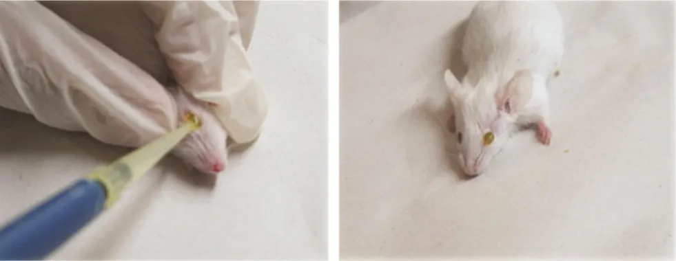
anesthetized with a mixture of Ketamine 10% (2-(2-chlorophenyl)-2-(methylamino)-cyclohexanone) (50 mg/kg) and Xylazine 2% (N-(2,6-Dimethylphenyl)-5,6-dihydro-4H -1,3-thiazin-2-amine) (10 mg/kg) prior to instillation. In each group, 10 µl of the virus solution was instilled into eyes of mice using a sterile pipette tip (Figure 1). In the first group, 1 LD50 of CVS stock (dilution= 1:3,200,000), in the second
group, 25 LD50 (dilution= 1:126,000) and in the third group,
50 LD50 (dilution= 1:63,000) were used. The mice in all
groups were kept in ventral recumbency position for 15 min for extending the contact time of viruses with cornea and increasing the possible infection (Figure 2). Mice of the control group were received 10 µl of normal saline into each eye after they were anesthetized.
Experiment2. The experiment 2 was performed with a higher dose of CVS and sample size. SRV was also used as the wild-type strain of rabies virus in this experiment. A total of 46 mice were divided into 2 groups. Ten µl of 20% suspension of CVS strain containing 106.5 MLD
50 and the
same volume of 10% suspension of SRV strain containing 104.7 MLD
50 were dropped in both eyes of each animal in
group 1 and group 2, respectively. Five mice were considered as negative control group, which received 10 µl of normal saline into each eye and were kept under daily observation for 3 months to ensure sufficient incubation time for street virus.
Table 1. Results of experiment 1
Group No. of mice Virus LD50/0.03 ml Deaths/ Exposed
1 5 CVS 1 0/5 (0%)
2 5 CVS 25 0/5 (0%)
3 5 CVS 50 0/5 (0%)
4 5 Normal Saline - 0/5 (0%)
Note: Various Lethal Doses of fixed rabies virus CVS were used to infect mice by ocular instillation. In control group, normal saline was used. No deaths were observed either due to rabies or accident during surveillance period.
Table 2. Results of experiment 2
Group No. of mice Virus LD50/0.03 ml Deaths/Exposed
1 23 CVS Stock (20% suspension) 106.5 0/23 (0%)
2 23 SRV Stock (10% suspension) 104.7 0/23 (0%)
3 5 Normal Saline - 0/5 (0%)
Note: In both groups of mice under study, no deaths were observed either due to rabies or accidental during three months of observation.
Fig. 1. Intraocular instillation of virus suspension into eye of an anaesthetized mouse using a sterile pipette
Fig. 2. Mouse remaining in ventral recumbency for 15 min after instillation
Fluorescent Antibody Test (FAT). After 3 months of observation, 2 mice of each group in experiment 1, and 5 mice of each group in experiment 2 were selected randomly and euthanized. FAT was performed on brain samples of mice based on a technique previously described by DJ Dean
et al. [8]. Briefly, smears of mice brain sample were prepared by the impression method on a clean slide and fixed for 30 min in cold acetone. The slides were covered with anti-rabies nucleocapsid rabbit immunoglobulin G conjugated with
fluorescein isothiocyanate (BIO-RAD,
Marnes-La-Coquette-France) and incubated at 37°C in a humid chamber for 1 h. Then, slides were washed twice with phosphate-buffered saline and observed by fluorescent microscope (Nikon Eclipse TE200; Nikon Corp, Tokyo, Japan).
RESULTS
Virus titration experiments determined the LD50s of CVS
and SRV suspensions as 106.5 and 104.7, respectively. In
experiment 1, none of the infected mice developed rabies following ocular instillation of different LD50s of CVS
(Table 1). During 3 months of observation, no signs of rabies were observed in mice inoculated either with CVS or SRV. The second experiment gave similar results comparable to the first experiment. All mice in both experiments, including control and inoculated groups were alive and healthy. FAT results were negative for mice brains in both experiments. No viral antigens were detected in brain samples of mice.
DISCUSSION
Salivary glands of experimentally infected dogs and cats with detectable virus which contain geometric mean titers ranging from 3,400 to 386,000 mouse LD50/g. Saliva of rabid
animal could be highly infectious if it comes to contact with sensitive area, such as mucosa or broken skin [9]. Therefore, it could be potentially dangerous to cause rabies infection through these routes as it has been mentioned previously. In this study, we have designed an experiment to test whether
contamination of eye with rabies virus could be a route of infection in mouse model. We hypothesized whether rabies virus can enter and penetrate into eye neurons through any part of intact eye, including cornea or mucosa. In experiment 2, certain conditions of the experiment 1 were applied to repeat the research with SRV and check the dose dependency of the CVS. The results of this study showed that the suspension containing 104.7 LD
50/0.03 ml of SRV from a
wild type strain originated from a rabid wolf or 106.5
LD50/0.03 ml of the fixed virus strain CVS could not infect
mice by absorption via ocular instillation. Previous reports have indicated that high concentrations of virus are necessary to infect mouse through oral or nasal routes [7]. However, the results of the present study showed that ocular instillation, even with high concentrations of CVS, did not cause clinical manifestations of rabies, even after 3 months. These findings are in agreement with unpublished data from Hanlon experiments, which showed that ocular instillation by drops of a virulent canine isolate of virus did not cause rabies in Syrian hamsters [5]. Although, there are studies that show the possibility of rabies infection through oral route. Charlton and Casey indicated that absorption of CVS through the oral mucosa is minimal in mice and skunks [3]. It has been shown that the lyophilized SAG2 oral rabies vaccine is effective in immunizing captive arctic foxes [10]. There are different studies showing that there is no viral amplification or penetration in animal tissues following oral vaccination, which implies the safety of this live rabies vaccine. However, SAG2 carries a double mutant attenuated virus strain that could not totally repeal the possibility of street virus penetration through oral rout [11]. In our study, both fixed and street viruses were tested, and the results in mice suggest that contamination of ocular mucosa could not cause rabies infection. Lafay et al. demonstrated that olfactory neuroreceptors can be directly infected by CVS following its instillation in the nasal cavity, and CVS spreads into brain along the olfactory pathway [4]. So, in our study, probably nerve endings in intact cornea and conjunctiva are not as easily accessible to the virus as intranasal route.
and corneal epithelial cells are in close contact with the nervous system and the nerve connection between the cornea and its corresponding CNS segments are short. Although, infection of the cornea with the rabies virus has been well indicated in the centrifugal spread of the virus [15], the results of this study revealed that the intact cornea does not serve as an entry site for the virus.
Yoned et al. showed that anti-rabies virus antibody titer in mice intranasally immunized with concentrated rabies virus antigen (CRV) plus cholera toxin (CT), was comparable to that of mice intraperitoneally immunized twice with the same amount of CRV. High levels of IgA and IgG were detected in mice immunized intranasally with CRV/CT [16]. Mucosal vaccination stimulates sIgA production in mucosal tissues and IgG antibodies in serum [16]. The mucosal immunization has been proven to be efficient against various infectious pathogens, such as influenza virus, Newcastle disease virus, foot and mouth disease virus, Aujeszky’s disease virus, HIV and Ascaris suum [17-21]. In the present study, rabies virus was instilled into eye. Conjunctiva, is a part of eye mucosa, which its underlying structures are known as conjunctiva-associated lymphoid tissue (CALT). In the CALT, antigens are taken up by the follicles and presented to lymphocytes by antigen presenting cells. This leads to activation of B and T cells, which carry out the immune reaction [12, 22, 23]. In this study, CALT might have an interfering function in neutralization of rabies virus and prevention of virus entrance to the CNS of mice, since contact of conjunctiva with virus containing suspension is inevitable.
Davis et al. showed the presence of rabies virus neutralizing antibody (VNA) in bats and mice exposed through aerosol, to 3 variants of bat rabies virus. In this exposure, all bats and certain numbers of mice survived and produced detectable rabies VNA [24]. In our study, all mice survived, which were exposed to rabies virus by ocular route. Further independent experiments are necessary to demonstrate whether or not survival of mice would be related to immune response and VNA production.
Age, immune status of the host, and factors involved in species spillover have been proved to be important factors in viral neurovirulence [5]. Further investigations would be necessary to rule out the ocular mucosa as a site of virus penetration. As the mouse eye is too small and difficult to instill virus in it, rabbit eye could act as a better model for
CONFLICT OF INTEREST
The authors declare that there are no conflicts of interest associated with this manuscript.
REFERENCES
1. World Health Organization [Homepage of internet]. Human and animal rabies. Available from: http://www.who.int/rabies/en/ [cited 2013 January 20].
2. Centers for Disease Control and Prevention. [Homepage of internet]. Rabies. Available from: http://www.cdc.gov/rabies/, [cited 2014 April 26].
3. Charlton KM, Casey GA. Experimental oral and nasal transmission of rabies virus in mice. Can J Comp Med. 1979; 43 (1): 10-5.
4. Lafay F, Coulon P,Astic L, Saucier D, Riche D, Holley A,
Flamand A. Spread of the CVS strain of rabies virus and of the avirulent mutant AvO1 along the olfactory pathways of the mouse
after intranasal inoculation. Virology. 1985; 183(1):320-30.
5. Jackson AC, Wunner WH. Rabies. 3rd ed. Elsevier Science. 2013; 303.
6. Fischman HR, Ward FE 3rd. Oral transmission of rabies virus in experimental animals. Am J Epidemiol. 1968; 88 (1): 132-8.
7. Correa-Giron EP, Allen R, Sulkin SE. The infectivity and pathogenesis of Rabies virus administered orally. Am J Epidemiol. 1970; 91 (2): 203-15.
8. Meslin FX, Kaplan MM, Koprowski H. Laboratory technique in rabies. 4th ed. Geneva: World Health Organization. 1996; 80-93.
9. Gongala G, Mudhusudanab SM, Sudarshanc MK, Mahendra BJ, Hemachudhae T, Wildee H. What is the risk of rabies transmission from patients to health care staff?. Asian Biomed. 2012; 6 (6): 937-9.
10. Follmann EH, Ritter DG, Donald WH. Oral vaccination of captive arctic foxes with lyophilized SAG2 rabies vaccine. J Wildl Dis. 2004; 40 (2): 328-34.
11. Cliquet F, Gurbuxani JP, Pradhan HK, Pattnaik B, Patil SS, Regnault A, Begouen H, Guiot AL, Sood R, Mahl P, Singh R, Meslin FX, et al. The safety and efficacy of the oral rabies vaccine SAG2 in Indian stray dogs. Vaccine. 2007; 25 (17): 3409-18.
12. Kageyama M, Nakatsuka K, Yamaguchi T, Owen RL, Shimada T. Ocular defense mechanisms with special reference to the demonstration and functional morphology of the conjunctiva-associated lymphoid tissue in Japanese monkeys. Arch Histol Cytol. 2006; 69 (5): 311-22.
13. Niederkon JY, Pleer JS, Mellon J. Phagocytosis of particular antigens by corneal epithelial cells stimulates interleukin-1 secretion and migration of Langerhans cells into the central cornea. Reg Immunol. 1989; 2 (2): 83-90.
14. Kucera P, Dolivo M, Coulon P. Pathways of the early propagation of virulent and avirulent rabies strains from the eye to the brain. J Virol. 1985; 55 (1): 158-62
15. Schneider LG. The Cornea Test; a New Method for the Intra-vitam Diagnosis of Rabies. Zentralbl Veterinarmed B. 1969; 16 (1): 24-31.
16. Yoneda A, Tuchiya K, Takashima Y, Arakawa T, Tsuji N, Hayashi Y, Matsumoto Y. Protection of Mice from Rabies by Intranasal Immunization with Inactivated Rabies Virus. Exp Anim. 2008; 57 (1): 1-9.
17. Song H, Wang Z, Zheng D, Fang W, Li Y, Liu Y, Niu Z, Qiu B. A novel mucosal vaccine against foot-and-mouth disease virus induces protection in mice and swine. Biotechnol Lett. 2005; 27 (21): 1669-74.
18. Takada A, Shimizu Y, Kida H. Protection of mice against Aujeszky's disease virus infection by intranasal vaccination with inactivated virus. J Vet Med Sci. 1994; 56 (4): 633-7.
19.Tsuji N, Suzuki K, Kasuga-Aoki H, Matsumoto Y, Arakawa T, Ishiwata K, Isobe T. Intranasal immunization with recombinant
Ascaris suum 14-kilodalton antigen coupled with cholera toxin B subunit induces protective immunity to A. suum infection in mice. Infec Immun, 2001; 69 (12): 7285-92.
20. Tsuji N, Suzuki K, Kasuga-Aoki H, Isobe T, Arakawa T, Matsumoto Y. Mice intranasally immunized with a recombinant 16-kilodalton antigen from roundworm Ascaris parasites are protected against larval migration of Ascaris suum. Infect Immun. 2003; 71 (9): 5314-23.
21. Tsuji N1, Miyoshi T, Islam MK, Isobe T, Yoshihara S, Arakawa T, Matsumoto Y, Yokomizo Y. Recombinant Ascaris 16-Kilodalton protein-induced protection against Ascaris suum larval migration after intranasal vaccination in pigs. J Infect Dis.
2004; 190(10): 1812-20.
22. Knop E, Knop N. Eye-associated lymphoid tissue (EALT) is continuously spread throughout the ocular surface from the lacrimal gland to the lacrimal drainage system. Ophthalmologe. 2003; 100 (11): 929-42.
23. Steven P, Gebert A. Conjunctiva-associated lymphoid tissue - current knowledge, animal models and experimental prospects. Ophthalmic Res. 2009; 42 (1): 2-8.
24. Davis AD, Rudd RJ, Bowen RA. Effects of Aerosolized Rabies Virus Exposure on Bats and Mice. J Infect Dis. 2007; 195 (8): 1144-50.
25. Gwon A. The Rabbit in Cataract/IOL Surgery. In: Animal
model in eye research. Tsonis PA editor. 1st ed. Elsevier Ltd.
2008; 184-204.