R E S E A R C H
Open Access
Laser microsampling of soil microbial
community
M. V. Gorlenko
1, E. A. Chutko
2, E. S. Churbanova
2, N. V. Minaev
2*, K. I. Kachesov
1, L. V. Lysak
1, S. A. Evlashin
6,
V. S. Cheptsov
1, A. O. Rybaltovskiy
3, V. I. Yusupov
2, V. S. Zhigarkov
2, G. A. Davydova
4, B. N. Chichkov
2,5and
V. N. Bagratashvili
2Abstract
Standard microorganism isolating technology applied for complex multiphase environmental samples such as soil or sediment needs pre-treatment steps to remove living cells from their mixed-phase microniche, by creating a liquid-phase sample. This process removes synergetic relationships, which help to maintain viability of yet-to-be-cultured and hard-to-culture bacteria. In this paper we demonstrate a high throughput Laser Micro-Sampling (LMS) technology for direct isolation of pure microbial cultures and microbial consortia from soil. This technology is based on laser printing of soil microparticles by focusing near-infrared laser pulses on specially prepared samples of a soil/ gel mixture spread onto a gold-coated glass plate. Microsamples of soil are printed on glucose-peptone-yeast agar plates, to estimate the LMS process influence on functional and taxonomic microbial diversity, and on «Eco-log» sole carbon sources microplates, to investigate functional diversity by“metabolic fingerprinting”. The obtained results are compared with traditionally treated soil samples. It was shown that LMS treatment leads to increasing of cultured biodiversity and modifies the functional diversity. The strain of rare genusNonomuraeawas isolated by LMS from complex natural environment without using media selective for this genus.
Keywords:Microbe isolation, Unculturable, Laser cell printing, Biodiversity, Metabolic fingerprinting,Nonomuraea
Introduction
According to recent research, over 90% of bacteria from environmental samples remain uncultivable while using standard cultivation methods on trivial media [1–3]. So-called «yet-to-be-cultured» microorganisms cannot be isolated from native environment using common cultivation procedures [2, 3]. The variety of yet-to-be-cultured bacteria, especially the soil ones, seems to be a largest native prokaryotic gene pool on the Earth. It is remarkably significant for the phylogen-etic studies and biotechnological research, in particular in context of new antibiotic producer findings [4].
In bacteriological studies, the standard soil treatment procedures, such as an ultrasonic or vortex processing, imply dispersing and homogenization of a soil sample in liquids that assumes disintegration of soil microaggregates and particles, leads to desorption of microorganisms and
destruction of biofilms. The resulting soil slurry is used to spread on a growth nutrient media surface during the standard germ cultivation procedure. The main unculturability phenomena appear due to the following reasons [5, 6]:
Competition for resources between co-cultivated mi-croorganisms, resulting in rapid development of dominant fast growing strains and in the lack of re-sources for slow growing microorganisms with chemical inhibition of their growth due to antibiotic production;
In absence of growth on agar, application of synthetic media not fulfilling the growth requirements for a given organism and/or
impossibility for now to select proper artificial media for some highly associated groups, such as
symbionts or pathogens;
Breaking vital liaisons existing in the soil micro microzones peculiar to some organisms, served to sustain the regulation and metabolism.
* Correspondence:[email protected]
2Research Center“Crystallography and Photonics”RAS, Institute of Photonic
Technologies, 142190, Troitsk, Moscow, Russia
Full list of author information is available at the end of the article
Some new methods of isolating “yet-to-be-cultured” microorganisms by using simulated natural environment as the Soil Diffusion System have been developed in re-cent years [7, 8]. A significant progress in the field of cultivation “yet-to-be-cultured” microorganisms has been achieved, by involving cell isolation micro-devices such as hollow fiber membranes [9] and isolation chips (Ichip) [10]. Using these methods, a new super-antibiotic (teixobactin) producer has been discovered [11]. An-other new technology for soil microparticle separation, based on laser bioprinting, has been recently demon-strated [12]. In contrast to wide-spread bioprinting pro-cesses dealing with a liquid transfer [13–16], soil microparticle printing has been performed in dry or slightly wet conditions (with glycerin addition), resulting in a relatively high degree of microparticle spraying [12].
In the methods of laser bioprinting that has been de-scribed earlier, a hydrogel containing living cells and bio-molecules is spread on a metal coated donor glass plate. Laser irradiation leads to the metal layer evaporation with a bubble generation [17]. The bubble expansion and col-lapse results in the formation of a liquid jet [13,17] trans-ferring a microdroplet containing living cells to the parallel acceptor glass plate. Gel laser bioprinting of eukaryotic cells is characterized by almost 100% cells sur-vival and viability [14]. The viability of bacterial cells after the dry soil printing process still remains unknown [12].
The present work is dedicated to the development of laser soil printing as a powerful laser microsampling (LMS) technique capable to achieve maximum spatial sep-aration of soil microzones, minimizing the microaggregate breakage, and maintaining high cell survival level. Differ-ent to the dry soil printing procedure, described in [12], we use a liquid soil/gel mixture to make laser printing of soil micro-aggregates more gentle and precise. This allows preserving original connectionin microbial consortia and reduces the concurrent interactions avoiding some of the major unculturability reasons.
Results and discussion
Laser printing of soil microparticles
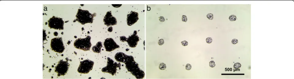
Laser printing of simple water-soil mix (without a gel addition) results in severe spraying, uneven form, and large deviations in the diameter of the transferred drop-lets (see Fig. 1a). In this figure, printing results of the dense soil slurry are shown. At the same time, printing of droplets containing only single soil particles requires heavy diluted mixes. In this case, single cell printing is possible, allowing separation of soil microbe cells, or printing of a small number of cells in consortia, which can be sufficient for effective suppression of antagonistic influence of the competing cell types, while keeping syn-ergetic bonds. Unfortunately, decreasing the viscosity of the water soil slurry by dilution leads to even stronger spraying and uncontrollable printing results.
Using water-soil slurry mixed with a gel in proportion 1:2 by volume considerably increases viscosity of the mix. Mixed droplets have less spreading on the donor slide and have no spraying (see Fig. 1b). Moreover, addition of gel allows to operate within a wide range of concentrations of soil particles in the mix and to print almost any type of soil.
A similar behavior can be observed during the soil print-ing onto agar plates. After laser printprint-ing of soil-water mix without a gel, chaotic growth of colonies with overlapping effects has been observed (see Fig. 2a). In case of gel addition, laser printing of soil microparticles resulted in regular growth patterns of colonies (see Fig.2b).
Effects of laser pulse energy, focusing conditions, and viscosity of hydrogel
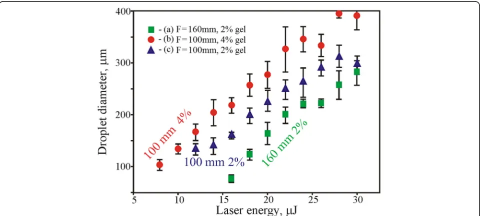
The laser pulse energy and beam focusing conditions have a great influence on the LMS process. At tight focusing of the laser beam, less pulse energy is required for soil drop-lets transfer (Fig.4). The value of the threshold energy for a lens with the focal distanceF= 160 mm isEth= 16μJ (2% gel), and for a lens withF= 100 mm it isEth= 8μJ (2% gel).
The laser transfer threshold depends also on the gel viscosity; more viscous 4% gel can be transferred at larger energy than less viscous 2% gel. Among differ-ent factors presdiffer-ented in Fig. 3, the combination of lens distance F = 160 mm and soil mix with 2% gel has been recognized as optimal. Using the combin-ation of lens with F = 100 mm and soil mix with 2% gel, strong spraying and instability of droplet transfer was observed. While the soil mix with 4% gel has quicker dried out and blade coating on the donor glass plate was less reproducible. Higher laser pulse energies resulted in a bigger drop sizes with a
non-rounded shape (see Figs. 3 and 4) and led to droplet spraying.
As it was shown in [15], such behavior can be ex-plained by an increased laser-induced jet speed and vio-lation of its laminarity at high laser pulse energies. At high energies, transfer of droplets leads to their enrich-ing with soil particles. In this case, the number of large particles inside the transferred droplets increases to-gether with their average size (Fig.4).
The volume of droplets with the growing laser pulse en-ergy varied from 200 to 1000 pl, respectively. On the basis of our experimental studies, optimum laser pulse energies
Fig. 2Laser printing of soil microparticle containing 6 × 6 droplet arrays onto agar substrate in cases of soil mix with water (a) and with the addition of 2% gel (b)
for the LMS of soil-gel mix with 2% gel concentration and lens focusF= 160 mm are in the range of 20 to 24μJ.
Microbial growth in Petri’s dishes on agar plates of a glucose-peptone-yeast agar
Experiments with Mollisol
After the LMS procedure, microorganisms were culti-vated on a solid surface of nutrient media (glucose-pep-tone-yeast agar) in Petri dishes. 400 colonies germinated after LMS were compared on genera level with 250 ones grown after the common plating. All of them have been analyzed. We consider that number of colonies analyzed was enough to cover the bacterial diversity (culturable in the conditions used) due to multiple detection of the same morphotypes and genera. Good’s coverage [18] cal-culated based on genera identification was 99.8 and
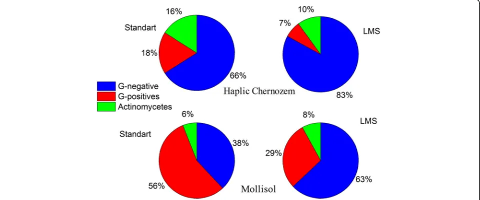
100% for LMS and common plating, respectively. All colonies were described and identified on the basis of phenotype analysis and potassium hydroxide test. The percentage distribution of Gram-positive (GP) and Gram-negative (GN) bacteria and actinomycetes was de-termined (see Fig. 5). Application of the LMS method increased the abundance of GN bacteria from 38 to 63%. It is well-known that the GN bacteria have thinner cell wall compared to that of GP bacteria which explains higher sensitivity of the GN bacteria to external influ-ences. In particular, destruction of some cells at cells de-sorption procedures (vortexing or ultrasound treatment) can be assumed. The observed bacteria abundance dem-onstrates that the LMS procedure is friendlier for the GN bacteria compared to the standard inoculation method. In the presence of stronger breaking influences
Fig. 4Microphotographs of soil mix drops with 2% gel printed at different laser pulse energies (F= 160 mm)
characteristic to the standard soil pre-treatment, domin-ance of more resistant GP spore forming bacteria is observed.
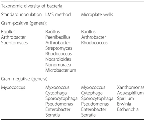
More detailed taxonomical variety on genera level of cultivated bacteria is presented in Table 1. The standard inoculation procedure allowed cultivating only three genera of GP bacteria namely Bacillus, Arthrobacter, Streptomyces and one GN genera Myxococcus, which is able to form microspore with high stress resistance. The LMS procedure leads to an increasing number of GP cultivated genera, up to 8 adding Paenibacillus, Rhodo-coccus, Nocardioides, Microbacterium and Nonomuraea which were not found by the standard inoculation method. The number of GN cultivated genera grew up from one to six due to the LMS application. More sig-nificant increase in GN bacteria abundance was achieved using sequential inoculation of microbial biomass grown up on monosubstrate liquid media from wells of “Eco-Log©” microplate after the LMS germination to universal solid growth media. This method allowed culti-vating ofXanthomonas,Aquaspirillum,Spirillum,Erwinia andEscherichia. The described improvements in the isola-tion procedure increased the number of GN isolates up to 11 genera. Because of the substrate composition and base media in“Eco-Log©”plates focused on GN cultivation, no such effects have been observed for GP bacteria.
A special attention should be paid to the 3–1 Str strain (GenBank accession number KY363604), which is re-lated toNonomuraeagenus on the basis of phylogenetic identification and was isolated by the LMS method with-out creation of any unique cultivation conditions. The genus Nonomuraea is a rare genus of actinomycetes which isolation by standard methods requires applying of selective media and addition of antibiotics cocktails
[19, 20]. It is important that Nonomuraea genus mem-bers can form a number of bioactive compounds which can be applied for medical, pharmaceutical, and agricul-tural purposes [19]. In connection with the trends in the search for new metabolites ofNonomuraea, it is planned to study the biological activity and to provide proper species identification of the strain isolated by us.
Experiments with haplic Chernozem
Culturing from this sample was carried out during suc-cession of microbial community initiated by wetting with sterile DI water, due to the fact that a low variety of bac-teria was initially cultured from the sample. For this soil we observed about 100 colony morphotypes, germinated after LMS, and about 100 ones after the standard inocu-lation. Haplic Chernozem sample shown lower cultur-able biodiversity than Mollisol, but at the same time it contained high proportion of morphotypes difficult for phenotypic identification. Due to this the representatives of each morphotype were identified on genera level using 16S rRNA genes sequencing. Representatives of each genera listed below were found as minimum twice, and Good’s coverage was 100% for LMS and standard inoculation both. During the first 4 weeks, there was no difference in the taxonomic composition of bacteria cul-tured by standard inoculation and LMS. Representatives of the genera Arthrobacter, Paenibacillus, Bacillus, Streptomyces, Sphingomonas, Burkholderia, Paracoccus, Methylobacterium, and Cytophagawere found. However, in the later stages of succession, a twofold greater variety of bacteria was cultured by LMS compared to standard in-oculation. Using LMS, representatives of genera of both GP (Staphylococcus,Microbacterium) and GN (Cupriavi-dus, Tardiphaga) bacteria were found, which previously have not been detected in this sample (Table2).
Substrate utilization
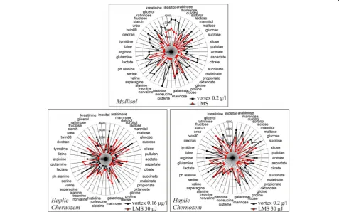
In Fig.6, the substrate utilization spectra of Mollisol and Haplic Chernozem microbial communities are pre-sented. These spectra were obtained from “Eco-Log©” microplates (4 reps averaged) inoculated using the standard and LMS methods. In case of Mollisol, the standard inoculation regime was used as a vortexed soil-water suspension in one concentration 0.2 g/l. In further experiments, the Haplic Chernozem in two concentra-tions was inoculated. We used soil-water-gel mix to equalize conditions in both experimental cases. The highest concentration been used was 0.2 g/l and the low-est one –0.16μg/l. In case of using the highest dilution for standard inoculation, the amount of soil transferred in each well of the microplate was approximately equal to the soil amount transferred by microdroplets during the LMS procedure. As demonstrated in Fig.6, for both soils, substrate utilization patterns obtained by the Table 1A taxonomic variety of bacteria isolated from Mollisol
using the standard method of inoculation, LMS procedure, and microplate wells
Taxonomic diversity of bacteria
Standard inoculation LMS method Microplate wells
classic way significantly differ from the spectrum re-ceived after the LMS procedure. It is important to note that for both soils on low diluted soil-water suspensions (0.2 g/l) narrowing of the substrate utilization spectra for the LMS samples has been observed. It can be ex-plained by the fact that the LMS procedure, in contrast to the standard technique of soil processing, allows to avoid mixing of microorganisms from different micro-zones. We select the parameters of the laser transfer in such a way that the effective transferred volume of the gel contains single micro particle of soil. Thus, LMS al-lows to “cut out” the narrower parts of the microbial community, increasing functional diversity by removing direct competition and separating the substrate niches in microplate wells. We suppose consortium of micro par-ticle carry out the metabolically fingerprint of entire community, that supports “functional doubling” of the
system. It means that the functional integrity of the sys-tem, supports by switching the units with different sus-tainability and growth requirements to environment but keep the same functionality meanwhile. We assume our microparticles are such a units. It is possible to assume that the lack of metabolic activity on some substrates oc-curs due to the declining number of sampled bacteria cells, which is the aim of the LMS procedure. Drastic re-duction of the sampled volume, when the droplet vol-ume approaches to 200 pl, plays also an important role. Therefore, the restriction of metabolic diversity, as a re-sult of the LMS procedure, may be considered as a good evidence for selective laser printing of particular microorganisms inhabiting soil microparticles. In con-trast to Mollisol, a highly-diluted Haplic Chernozem soil-water-gel mixes produce even broader substrate utilization spectra compared to the standard treatment, Table 2A taxonomic variety of bacteria isolated from Haplic Chernozem using the standard method of inoculation and LMS procedure
Time of succession Standard inoculation LMS method
6th week Bacillus
Streptomyces Burkholderia
Bacillus Streptomyces Staphylococcus
Burkholderia Cupriavidus Tardiphaga
8th week Streptomyces
Burkholderia Bordetella
Streptomyces Bacillus Microbacterium
Burkholderia Cupriavidus Tardiphaga
which could be explained by a possible laser activation of bacterial growth and metabolism (e.g. [21,22]).
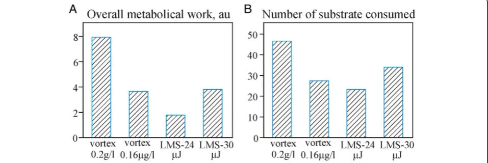
The analysis of overall metabolic work (presented by the total optical density of plate wells with consumed substrates) and the number of substrates been consumed (metabolic diversity) shows clear differences between the applied experimental regimes (Fig.7).
It is clear that 30μJ laser pulses demonstrate the high-est performance in biomass transfer. It is easy explain-able due to the larger droplet size.
Figure 8 demonstrates results of the cluster analysis (Euclidean distance, clustering by Ward) of the pre-sented multidimensional substrate utilization spectra.
By analyzing substrate utilization patterns of Mollisol, we can classify the cases and reveal the significant influ-ences and structural bonds. On the basis of the this den-drogram it is possible to draw a conclusion that the substrate utilization patterns of microbial consortia iso-lated by the LMS and standard treatments appear to be considerably different. At the same time, convergence of replicas in laser printing is slightly higher (about 10% dis-similarity), than that when the classical method of sample processing (vortexing) is used (30% dissimilarity). It means the increased homogeneity of the LMS samples. It seems LMS procedure allows transferring some “functional di-versity quantum”that remains stable in replicas. The ap-plication of LMS resulted in reduction of metabolic diversity. The observed reduction means that not the whole community was extracted but only an isolated part. Note that the isolated laser printing of cells and microcon-sortia is an ultimate goal of the demonstrated LMS method, so it is good evidence in this particular case.
Experiments with Haplic Chernozem demonstrate ap-proximately the same trend. The difference between the substrate utilization patterns produced at different LMS regimes is noticeable. We also have to highlight the fact that in vortex cases, in spite of impressive (106) dilution, substrate utilization patterns remain quite similar. It
demonstrates the remarkable stability of the“metabolical fingerprint”of soil microbial community.
Conclusions
A high throughput Laser Micro-Sampling (LMS) tech-nology for direct isolation from soil of pure microbial cultures and microbial consortia has been developed. This technology is based on laser printing of soil micro-particles by focusing near-infrared laser pulses on spe-cially prepared samples of a soil/gel mixture spread onto a gold-coated glass plate. Laser printing occurs in form of direct transfer of soil microparticles to agar surface or into microplate wells.
Different transfer modes of soil microparticle have been described and experimental protocol of laser printing of soil/gel droplets has been optimized. On the basis of tests with the Mollisol and Haplic Chernozem soils, the LMS method has been confirmed to be suitable for printing soil microparticles with maintaining sufficiently high viability of microbial cells. In cultivation experiments, a consider-able growth of germinated bacterial diversity after the LMS procedure has been observed. Application of the LMS method significantly increased the number of genus of iso-lated bacteria, both Gram-negative and Gram-positive groups, which were not cultivated from soil samples with the demonstrated abundance using the classical inocula-tion method, by putting soil suspensions on the surface of agar plates. A representative of the rare genus Nonomur-aeawas isolated due to the LMS technology without appli-cation of any complex selective media and addition of antibiotics cocktails.
Changes in functional diversity and narrowing of sub-strate utilization spectra induced by the LMS method have been demonstrated in contrast to low diluted sys-tems. The observed decrease in the number of substrates been consumed after the LMS treatment provides an evidence for microbial separating ability of this method. In case of comparison with highly diluted samples, LMS
leads to increased metabolic capability of the extracted microbial system that can be explained by possible laser activation. Combined cultivation technique, that in-cludes subsequent solid media cultivation of microbial biomass previously grown on sole carbon substrate media in wells of the LMS inoculated multisubstrate mi-croplate, dramatically increases the taxonomical abun-dance. This could be a new promising technology to achieve a higher level of rare or yet-to-be-cultured strains isolation. We hope that the LMS method will allow discovering an isolate rare and possibly new spe-cies of microorganisms.
In conclusion, we have to point out LMS is goes out of the frames of one narrow task such a soil transfer. It would be successfully used for wide variety of purposes such a bacterial consortia separation. In conclusion, it seems to us that the development of Laser Engineering of Microbial Systems (LEMS) and the study of its possi-bilities will be promising.
Materials and methods
Laser microsampling technique
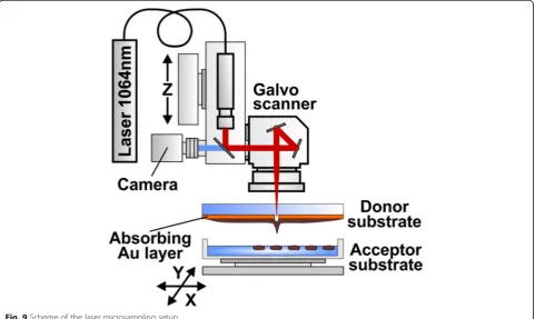
The experimental setup used for printing of soil micro-particles is shown in Fig. 9. Radiation of a nanosecond
Fig. 8Results of the cluster analysis of substrate utilization patterns after the LMS and ordinary vortex treatment. Ward’s clustering, Euclidean distances. Number of cases: 4 cases of 4 averaged repetitions for Mollisol; 6 cases of 4 averaged repetition for Haplic Chernozem. Number of variables (substrates): 47
ytterbium fiber laser YLPM-1-4 × 200–20-20 (IRE-Polus, Russia) with parameters λ= 1.06 μm, τ= 8 ns, ν= 105 KHz, E = 10–40μJ is focused by F-Theta lenses with the focal length F = 100 mm or F = 160 mm (Ronar-Smith, Singapore) onto a gold absorbing layer at the donor glass plate. Laser focus positioning in the target plane has been controlled by the scanning mirrors installed at the lens front. Printing of soil microparticles has been car-ried out from the donor slide to the collector glass plate located at a distance of 1 mm. The donor glass plate has been coated with a 50 nm thick gold layer covered with a layer of soil mix. Homogeneous coverage with the soil mix has been produced using a blade coater. In our ex-periments, thickness of the soil mix layer was 30μm and been regulated with the accuracy of 1μмby means of a micrometric vertical feed screw motion. Thinner layers (10–15μm) appeared to be quickly drying out and did not provide enough time to work with the samples. For culti-vation of microorganisms and their subsequent studies, laser printing of drops with a soil/gel mix has been carried out directly onto a surface of glucose-peptone-yeast agar in Petri’s dishes, and in 96-well“Eco-log” test plates. The “Eco-log” test plates have been used both as a functional diversity analysis tool and as a set of intermediate selective synthetic sole carbon substrate media.
Soil mix for laser microsampling
There were two soil samples have been used for soil mix preparation: top 10 cm layer of Mollisol (Belgorod region, Russia) and 19–32 cm layer of Haplic Chernozem (Saratov region, Russia). 0.2 g. of the dry soil has been mixed with 0.4 ml of water and 0.8 ml of hydrogel. The soil/gel mix has been gently homogenized by glass stick. In our experi-ments, polysaccharide gel based on low-molecular hyalur-onic acid (Hyalurhyalur-onic acid sodium salt fromStreptococcus equi, Sigma) has been used. A gel addition increases the mix viscosity and reduces spraying of printed drops on the collector surface [23]. However, the gel possibly could be used as a growth substrate by some microorganisms, lead-ing to a partial change of the cultured biodiversity. The gel preparation procedure includes addition of phosphate buffer solution (PBS) to the dry hyaluronic acid in propor-tions of 2 and 4% following cooling down at + 4 °C for 48 h to reach full dissolution.
The standard soil slurry has been used as a control: 5 g of this soil was mixed with 45 ml of deionized (DI) water and vortexed with subsequent differential centrifugation (1000 rpm for 1 min, 3000 rpm for 3 min) for fraction-ation; a tiny fraction has been separated and diluted with water to a required consistency. We concede possible to use DI water for soil samples dilution regarding the soil as a substance quite enriched with salts and electrolytes, so we do not expect a significant osmotic shock for Gram-negative (GN) bacteria. The experiments with
Haplic Chernozem implied using of soil/gel mixes in standard plating procedure. The Mollisol soil particle size distribution, recorded by Microtrac S3500 Particle Size Analyzer, demonstrated that the most of soil particles have sizes in the range of 5–50μm. For Haplic Chernozem the most of soil particles has sizes in the range of 0.3–60μm.
Microbiological methods
Cultivation and identification of bacteria
Direct laser printing on a surface of Petri’s dishes cov-ered by solid glucose-peptone-yeast media [24] was per-formed to estimate bacteria growth ability after the LMS process. Cultivation with the standard technique (plating using glass spatula) from vortexed soil slurry was also produced to estimate the effect of laser printing [25]. Similar as for LMS method soil dilution was used – soil\gel proportion 1:5. Moreover, for Halphic Cherno-zem, cultivation by the standard technique from the soil-gel mixture was carried out to avoid possible effects induced by osmotic shock and the gel presence. The dishes were incubated at a temperature of 28 °C for 5– 7 days and check the gel influence. There no significant effect of gel at all. The number of grown bacteria col-onies was counted with the main morphotypes detec-tion. Identification of bacteria cultures has been carried out on the basis of phenotypical, cultural, micromorpho-logical, and physiology-biochemical signs as key factors for phylogenetic identification of soil bacteria growth [26] and determinants of bacteria manuals [27].
Multisubstrate testing
The multisubstrate testing (MST) method, based on “Eco-Log©”technology, is a novel method in soil micro-biology [30] designed to estimate the functional diversity of microbial communities. It is an our realization of community level physiological profiling assay. The most known wide spread analog - a BIOLOG™technique [31]. This method allows estimating a capability of microbial community to consume аset of sole- carbone -sources media and obtaining the substrate utilization pattern -multidimensional “metabolic fingerprint” of a microbial system. That represents the ability to grow on different separate organic substrates and is described as microbio-logical functional diversity of environ. This method is useful for assessing the functional state of microbial communities, which reflects the influence of various ex-ternal factors, in particular, environmental pollution. In fact, the method allows possible to monitor the eco-logical situation. [32,33].
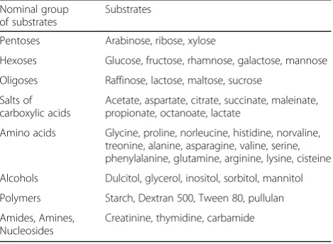
To obtain a substrate utilization pattern, microdroplets of the soil slurry have been transferred directly (by the LMS or standard method) into wells of 96th well test microplate“Eco-Log©”that contains a set of 47 test sub-strates in two replicates (each well contains one separate substrate, a mineral media, and the indicator of substrate consumption -tetrazolium bromide) (Table 3). The indi-cator of growth of microorganisms gets purple colored due to the cell respiration. Color intensity is propor-tional to the number of electrons passing through the membrane during respiration on a given substrate and correlate with the density of biomass grown on a test substrate. Test substrates used in this system include dif-ferent sugars, amino acids, polymers, nucleosides, salts of organic acids, alcohols, etc. Usually, in data processing two replicas plate data are averaged and treated as a 48-dimentional data vector. 2 plates with 2 reps were used for each experimental point (4 reps totally).
After the LMS procedure, microplates were incubated in a thermostat at 28°Сfor 72 h. At the end of incubation, the photometric reading of stained cell density (at 510 nm wave length) was carried out by hardware and software of the “Eco-Log©” system to estimate the intensity of sub-strate utilization [34]. A multidimensional data array was generated as a result of photometric measurements of values of optical density on all wells (all substrates). It rep-resents a substrate consumption spectrum of for this soil microbial system -“metabolic fingerprint”. For identifica-tion of the microorganisms grown in some wells, the mi-crobial biomass has been transferred from the wells to solid media for subsequent cultivation and identification.
The microplate technology was used for two purposes: 1) to estimate functional diversity of the transferred mi-crobial systems; 2) to use as sole carbon source selective media for precultivation that potentially reveals unusual niches for rare strains. We consider the microplate tech-nology as a model of soil microzones. It can provide ad-vantages for rare/new strains isolation by enlarging the logical space in our experiments.
Abbreviations
GN:Gram-negative; GP: Gram-positive; LMS: Laser Micro-Sampling
Acknowledgements
We thank Semyon Churbanov for technical support in the experiments. The authors thank anonymous reviewers for help in improving of the article.
Funding
This work was supported by the Russian Federal Agency of Scientific Organizations (Agreement No 007-GZ/C3363/26) in part of development of new method and systems for new 3d printer technologies of laser transfer; the Russian Foundation for Basic Research (RFBR) Project No. 16–02-00955 in part of microbiological research and Project No. 18–32-00607 in part research of laser transfer processes. S.A. Evlashin was granted by Scholarship of the President of the Russian Federation SP-1493.2016.4.
Availability of data and materials
The datasets used and/or analysed during the current study are available from the corresponding author on reasonable request.
Authors’contributions
MVG, VSC, KIK - preparation of culture plates with a set of different media, work with populated culture media; EAC, ESC, VSZ - development of transfer regimes, selection of parameters of gel media for soil and conducting a series of experiments on laser printing in microlates; NVM, AOR, SAE -development and assembly of the laser printing system, technical support of all the experiments conducted, selection of operating modes, research and production of donor substrates with absorbing coating; LVL, GAD - analysis of the obtained biological results, the morphological analysis of the isolated microorganisms; VIY, BNC, VNB - research management, organization of scientific work, analysis and execution of the results; All authors participated in the writing of the article, discussing and formalizing the results and preparing drawings.All authors read and approved the final manuscript.
Ethics approval and consent to participate
Not applicable.
Consent for publication
Not applicable.
Competing interests
The authors declare that they have no competing interests.
Table 3Sole carbon sources for the Multisubstrate Testing method
Nominal group of substrates
Substrates
Pentoses Arabinose, ribose, xylose
Hexoses Glucose, fructose, rhamnose, galactose, mannose
Oligoses Raffinose, lactose, maltose, sucrose
Salts of carboxylic acids
Acetate, aspartate, citrate, succinate, maleinate, propionate, octanoate, lactate
Amino acids Glycine, proline, norleucine, histidine, norvaline, treonine, alanine, asparagine, valine, serine, phenylalanine, glutamine, arginine, lysine, cisteine
Alcohols Dulcitol, glycerol, inositol, sorbitol, mannitol
Polymers Starch, Dextran 500, Tween 80, pullulan
Amides, Amines, Nucleosides
Publisher’s Note
Springer Nature remains neutral with regard to jurisdictional claims in published maps and institutional affiliations.
Author details
1Department of General Soil Science, Lomonosov Moscow State University,
119991 Moscow, Russia.2Research Center“Crystallography and Photonics”
RAS, Institute of Photonic Technologies, 142190, Troitsk, Moscow, Russia.
3Institute of Nuclear Physics, Lomonosov Moscow State University, 119991
Moscow, Russia.4Institute of Theoretical and Experimental Biophysics RAS,
142290, Puschino, Moscow Branch, Russia.5Leibniz Universität Hannover and
Laser Zentrum Hannover e.V, 30419 Hannover, Germany.6Center for Design, Manufacturing & Materials, Skolkovo Institute of Science and Technology, 143026, Skolkovo, Moscow, Russia.
Received: 3 April 2018 Accepted: 31 October 2018
References
1. Oliver JD. Recent findings on the viable but nonculturable state in pathogenic bacteria. FEMS Microbiol Rev. 2010;34:415–25.
2. Stewart EJ. Growing unculturable bacteria. Bacteriology. 2012;194:4151–60. 3. Colwell RR. Viable but non-culturable bacteria in the marine environment
and the biotechnological tools to detect them. In Encyclopedia of Life Support Systems. Oxford, UK: Eolss Publishers; 2008.
4. Riesenfeld CS, Goodman RM, Handelsman J. Uncultured soil bacteria are a reservoir of new antibiotic resistance genes. Environ Microbiol. 2004;6:981–9. 5. Kellenberger E. Exploring the unknown: the silent revolution of
microbiology. EMBO Rep. 2001;2:5–7.
6. Joint L, Mühling M, Querellou J. Culturing marine bacteria–an essential prerequisite for biodiscovery. Microb Biotechnol. 2010;3:564–75. 7. Kaeberlein T, Lewis K, Epstein SS. Isolating“uncultivable”microorganisms in
pure culture in a simulated natural environment. Science. 2002;296:1127–9. 8. Kakumanu ML, Williams MA. Soil diffusion system enriches the growth of
diverse and previously uncultivated bacterial taxa. Soil Sci Soc Am J. 2012; 76:463–74.
9. Aoi Y, Kinoshita T, Hata T, Ohta H, Obokata H, Tsuneda S. Hollow-fiber membrane chamber as a device for in situ environmental cultivation. Appl Environ Microbiol. 2009;75:3826–33.
10. Nichols D, Cahoon N, Trakhtenberg EM, Pham L, Mehta A, Belanger A, Kanigan T, Lewis K, Epstein SS. Use of ichip for high-throughput in situ cultivation of“uncultivable”microbial species. Appl Environ Microbiol. 2010; 76:2445–50.
11. Ling LL, Schneider T, Peoples AJ, Spoering AL, Engels I, Conlon BP, Mueller A, Schäberle TF, Hughes DE, Epstein S, Jones M, Lazarides L, Steadman VA, Cohen DR, Felix CR, Fetterman KA, Millett WP, Nitti AG, Zullo AM, Chen C, Lewis K. A new antibiotic kills pathogens without detectable resistance. Nature. 2015;517:455–9.
12. Ringeisen BR, Rincon K, Fitzgerald LA, Fulmer PA, Wu PK. Printing soil: a single-step, high-throughput method to isolate micro-organisms and near-neighbour microbial consortia from a complex environmental sample. Methods Ecol Evol. 2014;6:209–17.
13. Grüne M, Unger C, Koch L, Deiwick A, Chichkov B. Dispensing pico to nanoliter of a natural hydrogel by laser-assisted bioprinting. Biomed Eng Online. 2011;10:1–11.https://doi.org/10.1186/1475-925X-10-19.
14. Koch L, Kuhn S, Sorg H, Gruene M, Schlie S, Gaebel R, Polchow B, Reimers K, Stoelting S, Ma N, Vogt PM, Steinhoff G, Chichkov B. Laser printing of skin cells and human stem cells. Tissue Eng Part CMethods. 2010;16:847–54. 15. Ali M, Pages E, Ducom A, Fontaine A, Guillemot F. Controlling laser-induced
jet formation for bioprinting mesenchymal stem cells with high viability and high resolution. Biofabrication. 2014;6:045001.https://doi.org/10.1088/ 1758-5082/6/4/045001.
16. Lehner BA, Schmieden DT, Meyer AS. A straightforward approach for 3D bacterial printing. ACS Synth Biol. 2017;6:1124–30.
17. Yusupov VI, Zhigar'kov VS, Churbanova ES, Chutko EA, Evlashin SA, Gorlenko MV, Cheptsov VS, Minaev NV, Bagratashvili VN. Laser-induced transfer of gel microdroplets for cell printing. Quantum Electronics. 2017;47:1158–65. 18. Good IJ. The population frequencies of species and the estimation of
population parameters. Biometrika. 1953;40:237–64.
19. Sungthong R, Nakaew N. The genusNonomuraea: a review of a rare actinomycete taxon for novel metabolites. J Basic Microbiol. 2015;55:554–65.
20. Ara I, Kudo T, Matsumoto A, Takahashi Y, Omura S. Nonomuraea bangladeshensis sp. nov. and Nonomuraea coxensis sp. nov. Int J Syst Evol Microbiol. 2007;57:1504–9.
21. Nussbaum EL, Lilge L, Mazzulli T. Effects of 630-, 660-, 810-, and 905-nm laser irradiation delivering radiant exposure of 1-50 J/cm2 on three species of bacteria in vitro. J Clin Laser Med Sur. 2002;20:325–33.
22. Kohli R, Bose B, Gupta PK. Induction of phr gene expression in E. coli strain KY706/pPL-1 by he–ne laser (632.8 nm) irradiation. J Photochem Photobiol B. 2001;60:136–42.
23. Guillotin B, Souquet A, Catros S, Duocastella M, Pippenger B, Bellance S, Bareillea R, Rémya M, Bordenavea L, Amédéea J, Guillemot F. Laser assisted bioprinting of engineered tissue with high cell density and microscale organization. Biomaterials. 2010;31:7250–6.
24. Cheptsov VS, Vorobyova EA, Manucharova NA, Gorlenko MV, Pavlov AK, Vdovina MA, Lomasov VN, Bulat SA. 100 kGy gamma-affected microbial communities within the ancient Arctic permafrost under simulated Martian conditions. Extremophiles. 2017;21:1057–67.
25. Zvyagintsev, D. G. (1991) Methods of soil microbiology and biochemistry. MSU, Moscow (Book in Russian).
26. Lysak, L. V., Dobrovolskaya, T. G., Skvortsova, I. N. (2003) Methods of an assessment of a bacterial variety and identification of bacteria. MAX Press, Moscow (Book in Russian).
27. Bergey’s Manual of Systematic Bacteriology (2001-2012), 2nd ed. Springer, New York.
28. Marchesi JR, Sato T, Weightman AJ, Martin TA, Fry JC, Hiom SJ, Wade WG. Design and evaluation of useful bacterium-specific PCR primers that amplify genes coding for bacterial 16S rRNA. Appl Environ Microbiol. 1998;64:795–9. 29. Lane DJ. 16S/23S rRNA sequencing. In: Stackebrandt E, Goodfellow M,
editors. Nucleic acid techniques in bacterial systematic. West Sussex: John Wiley & Sons; 1991. p. 115–75.
30. PND FT (Federal Nature Protection Normative Document) no. 16.1.17–10 FR. 1.37.2010.08619: Method for Measuring the Test-Substrates Consumption Intensity by Soil Microbial Communities and Soil-Like Objects by Means of Photometric Method, Moscow, 2010. (In Russian).
31. Garland JL, Mills AL. Classification and characterization of heterotrophic microbial communities on the basis of patterns of community-level sole-carbon-source utilization. Appl. Environ. Microbiol. 1991;57:2351–9. 32. Gorlenko MV, Majorova TN, Kozhevin PA. Disturbances and their influence on
substrate utilization patterns in soil microbial communities. In: Insam H, Rangger A, editors. Microbial communities. Innsbruck: Springer; 1997. p. 84–93. 33. Vorobyova E, Soina V, Gorlenko M, Minkovskaya N, Zalinova N,
Mamukelashvili A, Gilichinsky D, Rivkina E, Vishnivetskaya T. The deep cold biosphere: facts and hypothesis. FEMS Microbiol Rev. 1997;20:277–90. 34. Gorlenko, M. V., Kozhevin, P. A., Terekhov, A. S. (2008) Way of multisubstrate