http://www.scirp.org/journal/jbm ISSN Online: 2327-509X
ISSN Print: 2327-5081
DOI: 10.4236/jbm.2018.62003 Feb. 26, 2018 23 Journal of Biosciences and Medicines
Association of Bax and Bcl-2 Functional
Polymorphisms and Protein Levels with
Posttraumatic Stress Disorder
Diana Avetyan
1, Arsen Arakelyan
1,2, Gohar Mkrtchyan
1*1Laboratory of Human Genomics and Immunomics, Institute of Molecular Biology, Armenian National Academy of Sciences,
Yerevan, Armenia
2Group of Bioinformatics, Institute of Molecular Biology, Armenian National Academy of Sciences, Yerevan, Armenia
Abstract
Background: Posttraumatic stress disorder (PTSD) is an anxiety disease in-fluenced by both environmental and genetic factors, which affects a patient’s quality of life and social stability. Recent studies have shown that the patho-genesis of PTSD is associated with apoptosis; however, the molecular me-chanisms that cause such damage are not well-understood. Also it is unclear whether these pathologic alterations are genetically determined or caused by other factors. The aim of this study was to investigate the genetic association of functional polymorphisms in genes coding for apoptosis-related Bcl-2 and Bax proteins with PTSD as well as proteins levels in the blood of affected subjects. Methods: The study groups consisted of 200 combat veterans with PTSD and an equal number of healthy subjects with no family- or past-history of any psychiatric disorders. Bax and Bcl-2 proteins levels in blood were measured by ELISA. DNA samples were genotyped for SNPs using PCR-SSP. Results: According to our results, PTSD patients are characte-rized by increased levels of apoptotic proteins and the imbalance in the Bax/Bcl-2 ratio compared to healthy subjects. Our results also demonstrate that rs956572*A minor allele of the BCL2gene was overrepresented in pa-tients with PTSD compared to healthy subjects. Conclusions: The results implicate Bcl-2 and Bax in pathogenesis of PTSD on genetic and protein le-vels, though further studies on enlarged cohort and in different populations are required.
Keywords
Posttraumatic Stress Disorder, Apoptosis, BCL2, BAX, Single Nucleotide Polymorphisms
How to cite this paper: Avetyan, D., Ara-kelyan, A. and Mkrtchyan, G. (2018) Associ-ation of Bax and Bcl-2 Functional Polymor-phisms and Protein Levels with Posttrau-matic Stress Disorder. Journal of Biosciences and Medicines, 6, 23-32.
https://doi.org/10.4236/jbm.2018.62003
Received: January 18, 2018 Accepted: February 23, 2018 Published: February 26, 2018
Copyright © 2018 by authors and Scientific Research Publishing Inc. This work is licensed under the Creative Commons Attribution International License (CC BY 4.0).
DOI: 10.4236/jbm.2018.62003 24 Journal of Biosciences and Medicines
1. Introduction
Posttraumatic stress disorder (PTSD; ICD-10-CM codes: F43.1; DSM-V code: 309.81) is an anxiety disease that develops as a result of a serious psychological trauma following an event of a threat of death or major injury [1] [2]. The diag-nosis of PTSD is based on the symptoms causing clinically significant suffering or impairment in social and/or professional dysfunction for a period of up to six months [3] [4].
Recently the number of risk factors contributing to the development of PTSD has dramatically increased worldwide. Therefore, one of the most common problems of health care nowadays is the development of efficient prognostic strategies and methods of early diagnostics and treatment of PTSD. Epidemio-logical, clinical, and experimental research data suggest that both environmental and genetic factors are involved in the pathogenesis of PTSD. Several studies suggest that PTSD is a complex disorder with polygenic inheritance [5] [6] [7].
PTSD is characterized by neuroendocrine abnormalities, particularly with dysfunction of the hypothalamic-pituitary-adrenal (HPA) axis, which is ex-pressed by low levels of cortisol and adrenocorticotropic hormone (ACTH) in plasma [8]. Many studies showed that the amygdala, hippocampus, and medial prefrontal cortex (mPFC) are responsible for the occurrence of PTSD. In partic-ular, mPFC is controlling stress and fear responses of amygdala and
hippocam-pus [9]. Recent neuro-imaging studies have shown that the volume of
hippo-campus decreases in PTSD patients compared to healthy subjects [10]. This atrophy can be associated with apoptosis, but these molecular mechanisms are yet to be understood [11].
mechan-DOI: 10.4236/jbm.2018.62003 25 Journal of Biosciences and Medicines
isms causing neuronal apoptosis in mPFC [20].
The aim of this study was the investigation of the potential role of anti-apoptotic protein Bcl-2 and pro-apoptotic proteins Bax in the pathogenesis of PTSD on the levels of functional polymorphism in coding genes and protein abundance.
2. Materials and Methods
2.1. Study Subjects
Study groups consisted of 200 combat veterans with PTSD (mean age: M ± SD = 54.52 ± 11.0 years) and an equal number of healthy subjects (HS) (mean age: M ± SD = 43.6 ± 9.1 years) with no family or past history of any psychological dis-orders. Clinical diagnosis was made according to the Clinician Administered PTSD Scale (CAPS) [4]. All subjects were Armenian nationality born and living in Armenia and Artsakh. Both, the informed consents from all study subjects and the approval of the Ethics Committee of the Institute of Molecular Biology of NAS RA (IRB #00004079) were received for these studies.
2.2. Blood Sampling and Genomic DNA Extraction
About 5 ml of peripheral venous blood was collected from each study subject and transferred to EDTA-containing tubes. Genomic DNA was isolated from the fresh blood samples according to Miller’s salting-out procedure [21] modifica-tion where proteinase K was omitted and a chloroform extracmodifica-tion phase was added and stored at −30˚C until further use.
2.3. Determination of Bax and Bcl-2 Levels by ELISA
Bax and Bcl-2 protein levels were determined with an ELISA Kit (USCN Life Science Inc., Wuhan, China) according to the manufacturer’s instructions.
2.4. Primer Design
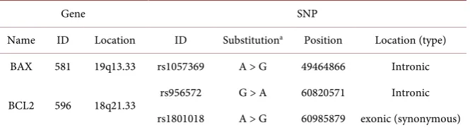
DNA samples were genotyped for BAX rs1057369, BCL2 rs956572 and rs1801018 functional SNPs (Table 1).
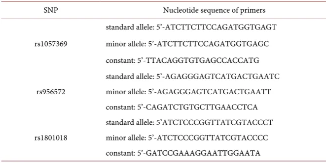
[image:3.595.208.541.611.703.2]All primers for PCR-SSP were designed using the genomic sequences in the Gen-Bank nucleotide sequence database (https://www.ncbi.nlm.nih.gov/genbank/) and are indicated in Table 2.
Table 1. Brief characteristics of selected genes and SNPs.
Gene SNP
Name ID Location ID Substitutiona Position Location (type)
BAX 581 19q13.33 rs1057369 A > G 49464866 Intronic
BCL2 596 18q21.33 rs956572 G > A 60820571 Intronic rs1801018 A > G 60985879 exonic (synonymous)
DOI: 10.4236/jbm.2018.62003 26 Journal of Biosciences and Medicines
Table 2. Primers designed for the selected SNPs.
SNP Nucleotide sequence of primers
rs1057369
standard allele: 5’-ATCTTCTTCCAGATGGTGAGT minor allele: 5’-ATCTTCTTCCAGATGGTGAGC constant: 5’-TTACAGGTGTGAGCCACCATG
rs956572
standard allele: 5’-AGAGGGAGTCATGACTGAATC minor allele: 5’-AGAGGGAGTCATGACTGAATT constant: 5’-CAGATCTGTGCTTGAACCTCA
rs1801018
standard allele: 5’ATCTCCCGGTTATCGTACCCT minor allele: 5’-ATCTCCCGGTTATCGTACCCC constant: 5’-GATCCGAAAGGAATTGGAATA
2.5. Polymerase Chain Reaction with Sequence Specific Primers
Genotyping was carried out by polymerase chain reaction with sequence-specific primers (PCR-SSP) according to protocol developed in Bunce et al. [22]. The presence/absence of allele-specific amplicons in the PCR products was visualized in 2% agarose gel stained with Ethidium Bromide fluorescent dye using DNA molecular weight markers as a reference. To check the reproducibility of results, randomly selected DNA samples (10% of total) were genotyped twice.
2.6. Data Analysis
The distributions of genotypes for all investigated SNPs were checked for cor-respondence to the Hardy-Weinberg (H-W) equilibrium. In order to find poten-tial relevance of the selected SNPs to PTSD, their genotype and allele frequencies and minor allele carriage rates in patients and HS were compared. The signific-ance of differences in genotype and allele frequencies and minor allele carriage between patients and HS was determined using Pearson’s Chi-square test. P-values less than 0.05 were considered statistically significant. P-values adjusted by Bonferroni multiple comparison correction is further indicated as pcorrected,
and those not adjusted as pnominal.
3. Results
3.1. Estimation of Bax and Bcl-2 Levels by ELISA
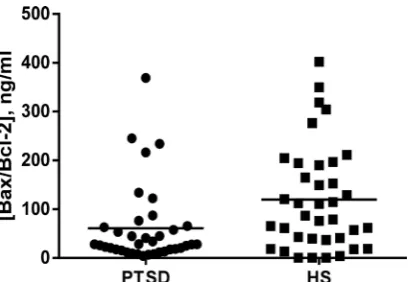
We evaluated Bax and Bcl-2 plasma levels of 39 HS and 40 patients with PTSD. The study indicates that Bax and Bcl-2 protein levels in the blood of patients with PTSD were increased compared to HS (Figure 1 and Figure 2).
The mean levels of Bax and Bcl-2 proteins, respectively, were significantly 1.6 (p = 0.002) and 2.1 (p = 0.0002) times higher among patients than among HS.
Bax is a pro-apoptotic, while Bcl-2 is an anti-apoptotic member of the Bcl-2 protein family. The relative amount or balance between the pro- and an-ti-apoptotic proteins influences the receptiveness to apoptosis.
DOI: 10.4236/jbm.2018.62003 27 Journal of Biosciences and Medicines
[image:5.595.271.476.239.378.2]Figure 1. Bax levels in PTSD patients and HS.
Figure 2. Bcl-2 levels in PTSD patients and HS.
Figure 3. Bax/Bcl-2 ratio in PTSD patients and HS.
ratio as compared with HS (1.96 time; p = 0.008).
3.2. Genotyping of
BAX
rs1057369 and
BCL
2 rs956572,
rs1801018 SNPs
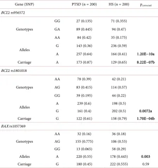
Distribution of BCL2 rs956572, rs1801018 and BAX rs1057369 variants in study groups is shown in Table 3. According to the data obtained, the rs956572*A minor allele of the BCL2 gene was overrepresented in patients with PTSD com-pared to HS (pnominal = 6.02E−11; OR = 2.59; 95%CI: 1.94 - 3.44). In addition, the
carriers of this allele were more in the group of patients compared to HS (pnominal
[image:5.595.272.477.406.547.2]DOI: 10.4236/jbm.2018.62003 28 Journal of Biosciences and Medicines
Table 3. Distribution of genotypes, alleles and carriage of minor alleles of BAX and BCL2
polymorphisms in patients with PTSD and HS.
Gene (SNP) PTSD (n = 200) HS (n = 200) pcorrected
BCL2 rs956572
Genotypes
GG 27 (0.135) 71 (0.355) GA 89 (0.445) 94 (0.47) AA 84 (0.42) 35 (0.175)
Alleles G 143 (0.36) 236 (0.59)
A 257 (0.64) 164 (0.41) 1.20E−10a
Carriage A 173 (0.87) 129 (0.65) 8.22E−07b
BCL2 rs1801018
Genotypes
AA 78 (0.39) 42 (0.21)
AG 83 (0.415) 114 (0.57) GG 39 (0.195) 44 (0.22)
Alleles A 239 (0.6) 198 (0.5)
G 161 (0.4) 202 (0.5) 0.0072a
Carriage G 122 (0.61) 158 (0.79) 1.70E−04b
BAX rs1057369
Genotypes
AA 32 (0.16) 36 (0.18)
AG 155 (0.775) 106 (0.53) GG 13 (0.065) 58 (0.29)
Alleles A 220 (0.55) 178 (0.445) 0.003
Carriage G 180 (0.45) 222 (0.555) 0.59
a. pcorrected values for comparison of mutant allele frequency between PTSD patients and controls. b. pcorrected
values for comparison of mutant allele carriage between PTSD patients and controls.
Further, we found that the frequency of the rs1801018*G minor allele of the
BCL2 gene was lower among patients compared to HS (pnominal = 0.0036; OR =
0.66, 95%CI: 0.5 - 0.87). Also, the carriers of the rs1801018*G minor allele was less frequent in patients than in HS (pnominal = 8.6E−5; OR = 0.42; 95%CI: 0.27 -
0.65). The rs1057369*G allele of the BAX gene was less frequent among patients than in HS (pnominal = 0.002; OR = 0.66, 95%CI: 0.5 - 0.87). Although there were
no significant differences of carriers of rs1057369*G minor allele in the group of patients compared to HS (pnominal = 0.6; OR = 1.15, 95%CI: 0.683 - 1.943). After
Bonferroni correction, difference in minor allele frequency between the patients and HS groups remained significant.
Thus the results of our study indicated positive association between the BCL2 gene rs956572 SNP and PTSD. There were no significant differences between selected SNPs and Bax and Bcl-2 proteins levels.
4. Discussion
DOI: 10.4236/jbm.2018.62003 29 Journal of Biosciences and Medicines
inducers of mitochondria-mediated cell death. In this study, we showed that both levels of pro-apoptotic Bax and anti-apoptotic Bcl-2 proteins were signifi-cantly increased in the plasma of PTSD patients. However, Bax/Bcl-2 ratio, which appears to be more informative parameter than individual protein levels
[23], was decreased in PTSD patients compared to healthy subjects (p = 0.008). The number of studies revealing the associations between apoptosis and PTSD in human subjects is limited and the mechanisms by which Bc1-2 and Bax de-velop their functions in PTSD are not well studied and mostly conducted on animal models. Thus, Li et al.’s study on the hippocampus of rats by using single prolong stress model, showed that apoptotic Bax, caspase-3 proteins, and the ra-tio of Bax/Bcl-2 in all stressed groups under 1 month were significantly in-creased, and the anti-apoptotic Bcl-2 protein was significantly decreased as compared with the healthy group [20] [24]. Alani et al. have shown that activa-tion of caspase-3 in the stress groups of rats indicates that it is one of the reasons of apoptosis inducing atrophy of hippocampus, and might play important role in the pathogenesis of PTSD [25].
While there is the evidence of apoptosis contribution to brain damage in PTSD, the opposite was noticed on systemic level. Our previous study showed that the blood level of annexin-А5 was significantly lower and the levels of TNF-α were significantly higher in PTSD patients. This low apoptosis rate may be one of the factors responsible for development of PTSD-associated low-grade inflammation [26]. Thus, our current research suggests that the low level of BAX/Bcl-2 ratio in peripheral blood can be explained by chronic low-grade in-flammatory response process in PTSD.
In the present study, we evaluated the association of SNPs of BAX rs1057369 (A > G; chromosome 19: 49464866; intron variant), BCL2 rs956572 (G > A; chromosome 18: 60820571, intron variant) and BCL2 rs1801018 (A > G; chro-mosome 18: 60985879, synonymous codon) with PTSD using PCR-SSP. The
BCL2 rs1801018 and rs956572 SNPs have been studied in different psychiatric disorders. The rs956572 SNP was shown to be associated with the risk of devel-oping bipolar disorder and can be modulating the expression of Bcl-2 protein, which leads to the increased cellular vulnerability to apoptosis [27]. This SNP was reported to affect the volume of gray matter in areas known to play key roles in the neurobiology of reward processes and emotion regulation, as well as in the pathophysiology of mood disorders [28]. It was demonstrated that the rs956572 SNP may modulate cognitive function and the volume of regional gray matter in non-demented elderly men, and affect language performance through its effect on the right middle temporal gyrus. The rs956572 SNP association with in-creased anterior cingulate cortical glutamate was demonstrated [29]. In addition, in patients with Bipolar disorder, abnormal BCL2 gene expression in the AA genotype of the rs956572 SNP contributes to dysfunctional Ca2+ homeostasis
[30].
DOI: 10.4236/jbm.2018.62003 30 Journal of Biosciences and Medicines
overrepresented in patients with PTSD compared to the healthy subjects, and rs1801018*G minor allele of the BCL2 gene was less frequent among PTSD pa-tients compared to healthy subjects.
The genetic understanding of PTSD through candidate gene studies is pre-mature at this point, although several genes hold promise as potential biomark-ers. Identifying and understanding the genetics of PTSD will enrich our ability of diagnosis of PTSD. Profound understanding of risks in PTSD is possible through classic and convergent genomic approaches and this will lead to development of targeted treatment and prevention approaches.
Limitations
The limitation of our study is a small sample size of the subjects studied. How-ever, all these subjects are homogeneous by their origin and traumatic event; they are Artsakh combat veterans, ethnic Armenians. Further research in other populations and ethnic groups is necessary to replicate these findings.
Acknowledgements
The authors express their gratitude to the administration and medical staff of the Stress Center of the ArtMed Medical Rehabilitation Center of the Republic of Armenia, Artsakh Scientific Center of the Republic of Artsakh and Erebouni Medical Center MH RA for selection of PTSD patients and healthy control sub-jects for this study.
This work made possible by a research grant from the Armenian National Science and Education Fund (ANSEF) based in New York, USA.
Declaration of Interest
The authors report no conflicts of interest.
References
[1] ICD-10-CM (2016) The International Statistical Classification of Diseases and Re-lated Health Problems. 10th Edition, World Health Organization, Geneva. [2] DSM-V (2013) Diagnostic and Statistical Manual of Mental Disorders by the
Amer-ican Psychiatric Association. 5th Edition, AmerAmer-ican Psychiatric Association Pub-lishing.
[3] American Psychiatric Association (2013) Diagnostic and Statistical Manual of Mental Disorders. American Psychiatric Association.
[4] Blake, D.D., Weathers, F.W., Nagy, L.M., Kaloupek, D.G., Gusman, F.D., Charney, D.S. and Keane, T.M. (1995) The Development of a Clinician Administered PTSD Scale. Journal of Traumatic Stress, 8, 75-90. https://doi.org/10.1002/jts.2490080106
[5] Afifi, T.O., Asmundson, G.J., Taylor, S. and Jang K.L. (2010) The Role of Genes and Environment on Trauma Exposure and Post-Traumatic Stress Disorder Symptoms: A Review of Twin Studies. Clinical Psychology Review, 30, 101-112.
https://doi.org/10.1016/j.cpr.2009.10.002
DOI: 10.4236/jbm.2018.62003 31 Journal of Biosciences and Medicines
Post-Traumatic Stress Disorder: Review and Recommendations for Genome-Wide Association Studies. Current Psychiatry Reports, 12, 313-326.
https://doi.org/10.1007/s11920-010-0126-6
[7] Boyajyan, A., Avetyan, D., Hovhannisyan, L. and Mkrtchyan, G. (2015) Genetics of Posttraumatic Stress Disorder: Candidate Genes and Their Implication in the Dis-ease-Associated Molecular Pathomechanisms. In: Durbano, F., Ed., A Fresh Look at Anxiety Disorders, InTech., London, 65-88. https://doi.org/10.5772/60443
[8] Shin, L.M., Rauch, S.L. and Pitman R.K. (2006) Amygdala, Medial Prefrontal Cor-tex, and Hippocampal Function in PTSD. Annals of the New York Academy of Sciences, 1071, 67-79. https://doi.org/10.1196/annals.1364.007
[9] Roozendaal, B., Griffith, Q.K., Buranday, J., De Quervain, D.J. and McGaugh, J.L. (2003) The Hippocampus Mediates Glucocorticoid Induced Impairment of Spatial Memory Retrieval: Dependence on the Basolateral Amygdale. Proceedings of the National Academy of Sciences of the United States of America, 100, 1328-1333.
https://doi.org/10.1073/pnas.0337480100
[10] Bonne, O., Brandes, D., Gilboa, A., Gomori, J.M., Shenton, M.E., Pitman, R.K. and Shalev, A.Y. (2001) Longitudinal MRI Study of Hippocampal Volume in Trauma Survivors with PTSD. American Journal of Psychiatry, 158, 1248-1251.
https://doi.org/10.1176/appi.ajp.158.8.1248
[11] Kitayama, N., Vaccarino, V., Kutner, M., Weiss, P. and Bremner, J.D. (2005) Mag-netic Resonance Imaging [MRI] Measurement of Hippocampal Volume in Post-traumatic Stress Disorder: A Meta-Analysis. Journal of Affective Disorders, 88, 79-86.https://doi.org/10.1016/j.jad.2005.05.014
[12] Skommer, J., Wlodkowic, D. and Deptala, A. (2007) Larger than Life: Mitochondria and the Bcl-2 Family. Leukemia Research, 31, 277-286.
https://doi.org/10.1016/j.leukres.2006.06.027
[13] Guicciardi, M.E. and Gores, G.J. (2009) Life and Death by Death Receptors. THE FASEB Journal, 23, 1625-1627.https://doi.org/10.1096/fj.08-111005
[14] Waldmeier, P.C. and Tatton, W.G. (2004) Interrupting Apoptosis in Neurodege-nerative Disease: Potential for Effective Therapy? Drug Discovery Today, 9, 210-208.https://doi.org/10.1016/S1359-6446(03)03000-9
[15] Bremner, J.D. (2006) Stress and Brain Atrophy. CNS & Neurological Disord-ers-Drug Targets, 5, 503-512.https://doi.org/10.2174/187152706778559309
[16] Shishkina, G.T., Kalinina, T.S., Berezova, I.V., Bulygina, V.V. and Dygalo, N.N. (2010) Resistance to the Development of Stress-Induced Behavioral Despair in the Forced Swim Test Associated with Elevated Hippocampal Bcl-xl Expression. Beha-vioural Brain Research, 213, 218-224.https://doi.org/10.1016/j.bbr.2010.05.003
[17] Panaretakis, T., Pokrovskaja, K., Shoshan, M.C. and Grandér, D. (2002) Activation of Bak, Bax, and BH3-Only Proteins in the Apoptotic Response to Doxorubicin. The Journal of Biological Chemistry, 277, 44317-44326.
https://doi.org/10.1074/jbc.M205273200
[18] Sun, F., Akazawa, S., Sugahara, K., Kamihira, S., Kawasaki, E., Eguchi, K. and Koji, T. (2002) Apoptosis in Normal Rat Embryo Tissues during Early Organogenesis: The Possible Involvement of Bax and Bcl-2. Archives of Histology and Cytology, 65, 145-157.https://doi.org/10.1679/aohc.65.145
[19] Li, X., Han, F., Liu, D. and Shi, Y. (2010) Changes of Bax, Bcl-2 and Apoptosis in Hippocampus in the Rat Model of Posttraumatic Stress Disorder. Neurological Re-search, 32, 579-586.https://doi.org/10.1179/016164110X12556180206194
Prefron-DOI: 10.4236/jbm.2018.62003 32 Journal of Biosciences and Medicines
tal Cortex Is Accompanied with Changes of Bcl-2 and Bax in a Rat Model of Post-Traumatic Stress Disorder. Journal of Molecular Neuroscience, 51, 127-137.
https://doi.org/10.1007/s12031-013-9965-z
[21] Miller, S.A., Dykes, D.D. and Polesky, H.F. (1988) A Simple Salting out Procedure for Extracting DNA from Human Nucleated Cells. Nucleic Acids Research, 16, 1215.https://doi.org/10.1093/nar/16.3.1215
[22] Bunce, M., Procter, J. and Welsh, K.I. (1999) A DNA Based Detection and Screen-ing System for IdentifyScreen-ing HLA Class I Expression Variants by Sequence-Specific Primers. Tissue Antigens, 53, 498-506.
https://doi.org/10.1034/j.1399-0039.1999.530506.x
[23] Strasser, A., O’Conner, L. and Dixit, V.M. (2000) Apoptosis Signaling. International Journal of Cancer, 69, 217-245.https://doi.org/10.1146/annurev.biochem.69.1.217
[24] Li, X.M., Han, F., Liu, J.D. and Shi, Y. (2010) Single-Prolonged Stress Induced Mi-tochondrial Dependent Apoptosis in Hippocampus in the Rat Model of Post-Traumatic Stress Disorder. Journal of Chemical Neuroanatomy, 40, 248-255.
https://doi.org/10.1016/j.jchemneu.2010.07.001
[25] Alani, B., Maghsoudi, N., Khatibi, A., Noureddini, M., Asefifar, F. and Shams, J. (2013) Study of the Variations in Apoptotic Factors in Hippocampus of Male Rats with Posttraumatic Stress Disorder. Advanced Biomedical Research, 2, 42.
https://doi.org/10.4103/2277-9175.109757
[26] Mkrtchian, G.M., Boiadzhian, A.S., Avetian, D.G. and Sukiasian, S.G. (2013) The Involvement of Abnormal Apoptosis in the Disturbance of Synaptic Plasticity in Posttraumatic Stress Disorder. Zhurnal Nevrologii I Psikhiatrii Imeni S.S. Korsako-va, 113, 26-29.
[27] Salvadore, G., Nugent, A.C., Chen, G., Akula, N., Yuan, P., Cannon, D.M., Zarate, C.A. Jr., McMahon, F.J., Manji, H.K. and Drevets, W.C. (2009) Bcl-2 Polymorphism Influences Gray Matter Volume in the Ventral Striatum in Healthy Humans. Bio-logical Psychiatry, 66, 804-807.https://doi.org/10.1016/j.biopsych.2009.05.025
[28] Yuan, P., Baum, A.E., Zhou, R., Wang, Y., Laje, G. and McMahon, F.J. (2008) Bcl-2 Polymorphisms Associated with Mood Disorders and Antidepressant-Responsiveness Regulate Bcl-2 Gene Expression and Cellular Resilience in Human Lymphoblastoid Cell Lines. Biological Psychiatry,63, 63S.
[29] Soeiro-de-Souza, M.G., Salvadore, G., Moreno, R.A., Otaduy, M.C., Chaim, K.T., Gattaz, W.F., Zarate, C.A. and Machado-Vieira, R. (2013) Bcl-2 rs956572 Polymor-phism Is Associated with Increased Anterior Cingulate Cortical Glutamate in Eu-thymic Bipolar I Disorder. Neuropsychopharmacology, 38, 468-475.
https://doi.org/10.1038/npp.2012.203
[30] Uemura, T., Green, M., Corson, T.W., Perova, T., Li, P.P. and Warsh, J.J. (2011) Bcl-2 SNP rs956572 Associates with Disrupted Intracellular Calcium Homeostasis in Bipolar I Disorder. Bipolar Disorder, 13, 41-51.