R E S E A R C H A R T I C L E
Open Access
Effects of palmitate on genome-wide mRNA
expression and DNA methylation patterns in
human pancreatic islets
Elin Hall
1, Petr Volkov
1, Tasnim Dayeh
1, Karl Bacos
1, Tina Rönn
1, Marloes Dekker Nitert
2and Charlotte Ling
1*Abstract
Background:Circulating free fatty acids are often elevated in patients with type 2 diabetes (T2D) and obese individuals. Chronic exposure to high levels of saturated fatty acids has detrimental effects on islet function and insulin secretion. Altered gene expression and epigenetics may contribute to T2D and obesity. However, there is limited information on whether fatty acids alter the genome-wide transcriptome profile in conjunction with DNA methylation patterns in human pancreatic islets. To dissect the molecular mechanisms linking lipotoxicity to impaired insulin secretion, we investigated the effects of a 48 h palmitate treatmentin vitroon genome-wide mRNA expression and DNA methylation patterns in human pancreatic islets.
Methods:Genome-wide mRNA expression was analyzed using Affymetrix GeneChip® Human Gene 1.0 ST whole transcript-based array (n = 13) and genome-wide DNA methylation was analyzed using Infinium HumanMethylation450K BeadChip (n = 13) in human pancreatic islets exposed to palmitate or control media for 48 h. A non-parametric paired Wilcoxon statistical test was used to analyze mRNA expression. Apoptosis was measured using Apo-ONE® Homogeneous Caspase-3/7 Assay (n = 4).
Results:While glucose-stimulated insulin secretion was decreased, there was no significant effect on apoptosis in human islets exposed to palmitate. We identified 1,860 differentially expressed genes in palmitate-treated human islets. These include candidate genes for T2D, such asTCF7L2, GLIS3, HNF1BandSLC30A8. Additionally, genes in glycolysis/ gluconeogenesis, pyruvate metabolism, fatty acid metabolism, glutathione metabolism and one carbon pool by folate were differentially expressed in palmitate-treated human islets. Palmitate treatment altered the global DNA methylation level and DNA methylation levels of CpG island shelves and shores, 5′UTR, 3′UTR and gene body regions in human islets. Moreover, 290 genes with differential expression had a corresponding change in DNA methylation, for example,TCF7L2 andGLIS3. Importantly, out of the genes differentially expressed due to palmitate treatment in human islets, 67 were also associated with BMI and 37 were differentially expressed in islets from T2D patients.
Conclusion:Our study demonstrates that palmitate treatment of human pancreatic islets gives rise to epigenetic modifications that together with altered gene expression may contribute to impaired insulin secretion and T2D.
Keywords:Palmitate, Human pancreatic islets, Type 2 diabetes, Lipotoxicity, DNA methylation, mRNA expression, Insulin secretion, Epigenetics
* Correspondence:[email protected]
1Epigenetics and Diabetes Unit, Department of Clinical Sciences, Lund
University Diabetes Centre, CRC, Lund University, Scania University Hospital, Malmö, Sweden
Full list of author information is available at the end of the article
© Hall et al.; licensee BioMed Central Ltd. This is an Open Access article distributed under the terms of the Creative Commons Attribution License (http://creativecommons.org/licenses/by/4.0), which permits unrestricted use, distribution, and reproduction in any medium, provided the original work is properly credited.
Background
The risk of developing type 2 diabetes (T2D) is influenced by both genetic and environmental factors. While genome-wide association studies (GWAS) have identified more than 60 single nucleotide polymorphisms (SNPs) as-sociated with an increased risk for T2D [1,2], obesity, physical inactivity and ageing represent non-genetic risk factors for the disease. Recent studies suggest that epigen-etic factors, such as DNA methylation, play a role in the pathogenesis of T2D [3-11]. Nevertheless, genome-wide human epigenetic studies linking altered DNA methyla-tion to diabetes remain scarce. In mammalian cells DNA methylation mainly occurs at the cytosine of CpG dinucle-otides. Methylated CpG sites can alter transcriptional ac-tivity by interfering with binding of transcription factors in promoter regions or by recruiting methyl binding pro-teins which in turn may recruit histone deacethylases and transcriptional co-repressors [3]. Increased DNA methyla-tion of beta-cell specific genes, such as PDX-1 and INS,
correlates negatively with the expression of respective genes in pancreatic islets from T2D patients [4,5].
Plasma levels of free fatty acids are often elevated in T2D patients and in obese individuals [12,13]. Chronic exposure to high levels of fatty acids has negative effects on beta-cell function [12,13]. The severity of this effect depends on the length and saturation of fatty acids. Long chain saturated fatty acids, for example, palmitate and stearate, are report-edly more cytotoxic than the long chain unsaturated fatty acid oleate [14-16], and long term treatment (≥48 h) with palmitate reduces glucose-stimulated insulin secretion in rodent islets and clonal beta-cells [17,18]. Moreover, pro-longed exposure to non-esterified fatty acids in vivo also resulted in impaired islets function and decreased glucose-stimulated insulin secretion in humans [19,20]. Addition-ally, transcriptome analyses of clonal beta-cells revealed differences in the gene expression pattern in cells treated with high palmitate concentrations. Specifically, palmitate exposure altered the expression of genes with a role in fatty acid metabolism and steroid biosynthesis [21,22]. In clonal beta-cells, palmitate exposure also altered his-tone modifications [22]. As most of the cell types in pan-creatic islets affect whole body energy homeostasis [23], it is essential to also study the impact of fatty acids on in-tact human islets. However, while some studies have ana-lyzed expression of specific genes in human islets exposed to palmitate [24-27], to our knowledge no previous study has analyzed the genome-wide expression profile in palmitate-treated human islets of more than five human donors [28,29]. Moreover, whether the genome-wide DNA methylation pattern is affected by fatty acids in hu-man islets remains unknown.
The aim of this study was therefore to investigate if treatment with palmitate for 48 h affects genome-wide mRNA expression and DNA methylation patterns in
human pancreatic islets and, consequently influences glucose-stimulated insulin secretion and/or apoptosis. To validate our in vitro findings, we related genome-wide gene expression in human islets to BMI in non-diabetic individuals and to T2D in a case-control cohort.
Methods
Human pancreatic islets
Pancreatic islets from 13 donors were included in the genome-wide RNA and DNA methylation array analyses. While pancreatic islets from eight donors were included in both the mRNA array analysis and the DNA methyla-tion array analysis, pancreatic islets from five donors were unique for each array (Table 1 and Additional file 1: Table S1). The impact of body mass index (BMI) on gene expression was studied in pancreatic islets from 87 non-diabetic donors (53 males and 34 females, BMI ranged be-tween 17.6 to 40.1 kg/m2, mean BMI = 25.8 ± 3.4 kg/m2, age = 56.7 ± 10.5 years). The impact of T2D on gene ex-pression was studied in pancreatic islets from 15 donors (10 males and 5 females, age = 59.5 ± 10.7 years and mean BMI = 28.3 ± 4.7 kg/m2) diagnosed with T2D and 34 non-diabetic donors (22 males and 12 females, age = 56.0 ± 9.0 years and mean BMI = 28.3 ± 4.7 kg/m2) with an HbA1c below 6.0%. Informed consent for organ donation for medical research was obtained from pancreatic donors or their relatives in accordance with the approval by the regional ethics committee in Lund, Sweden (Dnr 173/ 2007). This study was performed in agreement with the Helsinki Declaration.
Human pancreatic islets were prepared by collagenase digestion and density gradient purification. The islet purity was 80% ± 2.5%, as assessed by the ratio of expres-sion of islet (INS, GCG,and SST)and non-islet specific (AMY2A, PNLIP, CTRC) genes.
Preparation of medium containing palmitate
First, a stock solution of 10 mM palmitate and 10% fatty acid free BSA was created. A total of 128 mg palmitate was dissolved in 50 ml 99% ethanol and then 60μl 10 M NaOH was added. The solution was vacuum-dried and then resolved in 25 ml H2O during heating. Next, 6 g of
fatty acid free BSA was dissolved in 24 ml H2O and then
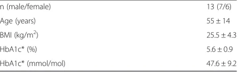
Table 1 Characteristics of human pancreatic donors included in the mRNA expression array analysis
n (male/female) 13 (7/6)
Age (years) 55 ± 14
BMI (kg/m2) 25.5 ± 4.3
HbA1c* (%) 5.6 ± 0.9
HbA1c* (mmol/mol) 47.6 ± 9.2
25 ml was taken and mixed with the 25 ml palmitate so-lution. The stock solution was then diluted to a final concentration of 1 mM palmitate and 1 weight % BSA (corresponding to 0.15 mM BSA) in the CMRL 1066 medium (ICN Biomedicals, Costa Mesa, CA, USA) sup-plemented with 10 mM nicotinamide (Sigma-Aldrich, Sweden, Stockholm), 10 mM HEPES buffer (GIBCO, BRL, Gaithersburg, MD, USA), 0.25 μg/ml fungizone (GIBCO), 50 μg/ml gentamicin, 2 mM L-glutamine (GIBCO), 10 μg/ml Ciprofloxacin (Bayer Healthcare, Leverkusen, Germany), 10% (v/v) heat-inactivated hu-man serum and 5.56 mM glucose. The molar (mmol/l) ratio of palmitate/BSA concentrations was 6.6:1 in the culture medium.
Palmitate treatment
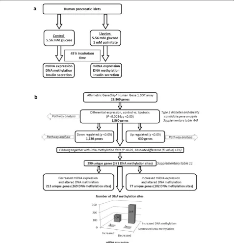
To study the impact of palmitate-induced lipotoxicity on human islets, approximately 1,000 islets from each donor (n = 13) were cultured for 48 h in CMRL 1066 medium (including 5.56 mM glucose) either with (lipotox) or with-out (control) 1 mM palmitate conjugated with 1% BSA (corresponding to 0.15 mM BSA) (Figure 1a). The same treatment time and palmitate/BSA ratio have been used in previous studies examining the impact of lipo-toxicity on islet function and was therefore selected in the present study [22,30]. Circulating non-esterified fatty acid levels have been reported to range between 0.59 to 0.83 mM for overweight, non-diabetic individ-uals (BMI approximately 26 kg/m2) and between 0.69 to 0.975 mM for overweight, diabetic individuals (BMI of approximately 29 kg/m2) [31]. The 1 mM palmitate used in the current study, which is close to the upper limit of the reference range, mimics the levels reported in overweight/obese individuals with diabetes. After 48 h DNA and RNA were extracted, glucose-stimulated insulin secretion was analyzed and/or apoptosis assays were performed.
RNA and DNA isolation
DNA and RNA were extracted from the human pancreatic islets using the AllPrep DNA/RNA kit (Qiagen, Hilden, Germany) according to the manufacturer’s instructions. RNA quantity and quality were assessed by Nanodrop (Nanodrop, Wilmington, DE, USA). The 260/280 ratios of all samples were between 1.98 and 2.16. The integrity and quality of the RNA was assessed using the Bioanalyzer (Agilent Technologies, Santa Clara, CA, USA). All RNA integrity number (RIN) values were≥7.4.
Microarray mRNA expression analysis
The Affymetrix GeneChip® Human Gene 1.0 ST whole transcript-based array (Affymetrix, Santa Clara, CA, USA) covering 28,869 genes was used to analyze mRNA ex-pression (Figure 1b) in pancreatic islets from 13 human
donors (Table 1) exposed to palmitate or control condi-tions (in total 26 samples) and in pancreatic islets from 87 diabetic donors as well as from diabetic and non-diabetic donors, according to the manufacturer’s recom-mendations. The Oligo package from Bioconductor was used to compute Robust Multichip Average expression measures [32].
Genome-wide DNA methylation analysis
A total of 500 ng genomic DNA from human pancreatic islets of 13 donors (Additional file 1: Table S1) exposed to palmitate or control conditions (in total 26 samples) was bisulfite-converted with the EZ DNA methylation kit (Zymo Research Corporation, Irvine, CA, USA). DNA methylation was analyzed by using the Infinium HumanMethylation450K BeadChip (Illumina, San Diego, CA, USA) which contains 485,577 probes and covers 99% of all RefSeq genes [33]. Bisulfite converted DNA was used to analyze DNA methylation with the Infi-nium® assay according to the standard Infinium HD Assay Methylation Protocol (Part # 15019519, Illumina). The Infinium HumanMethylation450K BeadChips were then imaged with the Illumina iScan. The raw methyla-tion score for each CpG site, which is represented as
β-value, was calculated using the GenomeStudio® methy-lation module software. Theβ-values were calculated as (β= intensity of the methylated allele (M)/(intensity of the Unmethylated allele (U) + intensity of the Methylated allele (M) + 100)). All samples passed GenomeStudio® quality control steps based on built-in control probes for staining, hybridization, extension and specificity, and dis-played high quality bisulfite conversion efficiency with an intensity signal above 4,000 [34]. Probes were filtered away from further analysis based on a mean detectionP-value >0.01. After quality control analysis, DNA methylation data were obtained for 483,844 probes.β-values were then converted to M-values (M = log2 (β/(1 - β))) for further bioinformatic and statistical analyses of the methylation data [35]. Background and quantile normalization was performed using the lumi package from Bioconductor [36]. Background correction was performed by subtracting the median M-value of the 600 built-in negative controls and methylation data were further normalized using quan-tile normalization [37]. ComBat was used to adjust for batch effects between arrays [38]. A linear regression model was used to identify differences in DNA methylation between control and palmitate-treated islets in a paired fashion as described elsewhere [39]. As β-values are bio-logically easier to interpret, M-values were reconverted to
KEGG pathway analysis
Kyoto Encyclopedia of Genes and Genomes (KEGG) pathway analysis of expression data was performed with the online tool WebGestalt [40,41] (accessed 27 March 2012 and 12 February 2014). For the pathway analysis of mRNA expression data, Affymetrix probe IDs were used to identify unique genes and Affymetrix
GeneChip® Human Gene 1.0 ST genes were used as background in this analysis. For the pathway analysis of the DNA methylation data, the gene symbol was used to identify unique genes and the human genome was used as background in this analysis. The Benjamini and Hochberg method was used to correct P-values for multiple testing.
Glucose-stimulated insulin secretion
Glucose-stimulated insulin secretion was analyzed in con-trol and palmitate-treated human islets from nine donors. After 48 h culture in control or palmitate-containing medium, 10 replicates of 10 human islets per culture con-dition (control and palmitate-treated) and donor were pre-incubated in HEPES-balanced salt solution (HBSS) containing (in mM) 114 NaCl, 4.7 KCl, 1.2 KH2PO4, 1.16
MgSO4, 20 HEPES, 25.5 NaHCO3, 2.5 CaCl2 at pH 7.2
with 0.575 BSA and 3.3 mM glucose (1.65 mM glucose for one sample) for 1 h at 37°C. Thereafter, for each donor, glucose was added to five of the replicates to a final con-centration of 16.7 mM glucose (15.05 mM glucose for one sample) to study glucose-stimulated insulin secretion and the other five replicates were kept in 3.3 mM glucose to study basal insulin secretion and the incubation was continued for one more hour. The supernatant was im-mediately removed and the insulin concentration in the medium was measured by radioimmunoassay (RIA) (Millipore, Uppsala, Sweden).
Assessment of apoptosis in human pancreatic islets
Apoptosis was measured in islets from four human do-nors with the Apo-ONE® Homogeneous Caspase-3/7 Assay (Promega, Madison, WI, USA) as described else-where [42]. The assay contains proflourescent rhodamin 110 (Z-DEVD-R110) which serves as a substrate for both Caspase-3 and -7. Upon lysis of cells, the available Cas-pase -3/-7 in the sample will cleave Z-DEVD-R110 to fluorescent rhodamine 110, which is then measured. Subsequently, the assay measures the combined activity of Caspase-3 and -7. After 48 h incubation in control or palmitate medium, triplicates of 20 human pancreatic is-lets each were handpicked from each culture condition, washed and transferred to a plate containing HBSS. After 1.5 h, fluorescence was measured with a Tecan Infinite M200pro plate reader (Tecan Group Ltd., Männedorf, Switzerland) to determine the Caspase-3/7 activity.
Statistics
A non-parametric paired test (Wilcoxon) was used to identify differences in mRNA expression between con-trol and palmitate-treated human islets. A False Discov-ery Rate (FDR) analysis was performed to correct for multiple testing in the mRNA expression data. Genes exhibiting differential expression with a FDR below 5% (q <0.05) were considered significant. To find associa-tions between BMI and gene expression in human is-lets, a linear regression model was used including age, gender, HbA1c, islet purity and days of culture as co-variates. To identify differences in gene expression be-tween T2D and non-diabetic islets a linear regression model was used including gender, BMI, age, islet pur-ity and days of culture as covariates. Data are presented
as mean ± standard error of mean (sem), unless stated otherwise.
Results
Impaired insulin secretion in human islets exposed to palmitate
To investigate the physiological response to 1 mM palmi-tate treatment for 48 h, we measured glucose-stimulated insulin secretion in human islets cultured under control (5.56 mM glucose) or lipotoxic (5.56 mM glucose and 1 mM palmitate) conditions. We found decreased glucose-stimulated insulin secretion measured as fold change (insu-lin secretion at high glucose levels/insu(insu-lin secretion at low glucose levels) in the palmitate-treated compared with control-treated human islets (Figure 2a). We also evaluated the effect of the palmitate treatment on apop-tosis in human islets by measuring the combined activity of Caspase-3 and -7. Palmitate treatment did not alter islet cell apoptosis rates (P= 0.62, Figure 2b).
Palmitate exposure influences mRNA expression in human islets
To study the impact of palmitate-induced lipotoxicity on gene expression in human islets, mRNA expression array data were generated for pancreatic islets from 13 donors cultured in control or lipotoxic conditions (Figure 1a). The characteristics of these 13 donors are described in Table 1. We identified 1,860 individual genes that were differentially expressed in human islets exposed to palmitate compared with the control condition after correction for multiple testing using a FDR below 5% (q <0.05) (Additional file 2: Table S2). Out of these 1,860 genes, 1,230 were down-regulated and 630 genes were up-down-regulated due to palmi-tate treatment. The work flow for the mRNA expression data can be viewed in Figure 1b.
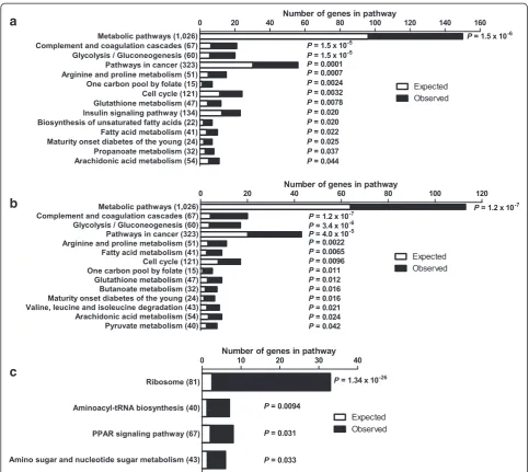
To test if genes in certain biological pathways were enriched among the differentially expressed genes in palmitate-treated human islets, KEGG pathway analyses were performed using WebGestalt. Pathway analyses were performed using either the list of all differentially expressed genes (q<0.05) or by dividing the genes based on down- or up-regulation in islets exposed to palmitate. A selection of the enriched pathways (Padjusted <0.05)
can be found in Figure 3 and all enriched pathways of possible relevance for lipotoxicity in human islets can be found in Additional file 3: Table S3, Additional file 4: Tables S4 and Additional file 5: Table S5. The metabolic pathways was the top KEGG pathway with down-regulated genes (Figure 3b and Additional file 4: Table S4) and it includes several genes encoding proteins involved in oxidative phosphorylation. Additionally, the glycolysis/ gluconeogenesis (Figures 3a, b, 4a and Additional file 3: Table S3 and Additional file 4: Table S4), fatty acid metab-olism, glutathione metabolism (Figures 3a, b, 4b and Additional file 3: Table S3 and Additional file 4: Table S4) and pyruvate metabolism (Figure 3b and Additional file 4: Table S4) pathways were enriched in human islets exposed to palmitate. Moreover, there was an enrich-ment of genes involved in the insulin signaling pathway (Figures 3a, 4c and Additional file 3: Table S3) and the biosynthesis of unsaturated fatty acids pathway (Figures 3a, 4d and Additional file 3: Table S3). Interest-ingly, the“one carbon pool by folate”was also enriched in the KEGG pathway analysis (Figure 3a, b and Additional file 3: Table S3 and Additional file 4: Table S4).
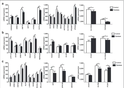
We further examined if candidate genes associated with either T2D, T2D-related traits or obesity in previ-ous GWAS were differentially expressed in human islets exposed to palmitate. Candidate gene lists were retrieved using the online GWAS SNP library [43,44] (accessed on 22 August 2012). Among these gene lists, we found 16 candidate genes for T2D out of a total of 86 genes (Figure 5a and Additional file 6: Table S6), 13 T2D-related trait genes out of a total of 76 (Figure 5b and
Additional file 7: Table S7) and 15 candidate genes for obesity out of a total of 127 (Figure 5c and Additional file 8: Table S8) that were differentially expressed palmitate-treated islets. However, since some candidate genes are associated with more than one trait, the differ-entially expressed candidate genes correspond to 38 unique genes out of a total of 262 genes.
Impact of palmitate on DNA methylation in human islets
To estimate the global DNA methylation in human is-lets, we calculated the average level of DNA methylation for all analyzed CpG sites on the Infinium Human-Methylation450K BeadChip array. The average level of genome-wide DNA methylation was slightly, but signifi-cantly, higher in palmitate-treated compared to control islets (44.9 ± 0.8 vs. 43.9 ± 1.2, P= 0.002). The analyzed DNA methylation sites on the array have been annotated to different gene and CpG island regions [33]. The anno-tated gene regions include TSS1500, TSS200, 5′UTR, 1st exon, gene body, 3′UTR and intergenic regions. Annota-tions were also made according to the location of the DNA methylation sites in relation to CpG islands, as previously defined [33]. The 2 kb sequences, directly up-and downstream of CpG islup-ands are called the northern and southern shore, respectively. The 2 kb sequences directly adjacent to the shores are called the northern and southern shelves. DNA methylation sites outside the CpG island regions are annotated as “open sea”. We then tested if palmitate exposure affects the average level of DNA methylation for any of these gene regions in hu-man islets. We found an increase in average DNA methylation in the palmitate-treated islets for all gene and CpG island regions except TSS200, 1st Exon and CpG islands (Figure 6 and Additional file 9: Table S9).
to palmitate exposure out of which 99 were located in 94 unique genes, and 30 were intergenic sites. The fold change for the 46,977 differentially methylated DNA methylation sites (P<0.05), calculated as DNA methyla-tion of palmitate-treated islets/DNA methylamethyla-tion of con-trol treated islets, ranged from 0.54 to 1.84. This corresponds to changes in DNA methylation from a 46% decrease to an 84% increase.
Overlapping changes in mRNA expression and DNA methylation in palmitate-treated human islets
Epigenetic modifications may regulate mRNA expression [3-5] and we therefore tested if any of the genes that
exhibit differential mRNA expression also exhibit differen-tial DNA methylation in islets exposed to palmitate. Sig-nificant mRNA expression data (q <0.05) were merged with DNA methylation sites withP<0.05 and an absolute difference in DNA methylation≥3%. We found 290 indi-vidual genes with differential mRNA expression (q <0.05) and a corresponding change in DNA methylation (in total, 371 DNA methylation sites) (Figure 1b and Additional file 11: Table S11). Out of these 290 genes, 213 had decreased mRNA expression together with altered DNA methylation (269 DNA methylation sites, whereof 264 sites had in-creased and 5 sites had dein-creased DNA methylation) in response to palmitate treatment. Additionally, 77 unique
a
b
c
genes had increased mRNA expression together with al-tered DNA methylation (102 DNA methylation sites, whereof 2 sites had decreased and 100 sites had increased DNA methylation) (Figure 1b). Furthermore, for some of the candidate genes for T2D, T2D related traits and obes-ity identified by GWAS, we found both differential mRNA expression and changes in DNA methylation in islets ex-posed to palmitate, for example,TCF7L2andGLIS3show decreased expression and increased DNA methylation (Additional file 6: Table S6, Additional file 7: Table S7 and Additional file 8: Table S8).
A KEGG pathway analysis was performed using Web-Gestalt to test if genes in biological pathways found in the mRNA expression pathway analysis (Additional file 3: Table S3, Additional file 4: Table S4 and Additional file 5: Table S5) also were enriched among the differen-tially methylated genes in palmitate-treated human islets.
The pathway analysis was performed using a list of all differentially methylated genes (P <0.05) (Additional file 10: Table S10). All 17 pathways with enrichment for both DNA methylation and gene expression are pre-sented in Additional file 12: Figure S1 and Additional file 13: Table S12.
The Infinium HumanMethylation450K BeadChip array has been reported to have probes with possible cross re-activity to other locations in the genome than their intended match [45]. Importantly, none of our reported probes withP <0.05 have a perfect match to other loca-tions in the genome. Furthermore, only 13 probes have a near-perfect match (Additional file 14: Table S13).
Impact of BMI and T2D on gene expression in human islets
Since it has been shown that T2D patients and obese individuals have elevated levels of free fatty acids
[12,13,46,47], we finally tested if increased BMI and/or T2D also affect islet expression in the same direction as any of the 1,860 genes that exhibit differential expression in human islets exposed to lipotoxicity for 48 h in vitro. The impact of BMI on expression of these 1,860 genes was examined in human islets from 87 non-diabetic do-nors with BMI spanning between 17.6 to 40.1 kg/m2. BMI was associated with differential expression of 67 of the 1,860 genes (Additional file 15: Table S14). The impact of T2D was examined in pancreatic islets from 15 donors with T2D and 34 non-diabetic donors. We found 37 genes differentially expressed (P<0.05) in islets from T2D versus non-diabetic donors overlapping with the 1,860 genes in the palmitate-exposed islets (Additional file 16: Table S15). The data for three of these genes (that is,CDKN1A, IL1RL2, TNFRSF10B) have been reported previously [48] and are hence not reported here. The top 10 genes show-ing differential expression in human islets due to both palmitate exposure and T2D are presented in Figure 7. Moreover, five genes, that is,RASGRP1, MIA2, CDKN1A,
TNFRSF103 and RAB7L1, were present among both the BMI- and T2D-associated genes.
Discussion
This study shows that palmitate exposure alters mRNA expression genome-wide in human islets in parallel with impaired insulin secretion, a defect often seen in T2D pa-tients. Several genes with altered expression in palmitate-treated human islets also exhibited differential expression in islets from patients with T2D. We also demonstrate for the first time that the genome-wide DNA methylation pat-tern in human islets was affected by palmitate treatment. Several genomic regions had significantly higher global DNA methylation levels in the palmitate-treated islets compared with control islets, although these differences were generally small. This may be the result of the rela-tively short treatment (48 h) and that DNA methylation changes of a larger magnitude may require longer expos-ure to hyperlipidemia, a condition seen in many T2D pa-tients. Also, since T2D is known to be a polygenic disease,
a
b
c
it is possible that a combination of several modest changes in DNA methylation might have a combined larger effect which together could contribute to the pathogenesis of the disease. In support of this hypothesis, previous studies have shown relatively modest differences of DNA methy-lation in non-cancerous tissues and cell types, ranging from 0.13% to 11% [9,49,50]. However, even an absolute change of only a few percent units can represent a large difference in relative terms, as evident by the findings in our study where the fold change of DNA methylation be-tween the treatment groups (palmitate treatment/control treatment) ranged from 0.54 to 1.84. This is in line with data from a recent study, where we found differential DNA methylation of 3,116 CpG sites in human pancreatic islets from subjects with T2D compared with non-diabetic controls with a fold change ranging from 0.58 to 1.61 when dividing the degree of methylation in diabetics with that in controls [48].
We also identified many genes with a difference in mRNA expression and a corresponding change in DNA methylation. This could suggest that altered DNA methy-lation influences the expression of the corresponding genes. Indeed, we have previously shown that increased
DNA methylation reduces the transcriptional activity in functionalin vitrostudies [5,8]. Interestingly, here we find decreased expression in parallel with increased DNA methylation of several candidate genes for T2D, such as
TCF7L2 and GLIS3 [51], in palmitate-treated human is-lets, suggesting that lipid-induced epigenetic modifications may affect the risk for diabetes. The fact that many of the up-regulated genes have corresponding increased DNA methylation could be due to the location of these CpG sites in the gene body. Indeed, DNA methylation of the gene body has been demonstrated to have a positive effect on gene expression [52]. The gene regions with differential gene expression but without any change in DNA methyla-tion could be targets for other forms of transcripmethyla-tional regulation, such as histone modifications and/or altered activation by transcription factors. Also, genetic and epi-genetic variation may interact to affect gene expression and subsequently contribute to the development of complex metabolic disease, such as obesity and T2D. In-deed, it has previously been shown that SNPs that
Figure 6Impact of palmitate treatment on global DNA methylation in human islets.Average DNA methylation levels ofa)gene regions andb)CpG island regions in control and lipotoxic treated human islets. All data are presented as mean ± sem. *q<0.05.
introduce or remove a CpG site, so called CpG-SNPs, can influence the expression of target genes by interfer-ing with certain proteins [53]. Moreover, we recently showed that approximately 50% of SNPs associated with T2D are CpG-SNPs, which affect the degree of DNA methylation in the SNP site as well as gene expression and alternative splicing events in human pancreatic islets [7]. It has been hypothesized that since DNA methylation can affect the regulation of splicing, CpG-SNPs can possibly affect alternative splicing events [54].
There is an increased risk for obesity and T2D among children with obese and/or diabetic parents [55,56]. Add-itionally, rodent studies demonstrate that an altered intra-uterine environment gives rise to epigenetic changes, which later in life can predispose the offspring to impaired metabolism and T2D [57-59]. These data suggest that epi-genetic modifications contribute to the pathogenesis of T2D. Based on the results from our study, we speculate that early exposure to palmitate may affect the epigenetic patterns of genes which are known to affect the risk of T2D. This may increase the risk of disease later in life. However, we cannot exclude that epigenetic changes seen in patients with T2D are secondary to the disease [4,5,48,60,61].
Our human insulin secretion data are in concordance with previous rodent studies, where palmitate treatment was found to lower glucose-stimulated insulin secretion in rodent pancreatic islets [17,18]. A tight coupling of gly-colysis to mitochondrial respiration and ATP production is essential for proper beta-cell function and glucose-stimulated insulin secretion. Palmitate treatment of hu-man islets resulted in altered expression of individual metabolic genes as well as of genes in metabolic pathways such as glycolysis/gluconeogenesis, pyruvate metabolism and biosynthesis of unsaturated fatty acids. Additionally, several down-regulated genes in the enriched metabolic pathways encode proteins which are part of the respira-tory chain, for example, NDUFA4, NDUFB5, NDUFS1,
NDUFS2, SDHA and UQCRB. Decreased expression of these genes may contribute to decreased oxidative phos-phorylation and subsequently decreased ATP production and insulin secretion in islets exposed to lipotoxicity. In-deed, our previous study showed that decreased expres-sion of genes involved in oxidative phosphorylation results in impaired insulin secretion [62].
While some studies have found decreased beta-cell number in T2D islets, others do not find an altered cell composition in diabetic islets [10,63-65]. In the present study, palmitate had no significant effect on apoptosis in human islets and it is hence unlikely that the beta-cell number is significantly decreased. As the majority of cell types in human islets have important effects on whole body glucose homeostasis [23], it is physiologically war-ranted to study both whole human islets and cell-lines
representing the individual cell types in the pancreatic islets.
Additionally, the insulin signaling pathway was signifi-cantly enriched when performing a pathway analysis on all the significant expression data, including both up-and down-regulated genes. Interestingly, this pathway was also enriched when performing a pathway analysis on the differentially methylated genes. Previous studies have shown that insulin signaling contributes in the regulation of beta-cell mass and apoptosis as well as in-sulin synthesis and secretion [66] and here we show that this pathway is affected by palmitate treatment in human islets. This in turn could potentially affect insulin secre-tion in these islets. PPARGC1A (encoding PGC1α) is a part of the insulin signaling pathway and its expression was reduced in human islets exposed to palmitate. We have previously shown thatPPARGC1Aexpression is de-creased in islets from T2D patients compared to non-diabetics, andPPARGC1Aexpression correlated positively with insulin secretion in human islets [6]. PPARGC1A
encodes a transcriptional co-activator of mitochondrial genes involved in oxidative phosphorylation and silencing ofPPARGC1Ain human islets results in decreased insulin secretion [6]. Furthermore, SCD (encoding for stearoyl-CoA desaturase (delta-9-desaturase)) was up-regulated in human islets due to palmitate treatment.SCDis a compo-nent of the biosynthesis of unsaturated fatty acids path-way, which was enriched in the KEGG pathway analysis. Stearoyl-CoA desaturase catalyzes the conversion of satu-rated fatty acids to unsatusatu-rated fatty acids, and it has been shown to protect rodent and human beta-cells from palmitate-induced ER stress and apoptosis [67,68]. Our re-sult is in accordance with these previous studies and could provide an explanation for the absence of an increase in apoptosis in the palmitate-treated human islets.
Furthermore, the “one carbon pool by folate” pathway was enriched in the KEGG pathway analysis using both mRNA expression data and DNA methylation data. Al-tered expression of genes in this pathway may affect the amount of methyl donors, for example, S-adenosyl me-thionine in the islets exposed to palmitate and, thereby, contribute to differential DNA methylation. SHMT2
andMTHFD2 were both up-regulated due to palmitate exposure. The enzymes encoded by these genes are in-volved in the folate cycle which is linked to the methionine cycle, which in turn controls the amount of S-adenosyl methionine [69].
in vivo. While some previous studies have examined the impact of lipotoxicity on the expression of a limited number of candidate genes in human islets in vitro
[24-26], the present study is to our knowledge the first to perform a genome-wide analysis of gene expression in lipotoxic-treated human islets of more than five donors [28,29].
It is debated whether lipotoxicity can occur in the ab-sence of high glucose levels, a phenomenon known as glucolipotoxicity. However, previous in vivo studies in humans have shown that prolonged exposure (24 to 48 h) to free fatty acids, in the absence of elevated glu-cose levels, perturbs islet function [20]. Moreover, a re-cent study showed that the lipotoxic effect of palmitate occurs even at low concentrations of glucose in intact human islets [30]. Our findings provide further evidence that palmitate-induced lipotoxicity under normal glucose conditions results in extensive transcriptional changes and impaired insulin secretion in human islets. It is im-portant, however, to note that our study only examined the effects of palmitate on human islets, and it is known that different fatty acids can have divergent, and even opposite, effects on cell function. Also, the in vivo fatty acid composition in plasma contains several different fatty acids [71] where palmitate is one of the most abun-dant saturated fatty acids. We can therefore in our study not rule out that other types of fatty acids have add-itional effects on human islets. However, our study pro-vides evidence for palmitate-induced changes on gene expression, DNA methylation and insulin secretion which might be of relevance to phenotypes seen in obese individuals and T2D patients. Finally, as our previous studies have shown that the genome-wide methods used in the present study are robust and reproducible, we did not technically validate the array results in the present study [8,72-74].
Conclusion
In conclusion, we have identified novel genes and meta-bolic pathways that are affected by palmitate exposure in human pancreatic islets. Importantly, a number of these genes also show differential gene expression in islets from patients with T2D. We have also shown for the first time that there are both global and specific changes in the DNA methylation pattern in the palmitate-treated islets which may affect mRNA expression. Together, these changes may contribute to the impaired insulin se-cretion seen in palmitate-treated human islets.
Additional files
Additional file 1: Table S1.Characteristics of the human pancreatic donors included in the DNA methylation array analysis.
Additional file 2: Table S2.Differentially expressed genes in human islets exposed to palmitate compared with the control (q<0.05).
Additional file 3: Table S3.Biological pathways including all genes that exhibit differential mRNA expression (q<0.05) in human pancreatic islets exposed to palmitate compared to control treatment.
Additional file 4: Table S4.Biological pathways including genes that exhibit decreased mRNA expression (q<0.05) in human pancreatic islets exposed to palmitate compared to control treatmet.
Additional file 5: Table S5.Biological pathways including genes that exhibit increased mRNA expression (q<0.05) in human pancreatic islets exposed to palmitate compared to control treatmet.
Additional file 6: Table S6.Differential mRNA expression (q<0.05) of candidate genes for type 2 diabetes (T2D) in human pancreatic islets exposed to palmitate versus control. DNA methylation data are displayed if the absolute difference in DNA methylation≥3% andP<0.05.
Additional file 7: Table S7.Differential mRNA expression (q<0.05) of candidate genes for type 2 diabetes (T2D) related traits in human pancreatic islets exposed to palmitate versus control. DNA methylation data are displayed if the absolute difference in DNA methylation≥3% andP<0.05.
Additional file 8: Table S8.Differential mRNA expression (q<0.05) of candidate genes for obesity in human pancreatic islets exposed to palmitate versus control. DNA methylation data are displayed if the absolute difference in DNA methylation≥3% andP<0.05.
Additional file 9: Table S9.Global DNA methylation in human pancreatic islets exposed to palmitate compared to control treatment. Average DNA methylation (%) in gene- and CpG-regions in human pancreatic islets treated for 48 h with control media or palmitate-containing media.
Additional file 10: Table S10.CpG sites with differential DNA methylation in human pancreatic islets exposed to palmitate compared to control treatmentP<0.05.
Additional file 11: Table S11.Differential mRNA expression (q<0.05) of genes with a corresponding change in DNA methylation (P<0.05 and absolute DNA methylation difference≥3%) of the nearest gene, in human pancreatic islets exposed to palmitate versus control.
Additional file 12: Figure S1.Results from KEGG pathway analysis using genes that exhibit differential DNA methylation (P<0.05) in human pancreatic islets exposed to palmitate compared to control treatment. It includes only pathways which also displayed enrichment for genes with differential expression after palmitate treatment in human pancreatic islets.
Additional file 13: Table S12.Biological pathways including genes that exhibit differential DNA methylation (P<0.05) in human pancreatic islets exposed to palmitate compared to control teatment. It includes only pathways which also displayed enrichment for genes with differential expression after palmitate treatment in human pancreatic islets.
Additional file 14: Table S13.Infinium HumanMethylation450K BeadChip array probes with possible cross reactivity to other locations in the genome.
Additional file 15: Table S14.Genes displaying significant difference in mRNA expression between control and palmitate-treated human pancreatic islets (q<0.05) as well as significant correlation between BMI and mRNA expression in human pancreatic islets (P<0.05).
Additional file 16: Table S15.Differentially expressed genes (P<0.05) in islets from T2D versus non-diabetic donors overlapping with the 1,860 genes with altered expression in the palmitate-exposed islets.
Abbreviations
Competing interests
The authors declare that there is no duality of interest associated with this manuscript.
Authors’contributions
EH designed and conducted the study, performed lab work, collected, analyzed and interpreted data, and wrote the manuscript. PV analyzed and interpreted data and reviewed and edited the manuscript. TD interpreted data and reviewed and edited the manuscript. KB performed lab work and reviewed and edited the manuscript. TR interpreted data and reviewed and edited the manuscript. MDN designed and conducted the study, collected and interpreted data, and reviewed and edited the manuscript. CL designed and conducted the study, interpreted data, and reviewed and edited the manuscript. EH, MDN and CL are guarantors of this work and, as such, had full access to all of the data in the study and take responsibility for the integrity of the data. All authors read and approved the final version of the manuscript.
Acknowledgements
We thank SCIBLU at Lund University for analyzing mRNA expression. We thank the Nordic Network for Clinical Islet Transplantation (JDRF award 31-2008-413), the tissue isolation teams and Human Tissue Laboratory within EXODIAB/Lund University Diabetes Centre. We also thank Professor Claes Wollheim (Lund University, Department of Clinical Sciences, Malmö, Sweden) for helpful comments on the manuscript. Thanks to Siri Malmgren (Lund University, Department of Clinical Sciences, Lund, Sweden) and Malin Fex (Lund University, Department of Clinical Sciences, Malmö, Sweden) for dedicating their time and knowledge. This work was supported by grants from the Swedish Research Council, Region Skåne, Knut and Alice Wallenberg Foundation, Novo Nordisk foundation, Söderberg, Diabetes Fonden, Påhlsson, Tore Nilsson Foundation, Thurings Foundation, Nilsson-Ehle Foundation, Linné grant (B31 5631/2006) and EXODIAB.
Author details
1Epigenetics and Diabetes Unit, Department of Clinical Sciences, Lund
University Diabetes Centre, CRC, Lund University, Scania University Hospital, Malmö, Sweden.2School of Medicine, Royal Brisbane Clinical School, The
University of Queensland, Herston, QLD 4006, Australia.
Received: 4 December 2013 Accepted: 25 April 2014 Published:
References
1. McCarthy MI:Genomics, type 2 diabetes, and obesity.N Engl J Med2010, 363:2339–2350.
2. Voight BF, Scott LJ, Steinthorsdottir V, Morris AP, Dina C, Welch RP, Zeggini E, Huth C, Aulchenko YS, Thorleifsson G, McCulloch LJ, Ferreira T, Grallert H, Amin N, Wu G, Willer CJ, Raychaudhuri S, McCarroll SA, Langenberg C, Hofmann OM, Dupuis J, Qi L, Segrè AV, van Hoek M, Navarro P, Ardlie K, Balkau B, Benediktsson R, Bennett AJ, Blagieva R,et al:Twelve type 2 diabetes susceptibility loci identified through large-scale association analysis.Nat Genet2010,42:579–589.
3. Ling C, Groop L:Epigenetics: a molecular link between environmental factors and type 2 diabetes.Diabetes2009,58:2718–2725.
4. Yang BT, Dayeh TA, Kirkpatrick CL, Taneera J, Kumar R, Groop L, Wollheim CB, Dekker Nitert M, Ling C:Insulin promoter DNA methylation correlates negatively with insulin gene expression and positively with HbA(1c) levels in human pancreatic islets.Diabetologia2011,54:360–367. 5. Yang BT, Dayeh TA, Volkov PA, Kirkpatrick CL, Malmgren S, Jing X, Renstrom
E, Wollheim CB, Dekker Nitert M, Ling C:Increased DNA methylation and decreased expression of PDX-1 in pancreatic islets from patients with type 2 diabetes.Mol Endocrinol2012,26:1203–1212.
6. Ling C, Del Guerra S, Lupi R, Rönn T, Granhall C, Luthman H, Masiello P, Marchetti P, Groop L, Del Prato S:Epigenetic regulation of PPARGC1A in human type 2 diabetic islets and effect on insulin secretion.Diabetologia 2008,51:615–622.
7. Dayeh TA, Olsson AH, Volkov P, Almgren P, Rönn T, Ling C:Identification of CpG-SNPs associated with type 2 diabetes and differential DNA methylation in human pancreatic islets.Diabetologia2013,56:1036–1046. 8. Nitert MD, Dayeh T, Volkov P, Elgzyri T, Hall E, Nilsson E, Yang BT, Lang S,
Parikh H, Wessman Y, Weishaupt H, Attema J, Abels M, Wierup N, Almgren
P, Jansson PA, Rönn T, Hansson O, Eriksson KF, Groop L, Ling C:Impact of an exercise intervention on DNA methylation in skeletal muscle from first-degree relatives of patients with type 2 diabetes.Diabetes2012, 61:3322–3332.
9. Ribel-Madsen R, Fraga MF, Jacobsen S, Bork-Jensen J, Lara E, Calvanese V, Fernandez AF, Friedrichsen M, Vind BF, Hojlund K, Beck-Nielsen H, Esteller M, Vaag A, Poulsen P:Genome-wide analysis of DNA methylation differences in muscle and fat from monozygotic twins discordant for type 2 diabetes. PLoS One2012,7:e51302.
10. Volkmar M, Dedeurwaerder S, Cunha DA, Ndlovu MN, Defrance M, Deplus R, Calonne E, Volkmar U, Igoillo-Esteve M, Naamane N, Del Guerra S, Masini M, Bugliani M, Marchetti P, Cnop M, Eizirik DL, Fuks F:DNA methylation profiling identifies epigenetic dysregulation in pancreatic islets from type 2 diabetic patients.EMBO J2012,31:1405–1426.
11. Barres R, Osler ME, Yan J, Rune A, Fritz T, Caidahl K, Krook A, Zierath JR: Non-CpG methylation of the PGC-1alpha promoter through DNMT3B controls mitochondrial density.Cell Metab2009,10:189–198. 12. Boden G, Shulman GI:Free fatty acids in obesity and type 2 diabetes:
defining their role in the development of insulin resistance and beta-cell dysfunction.Eur J Clin Invest2002,32:14–23.
13. Kahn SE, Hull RL, Utzschneider KM:Mechanisms linking obesity to insulin resistance and type 2 diabetes.Nature2006,444:840–846.
14. Maedler K, Oberholzer J, Bucher P, Spinas GA, Donath MY:
Monounsaturated fatty acids prevent the deleterious effects of palmitate and high glucose on human pancreatic beta-cell turnover and function. Diabetes2003,52:726–733.
15. Morgan NG:Fatty acids and beta-cell toxicity.Curr Opin Clin Nutr Metab Care2009,12:117–122.
16. Cnop M, Hannaert JC, Hoorens A, Eizirik DL, Pipeleers DG:Inverse relationship between cytotoxicity of free fatty acids in pancreatic islet cells and cellular triglyceride accumulation.Diabetes2001,50:1771–1777. 17. Zhou YP, Grill VE:Long-term exposure of rat pancreatic islets to fatty
acids inhibits glucose-induced insulin secretion and biosynthesis through a glucose fatty acid cycle.J Clin Invest1994,93:870–876. 18. Olofsson CS, Collins S, Bengtsson M, Eliasson L, Salehi A, Shimomura K,
Tarasov A, Holm C, Ashcroft F, Rorsman P:Long-term exposure to glucose and lipids inhibits glucose-induced insulin secretion downstream of granule fusion with plasma membrane.Diabetes2007,56:1888–1897. 19. Carpentier A, Mittelman SD, Lamarche B, Bergman RN, Giacca A, Lewis GF:
Acute enhancement of insulin secretion by FFA in humans is lost with prolonged FFA elevation.Am J Physiol1999,276:E1055–E1066. 20. Leung N, Sakaue T, Carpentier A, Uffelman K, Giacca A, Lewis GF:
Prolonged increase of plasma non-esterified fatty acids fully abolishes the stimulatory effect of 24 hours of moderate hyperglycaemia on insulin sensitivity and pancreatic beta-cell function in obese men. Diabetologia2004,47:204–213.
21. Busch AK, Cordery D, Denyer GS, Biden TJ:Expression profiling of palmitate- and oleate-regulated genes provides novel insights into the effects of chronic lipid exposure on pancreatic beta-cell function. Diabetes2002,51:977–987.
22. Malmgren S, Spégel P, Danielsson AP, Nagorny CL, Andersson LE, Dekker Nitert M, Ridderstråle M, Mulder H, Ling C:Coordinate changes in histone modifications, mRNA levels and metabolite profiles in clonal INS-1 832/13 beta-cells accompany functional adaptations to lipotoxicity. J Biol Chem2013,288:11973–11987.
23. Woods SC, Lutz TA, Geary N, Langhans W:Pancreatic signals controlling food intake; insulin, glucagon and amylin.Philos Trans R Soc Lond B Biol Sci2006,361:1219–1235.
24. Igoillo-Esteve M, Marselli L, Cunha DA, Ladrière L, Ortis F, Grieco FA, Dotta F, Weir GC, Marchetti P, Eizirik DL, Cnop M:Palmitate induces a pro-inflammatory response in human pancreatic islets that mimics CCL2 expression by beta cells in type 2 diabetes.Diabetologia2010, 53:1395–1405.
25. Cunha DA, Igoillo-Esteve M, Gurzov EN, Germano CM, Naamane N, Marhfour I, Fukaya M, Vanderwinden JM, Gysemans C, Mathieu C, Marselli L, Marchetti P, Harding HP, Ron D, Eizirik DL, Cnop M:Death protein 5 and p53-upregulated modulator of apoptosis mediate the endoplasmic reticulum stress-mitochondrial dialog triggering lipotoxic rodent and human beta-cell apoptosis.Diabetes2012,61:2763–2775. 26. Lefebvre B, Vandewalle B, Balavoine AS, Queniat G, Moerman E, Vantyghem
and functional effects of ZNT8 in human pancreatic islets.J Endocrinol 2012,214:225–232.
27. Natalicchio A, Labarbuta R, Tortosa F, Biondi G, Marrano N, Peschechera A, Carchia E, Orlando MR, Leonardini A, Cignarelli A, Marchetti P, Perrini S, Laviola L, Giorgino F:Exendin-4 protects pancreatic beta cells from palmitate-induced apoptosis by interfering with GPR40 and the MKK4/7 stress kinase signalling pathway.Diabetologia2013,56:2456–2466. 28. Ghanaat-Pour H, Sjöholm Å:Exenatide and pioglitazone regulate fatty
acid-induced gene expression in normal and diabetic human islets. Metabolomics2011,1:102.
29. Cnop M, Abdulkarim B, Bottu G, Cunha DA, Igoillo-Esteve M, Masini M, Turatsinze JV, Griebel T, Villate O, Santin I, Bugliani M, Ladriere L, Marselli L, McCarthy MI, Marchetti P:Sammeth M.Eizirik DL: RNA-sequencing identifies dysregulation of the human pancreatic islet transcriptome by the saturated fatty acid palmitate. Diabetes; 2013 [Epub ahead of print].
30. Sargsyan E, Bergsten P:Lipotoxicity is glucose-dependent in INS-1E cells but not in human islets and MIN6 cells.Lipids Health Dis2011,10:115. 31. Carlsson M, Wessman Y, Almgren P, Groop L:High levels of nonesterified
fatty acids are associated with increased familial risk of cardiovascular disease.Arterioscler Thromb Vasc Biol2000,20:1588–1594.
32. Irizarry RA, Bolstad BM, Collin F, Cope LM, Hobbs B, Speed TP:Summaries of Affymetrix GeneChip probe level data.Nucleic Acids Res2003,31:e15. 33. Bibikova M, Barnes B, Tsan C, Ho V, Klotzle B, Le JM, Delano D, Zhang L,
Schroth GP, Gunderson KL, Fan JB, Shen R:High density DNA methylation array with single CpG site resolution.Genomics2011,98:288–295. 34. Teschendorff AE, Menon U, Gentry-Maharaj A, Ramus SJ, Gayther SA,
Apostolidou S, Jones A, Lechner M, Beck S, Jacobs IJ, Widschwendter M:An epigenetic signature in peripheral blood predicts active ovarian cancer. PLoS One2009,4:e8274.
35. Du P, Zhang X, Huang CC, Jafari N, Kibbe WA, Hou L, Lin SM:Comparison of Beta-value and M-value methods for quantifying methylation levels by microarray analysis.BMC Bioinforma2010,11:587.
36. Du P, Kibbe WA, Lin SM:Lumi: a pipeline for processing Illumina microarray.Bioinformatics2008,24:1547–1548.
37. Bolstad BM, Irizarry RA, Astrand M, Speed TP:A comparison of normalization methods for high density oligonucleotide array data based on variance and bias.Bioinformatics2003,19:185–193. 38. Chen C, Grennan K, Badner J, Zhang D, Gershon E, Jin L, Liu C:Removing
batch effects in analysis of expression microarray data: an evaluation of six batch adjustment methods.PLoS One2011,6:e17238.
39. Smyth GK:Limma: linear models for microarray data.InBioinformatics and Computational Biology Solutions Using R and Bioconductor.New York: Springer; 2005:397–420.
40. Zhang B, Kirov S, Snoddy J:WebGestalt: an integrated system for exploring gene sets in various biological contexts.Nucleic Acids Res2005,33:W741–W748. 41. WEB-based GEne SeT AnaLysis Toolkit: translating gene lists into
biological insights.[http://bioinfo.vanderbilt.edu/webgestalt/] 42. Scholz H, Lund T, Dahle MK, Collins JL, Korsgren O, Wang JE, Foss A:
The synthetic liver X receptor agonist GW3965 reduces tissue factor production and inflammatory responses in human isletsin vitro. Diabetologia2009,52:1352–1362.
43. National Human Genome Research Institute.[www.genome.gov/ gwastudies].
44. Hindorff LA, Sethupathy P, Junkins HA, Ramos EM, Mehta JP, Collins FS, Manolio TA:Potential etiologic and functional implications of genome-wide association loci for human diseases and traits. Proc Natl Acad Sci U S A2009,106:9362–9367.
45. Chen YA, Lemire M, Choufani S, Butcher DT, Grafodatskaya D, Zanke BW, Gallinger S, Hudson TJ, Weksberg R:Discovery of cross-reactive probes and polymorphic CpGs in the Illumina Infinium HumanMethylation450 microarray.Epigenetics2013,8:203–209.
46. Kankaanpaa M, Lehto HR, Parkka JP, Komu M, Viljanen A, Ferrannini E, Knuuti J, Nuutila P, Parkkola R, Iozzo P:Myocardial triglyceride content and epicardial fat mass in human obesity: relationship to left ventricular function and serum free fatty acid levels.J Clin Endocrinol Metab2006, 91:4689–4695.
47. Clore JN, Allred J, White D, Li J, Stillman J:The role of plasma fatty acid composition in endogenous glucose production in patients with type 2 diabetes mellitus.Metabolism2002,51:1471–1477.
48. Dayeh T, Volkov P, Salö S, Hall E, Nilsson E, Olsson AH, Kirkpatrick CL, Wollheim CB, Eliasson L, Rönn T, Bacos K, Ling C:Genome-wide DNA
methylation analysis of human pancreatic islets from type 2 diabetic and non-diabetic donors identifies candidate genes that influence insulin secretion.PLoS Genet2014,10:e1004160.
49. Rakyan VK, Beyan H, Down TA, Hawa MI, Maslau S, Aden D, Daunay A, Busato F, Mein CA, Manfras B, Dias KR, Bell CG, Tost J, Boehm BO, Beck S, Leslie RD: Identification of type 1 diabetes-associated DNA methylation variable positions that precede disease diagnosis.PLoS Genet2011,7:e1002300. 50. Toperoff G, Aran D, Kark JD, Rosenberg M, Dubnikov T, Nissan B, Wainstein J,
Friedlander Y, Levy-Lahad E, Glaser B, Hellman A:Genome-wide survey reveals predisposing diabetes type 2-related DNA methylation variations in human peripheral blood.Hum Mol Genet2012,21:371–383.
51. Nogueira TC, Paula FM, Villate O, Colli ML, Moura RF, Cunha DA, Marselli L, Marchetti P, Cnop M, Julier C, Eizirik DL:GLIS3, a susceptibility gene for type 1 and type 2 diabetes, modulates pancreatic beta cell apoptosis via regulation of a splice variant of the BH3-Only protein bim.PLoS Genet 2013,9:e1003532.
52. Jones PA:Functions of DNA methylation: islands, start sites, gene bodies and beyond.Nat Rev Genet2012,13:484–492.
53. Taqi MM, Bazov I, Watanabe H, Sheedy D, Harper C, Alkass K, Druid H, Wentzel P, Nyberg F, Yakovleva T, Bakalkin G:Prodynorphin CpG-SNPs associated with alcohol dependence: elevated methylation in the brain of human alcoholics.Addict Biol2011,16:499–509.
54. Shukla S, Kavak E, Gregory M, Imashimizu M, Shutinoski B, Kashlev M, Oberdoerffer P, Sandberg R, Oberdoerffer S:CTCF-promoted RNA polymerase II pausing links DNA methylation to splicing.Nature2011, 479:74–79.
55. Whitaker RC, Wright JA, Pepe MS, Seidel KD, Dietz WH:Predicting obesity in young adulthood from childhood and parental obesity.N Engl J Med 1997,337:869–873.
56. Köbberling J, Tillil H:Empirical risk figures for first-degree relatives of non-insulin dependent diabetics. InThe Genetics of Diabetes Mellitus. London: Academic Press; 1982:201–209.
57. Park JH, Stoffers DA, Nicholls RD, Simmons RA:Development of type 2 diabetes following intrauterine growth retardation in rats is associated with progressive epigenetic silencing of Pdx1.J Clin Invest2008, 118:2316–2324.
58. Pinney SE, Jaeckle Santos LJ, Han Y, Stoffers DA, Simmons RA:Exendin-4 increases histone acetylase activity and reverses epigenetic
modifications that silence Pdx1 in the intrauterine growth retarded rat. Diabetologia2011,54:2606–2614.
59. Thompson RF, Fazzari MJ, Niu H, Barzilai N, Simmons RA, Greally JM: Experimental intrauterine growth restriction induces alterations in DNA methylation and gene expression in pancreatic islets of rats.J Biol Chem 2010,285:15111–15118.
60. Rakyan VK, Down TA, Balding DJ, Beck S:Epigenome-wide association studies for common human diseases.Nat Rev Genet2011,12:529–541. 61. Hall E, Dayeh T, Kirkpatrick CL, Wollheim CB, Dekker Nitert M, Ling C:DNA
methylation of the glucagon-like peptide 1 receptor (GLP1R) in human pancreatic islets.BMC Med Genet2013,14:76.
62. Koeck T, Olsson AH, Dekker Nitert M, Sharoyko VV, Ladenvall C, Kotova O, Reiling E, Rönn T, Parikh H, Taneera J, Eriksson JG, Metodiev MD, Larsson NG, Balhuizen A, Luthman H, Stančáková A, Kuusisto J, Laakso M, Poulsen P, Vaag A, Groop L, Lyssenko V, Mulder H, Ling C:A common variant in TFB1M is associated with reduced insulin secretion and increased future risk of type 2 diabetes.Cell Metab2011,13:80–91.
63. Butler AE, Janson J, Bonner-Weir S, Ritzel R, Rizza RA, Butler PC:Beta-cell deficit and increased beta-cell apoptosis in humans with type 2 diabetes.Diabetes2003,52:102–110.
64. Clark A, Jones LC, de Koning E, Hansen BC, Matthews DR:Decreased insulin secretion in type 2 diabetes: a problem of cellular mass or function? Diabetes2001,50:S169–S171.
65. Henquin JC, Rahier J:Pancreatic alpha cell mass in European subjects with type 2 diabetes.Diabetologia2011,54:1720–1725.
66. Leibiger IB, Leibiger B, Berggren PO:Insulin signaling in the pancreatic beta-cell.Annu Rev Nutr2008,28:233–251.
67. Hellemans KH, Hannaert JC, Denys B, Steffensen KR, Raemdonck C, Martens GA, Van Veldhoven PP, Gustafsson JA, Pipeleers D:Susceptibility of pancreatic beta cells to fatty acids is regulated by LXR/PPARalpha-dependent stearoyl-coenzyme A desaturase.PLoS One2009,4:e7266.
stearoyl coenzyme A desaturase protects pancreatic beta-cells from lipoapoptosis.Diabetes2005,54:2917–2924.
69. Locasale JW:Serine, glycine and one-carbon units: cancer metabolism in full circle.Nat Rev Cancer2013,13:572–583.
70. Taneera J, Lang S, Sharma A, Fadista J, Zhou Y, Ahlqvist E, Jonsson A, Lyssenko V, Vikman P, Hansson O, Parikh H, Korsgren O, Soni A, Krus U, Zhang E, Jing XJ, Esguerra JL, Wollheim CB, Salehi A, Rosengren A, Renström E, Groop L:A systems genetics approach identifies genes and pathways for type 2 diabetes in human islets.Cell Metab2012, 16:122–134.
71. Watt MJ, Hoy AJ, Muoio DM, Coleman RA:Distinct roles of specific fatty acids in cellular processes: implications for interpreting and reporting experiments.Am J Physiol Endocrinol Metab2012,302:E1–E3. 72. Olsson AH, Yang BT, Hall E, Taneera J, Salehi A, Nitert MD, Ling C:
Decreased expression of genes involved in oxidative phosphorylation in human pancreatic islets from patients with type 2 diabetes.Eur J Endocrinol2011,165:589–595.
73. Rönn T, Volkov P, Davegårdh C, Dayeh T, Hall E, Olsson AH, Nilsson E, Tornberg A, Dekker Nitert M, Eriksson KF, Jones HA, Groop L, Ling C: A six months exercise intervention influences the genome-wide DNA methylation pattern in human adipose tissue.PLoS Genet2013, 9:e1003572.
74. Jacobsen SC, Brons C, Bork-Jensen J, Ribel-Madsen R, Yang B, Lara E, Hall E, Calvanese V, Nilsson E, Jorgensen SW, Mandrup S, Ling C, Fernandez AF, Fraga MF, Poulsen P, Vaag A:Effects of short-term high-fat overfeeding on genome-wide DNA methylation in the skeletal muscle of healthy young men.Diabetologia2012,55:3341–3349.
Cite this article as:Hallet al.:Effects of palmitate on genome-wide mRNA expression and DNA methylation patterns in human pancreatic islets.BMC Medicine
Submit your next manuscript to BioMed Central and take full advantage of:
• Convenient online submission
• Thorough peer review
• No space constraints or color figure charges
• Immediate publication on acceptance
• Inclusion in PubMed, CAS, Scopus and Google Scholar
• Research which is freely available for redistribution
Submit your manuscript at www.biomedcentral.com/submit 10.1186/1741-7015-12-103