R E S E A R C H
Open Access
Tissue source determines the
differentiation potentials of mesenchymal
stem cells: a comparative study of human
mesenchymal stem cells from bone
marrow and adipose tissue
Liangliang Xu
1,3†, Yamei Liu
2†, Yuxin Sun
3, Bin Wang
3, Yunpu Xiong
7, Weiping Lin
3, Qiushi Wei
1, Haibin Wang
1,
Wei He
1,9*, Bin Wang
7*and Gang Li
3,4,5,6,8*Abstract
Background:Mesenchymal stem cells (MSCs) possess intrinsic regeneration capacity as part of the repair process in response to injury, such as fracture or other tissue injury. Bone marrow and adipose tissue are the major sources of MSCs. However, which cell type is more effective and suitable for cell therapy remains to be answered. The intrinsic molecular mechanism supporting the assertion has also been lacking.
Methods:Human bone marrow-derived MSCs (BMSCs) and adipose tissue-derived MSCs (ATSCs) were isolated from bone marrow and adipose tissue obtained after total hip arthroplasty. ATSCs and BMSCs were incubated in standard growth medium. Trilineage differentiation including osteogenesis, adipogenesis, and chondrogenesis was performed by addition of relevant induction mediums. The expression levels of trilineage differentiation marker genes were evaluated by quantitative RT-PCR. The methylation status of CpG sites of Runx2, PPARγ, and Sox9 promoters were checked by bisulfite sequencing. In addition, ectopic bone formation and calvarial bone critical defect models were used to evaluate the bone regeneration ability of ATSCs and BMSCs in vivo. Results:The results showed that BMSCs possessed stronger osteogenic and lower adipogenic differentiation potentials compared to ATSCs. There was no significant difference in the chondrogenic differentiation potential. The CpG sites of Runx2 promoter in BMSCs were hypomethylated, while in ATSCs they were hypermethylated. The CpG sites of PPARγpromoter in ATSCs were hypomethylated, while in BMSCs they were hypermethylated. The methylation status of Sox9 promoter in BMSCs was only slightly lower than that in ATSCs.
(Continued on next page)
* Correspondence:[email protected];[email protected];
†Equal contributors
1Key Laboratory of Orthopaedics & Traumatology, The First Affiliated Hospital
of Guangzhou University of Chinese Medicine, The First Clinical Medical College, Guangzhou University of Chinese Medicine, Guangzhou, China
7Department of Traumatology, The Third Affiliated Hospital of Guangzhou
University of Traditional Chinese Medicine, Guangzhou, Guangdong 510240, People’s Republic of China
3Department of Orthopaedics & Traumatology, Faculty of Medicine, The
Chinese University of Hong Kong, Prince of Wales Hospital, Shatin, Hong Kong, Special Administrative Region of China
Full list of author information is available at the end of the article
(Continued from previous page)
Conclusions:The epigenetic memory obtained from either bone marrow or adipose tissue favored MSC differentiation along an osteoblastic or adipocytic lineage. The methylation status of the main transcription factors controlling MSC fate contributes to the differential differentiation capacities of different source-derived MSCs.
Keywords:Mesenchymal stem cells, Epigenetic regulation, Bone marrow-derived MSCs, Adipose tissue-derived MSCs
Background
Mesenchymal stem cells (MSCs) possess intrinsic regen-eration capacity as part of the repair process in response to injury, such as fracture or other tissue injury. Several characteristics of MSCs, such as the potential to differ-entiate into multiple lineages and the ability to be easily expanded ex vivo while retaining their original lineage differentiation commitment, make these cells very prom-ising targets for therapeutic use in regenerative medicine and tissue engineering [1]. However, low cell survival rate and differentiation capacity in vivo after MSC trans-plantation have significantly reduced the effectiveness of stem cell therapy [2–5]. Over the past decade, MSCs have been isolated from the umbilical cord, umbilical cord blood, bone marrow, adipose tissue, and many other adult tissues. To date, bone marrow-derived MSCs (BMSCs) and adipose tissue-derived MSCs (ATSCs) are still the main source of MSCs, especially in autologous cell-based therapies, due to ease of harvest and potential autologous application [6]. An important question, which type of cell is more effective and suitable for cell therapy remains unknown. Many studies have shown that BMSCs and ATSCs share similar features, including the morphology and cell surface markers, but significant biologic differences have been found concerning their proliferation and differentiation capacities [7–10]. Con-flicting results have been reported; some studies indicate that the clinical application potential of ATSCs is more effective than or as effective as that of BMSCs, while other studies conclude that BMSCs are superior to ATSCs [11–13]. In addition, significant differences in the cytokine secretome and chemokine receptor expres-sion between ATSCs and BMSCs have also been re-ported [14, 15]. Despite different gene expression and differentiation capacities have been observed among ATSCs and BMSCs, the underlying mechanisms re-garding epigenetic regulation are yet to be investigated.
Interestingly, recent studies from both our group and others have demonstrated that epigenetic regulation is an important factor to control MSC differentiation and proliferation [16]. Up to now, DNA methylation and his-tone modifications are the most important epigenetic regulations which possess the power to control the dif-ferentiation or maintain the self-renewal of MSCs [17]. Changes in the methylation states of the CpG islands in
the promoter regions or the first exon are known to be inversely responsible for expression of the corresponding genes. The bivalent loci in MSCs are often low in DNA methylation and can be further methylated or activated, which are distinct from those in the embryonic stem cells and differentiated cells [18]. Targeted DNA methy-lation within the Trip10 promoter has been shown to accelerate the MSCs to neuron or osteocyte differenti-ation [19].
In the present study, we determined the effect of epi-genic regulation of MSC fate. The results showed that epigenetic memory obtained from either bone marrow or adipose tissue favored their differentiation along an osteoblastic or adipocytic lineage. The CpG sites of Runx2 promoter in BMSCs were hypomethylated, while in ATSCs they were hypermethylated. The CpG sites of PPARγ promoter in ATSCs were hypomethylated, while in BMSCs they were hypermethylated. The methylation status of Sox9 promoter in BMSCs was only slightly lower than that in ATSCs. We concluded that the methylation status of the main transcription factors con-trolling MSC fate contributed to the differential differen-tiation capacities of different source-derived MSCs.
Methods
Isolation and culture of human BMSCs and ATSCs
ATSCs and BMSCs were prepared as described previ-ously [20, 21]. Briefly, the BMSCs were fractionated on a Ficoll density gradient (Ficoll-Paque™-PLUS; Amersham Pharmacia, Sweden) and the MSC-enriched fraction was washed, seeded in culture flasks, and maintained at 37 °C in a humidified atmosphere. The adipose tissue was washed exten-sively with equal volumes of phosphate-buffered saline (PBS), and the extracellular matrix was digested with 0.075% collagenase (type I; Sigma-Aldrich, St Louis, MO, USA) at 37 °C for 30 min. With α-MEM con-taining 10% FBS and antibiotics (100 U/ml penicillin G and 100 μg/ml streptomycin), the sample was cen-trifuged at 1200 ×g for 10 min. The cell pellet was resuspended in 160 mM NH4Cl and incubated at
2 weeks, and nearly all cells transformed into fibroblast-like cells, which are morphologically similar to BMSCs.
Phenotypic characterization of hMSCs
After reaching 80% confluence, the cells were rinsed twice with PBS and treated with 0.05% trypsin–EDTA for 2 min. Serum-containing medium was then immedi-ately added to the culture to end trypsinization. The fluid was then collected and centrifuged (800 ×g for 5 min). After discarding the supernatant, the precipitate was resuspended in staining buffer and incubated with fluorochrome-conjugated primary antibodies against CD34, CD44, CD45, CD73, CD90, and CD105 or corre-sponding isotype control (BD Biosciences, USA) at 4 °C for 30 min. The stained cells were immediately detected using flow cytometry (BD Biosciences, USA).
Osteogenic differentiation
MSCs were plated at 4 × 10³cells/cm2in a 12-well plate and cultured in the basal medium until the cells reached confluence. The cells were then incubated in osteogenic induction medium (OIM), which is basal medium sup-plemented with 1 nM dexamethasone, 50 μM ascorbic acid, and 20 mM β-glycerolphosphate (all from Sigma-Aldrich), at 37 °C, 5% CO2 as described previously [20,
21]. At day 14, the mineralization of MSCs was assessed by Alizarin Red S staining. Briefly, to evaluate the min-eralized nodule formation in vitro, the cell/matrix layer was washed with PBS, fixed with 70% ethanol for 10 min, and stained with 0.5% Alizarin Red S (pH 4.1; Sigma, St Louis, MO, USA) for 5 min.
Adipogenic differentiation
MSCs were plated at 4 × 10³ cells/cm2 in a six-well culture plate and cultured until the cells reached conflu-ence. The medium was then replaced with adipogenic induction medium (AIM), which is basal medium sup-plemented with 500 nM dexamethasone, 0.5 mM isobu-tylmethylxanthine, 50 mM indomethacin, and 10 mg/ml of insulin (all from Sigma-Aldrich). The cells were cul-tured for another 21 days, and then the cells were fixed with 70% ethanol for 10 min and stained with 0.3% fresh Oil Red O solution (Sigma-Aldrich) for 10 min. The wells were rinsed three times with distilled water and viewed using a LEICA Q500MC microscope (Leica Cambridge Ltd).
Chondrogenic differentiation
For chondrogenic differentiation, a micromass culture system was used. MSCs (in 5μl) at a centration of 1.6 × 10⁷ cells/ml were dropped in the centers of 24-well plates. The plates were placed in incubator at 37 °C, 5% CO2 without culture medium for 2 hours. These cells
were then cultured in chondrogenic induction medium (CIM), which is basal medium supplemented with 10 ng/ ml transforming growth factor-β3 (R&D Systems), 500 ng/ ml bone morphogenetic protein-2 (R&D Systems), 10–7M dexamethasone, 50 mg/ml ascorbate-2-phosphate, 40 mg/ ml proline, 100 mg/ml pyruvate (all from Sigma-Aldrich), and 1:100 diluted ITS + Premix (6.25 mg/ml insulin, 6.25 mg/ml transferrin, 6.25 mg/ml selenous acid, 1.25 mg/ml bovine serum albumin, and 5.35 mg/ml lino-leic acid) (Becton Dickinson). The chondrogenic medium was changed every 3 days.
Quantitative real-time RT-PCR
The cells were harvested and homogenized for RNA ex-traction with the RNeasy mini kit (Qiagen, Hilden, Germany). The mRNA was reverse-transcribed to cDNA by the PrimeScript First Strand cDNA Synthesis Kit (TaKaRa). Then 5μl of total cDNA from each sample was amplified in a final volume of 25 μl of reaction mixture containing Platinum SYBR Green, qPCR SuperMix-UDG ready-to-use reaction cocktail, and specific primers using the ABI StepOne Plus system (all from Applied Biosys-tems, CA, USA). The expression level of the target gene was normalized to that of the β-actin gene, which was shown to be stable in this study. Relative gene expression was calculated with the 2–△CTformula. The sequences of the primers were presented in Additional file 1: Table S1.
DNA isolation and bisulfite treatment
Genomic DNA was isolated from MSCs using the Pure-Link® Genomic DNA isolation kit following the manu-facturer’s instructions (Invitrogen). Bisulfite modification was done as described previously [22]. Briefly, about 2 μg of genomic DNA was denatured by NaOH (final concentration 0.2 mol/L) for 10 min at 37 °C. Hydro-quinone and sodium hydroxide were added, and samples were incubated at 50 °C for 16 hours. Modified DNA was purified using the Wizard DNA Clean-Up System following the manufacturer’s instructions (Promega) and eluted into 50μl of water. DNA was treated with NaOH (final concentration 0.3 mol/l) for 5 min at room temperature, ethanol precipitated, and resuspended in 20μl of water. Modified DNA was used immediately or stored at–20 °C.
Bisulfite sequencing
(TaKaRa). Purified bands were cloned using the pMD™19-T Vector Cloning Kit following the manufac-turer’s instructions (TaKaRa). Colonies were selected and grown overnight in Luria-Bertani medium contain-ing ampicillin (100 μg/ml) with shaking at 37 °C. Plas-mid DNA was isolated using the TaKaRa MiniBEST Agarose Gel DNA Extraction Kit following the manufac-turer’s instructions (TaKaRa). Plasmids were sequenced using the M13 universal reverse primer (BGI).
Ectopic bone formation
In-vivo studies were performed with the approval of the Animal Experimentation Ethics Committee of The Chinese University of Hong Kong. After anesthesia, an incision was made on the dorsum and a subcuta-neous pocket was created. 2.5 × 10⁶ ATSCs or BMSCs were seeded onto sterilized Skelite® resorbable Si-TCP bone graft substitute, and Si/TCP cubes with PBS served as the control group. The cells were then transplanted into the same mice. The wound was then closed in layers. At week 8, the scaffolds with cells were harvested for HE staining, as well as immohistochemical staining of osteocalcin (OCN). The osteoid matrix areas were measured using ImageJ software, and five microscopic fields were chosen from each sample and measured.
Calvarial bone critical defect model
Six nude mice (6 weeks old, body weight 50 g) were used. All animals were placed under general anesthesia with a dosage of 0.2 ml/100 g body weight via intraperitoneal injection of a combination of keta-mine, xylazine, and saline at a ratio of 3:2:3. The dor-sal part of animal’s cranium was shaved and disinfected with iodine solution. The skin and under-lying tissues including the periosteum were detached to expose the parietal bones on both sides. One piece of circular bone was removed in the middle region of the cranium using a hollow trephine bur with a 5-mm outer diameter. Continuous irrigation with sterile PBS was used to prevent overheating of the bone margins and to maintain moisture in the tissue. Any animal with evidence of meninges injury or continu-ous hemorrhaging was excluded. Then 50 μl of 2% hyaluronic acid hydrogel (5-mm-diameter cylinder) with 1 × 10⁵ human ATSCs or BMSCs was immedi-ately implanted into the defect cavity. The periosteum and scalp were closed by suture. Animals were allowed to move following recovery from the anesthesia and were sacrificed by overdose of pento-barbital 6 weeks after surgery. The defect sites were removed, including sufficient parietal bone and soft connective tissues surrounding the defect areas.
Micro-computed tomography imaging analysis
Micro-computed tomography (microCT) was used for quantitative evaluation of the bone formation. The samples were imaged using a high-resolution 70-kVp scan by microCT machine (VivaCT; Scanco Medical, Bassersdorf, Switzerland). The 3D reconstruction was performed using standardized segmentation parame-ters (sigma 0.8, threshold 160–1000), which were kept constant through the scan. Circular contour lines were drawn around the defect area (diameter = 5 mm) excluding the neighboring native bone. The 3D recon-structive images of samples were generated from 2D slices by machine built-in software. The bone volume within the selected circular defect was calculated using the quantitative 3D evaluation program in-cluded in the microCT software package.
Histology and immunohistochemistry
Immunohistochemical staining was performed as de-scribed previously [23]. The samples were washed in PBS, fixed in 4% paraformaldehyde, decalcified, dehy-drated, and embedded in paraffin. Sections were cut at a thickness of 5μm and were stained with HE after depar-affination. Endogenous peroxidase activity was quenched with 3% hydrogen peroxide for 20 min at room temperature. Antigen retrieval was then performed with citrate buffer at 80 °C for 10 min for immunohistochem-istry detection. Primary antibody against osteocalcin (1:100, sc-365797; Santa Cruz, CA, USA) was used. Don-key anti-goat IgG horseradish peroxidase (HRP)-conju-gated secondary antibody was then added for 1 hour, followed by 3,3′-diaminobenzidine tetrahydrochloride (DAKO, Glostrup, Denmark) in the presence of H2O2
for signal detection of osteocalcin. Afterward, the sec-tions were rinsed, counterstained in hematoxylin, dehy-drated with graded ethanol and xylene, and mounted with p-xylene-bis-pyridinium bromide (DPX) permount (Sigma-Aldrich). Primary antibody was replaced with blocking solution in the negative controls. All incubation times and conditions were strictly controlled. The sec-tions were examined under light microscopy (DMRXA2; Leica Microsystems Wetzlar GmbH, Germany).
Data analysis
All experiments were performed at least three times. All data were expressed as the mean ± SD. The data were analyzed by independent two-tailed Student’sttest using SPSS (version16.0; Chicago, IL, USA). p< 0.05 was regarded as statistically significant.
Results
Characterize ATSCs and BMSCs with flow cytometry
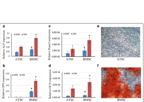
Fig. 2Difference in osteogenesis ability between ATSCs and BMSCs in vitro.a–dTotal RNA extracted from ATSCs and BMSCs or cells subjected to OIM for 7 days. Relative expression levels of ALP, Runx2, OPN, and OCN checked by qRT-PCR.β-actin as an internal control. Data expressed as mean ± SD (n= 3). *p< 0.05, compared with MSCs in OIM; #p< 0.05, compared with MSCs inα-MEM.e, fAlizarin Red S staining of calcium deposits formed by ATSCs and BMSCs. ATSCs and BMSCs cultured in OIM for 14 days, then cells fixed and stained with Alizarin Red S. ALP: alkaline phosphatase, ATSC: adipose tissue-derived MSC, BMSC: bone marrow-derived MSC, MEM: minimum essential medium, MSC: mesenchymal stem cell, OCN: Osteocal-cin, OIM: osteogenic induction medium, OPN: Osteopontin, Runx2: runt-related transcription factor 2
cells were positive for CD90, CD44, and CD73 and nega-tive for CD31 and CD45 (Fig. 1). The data showed that the cells expressed typical surface markers of MSCs and therefore were used for the experiments described in the following.
Compare osteogenesis of ATSCs and BMSCs in vitro
In order to compare the osteogenic differentiation potential capacities of ATSCs and BMSCs, MSCs were treated with OIM for several days and then the mRNA expression levels of genes related to osteogenesis were detected by quantitative real-time RT-PCR (qRT-PCR). As shown in Fig. 2a–d, the expression levels of alkaline phosphatase (ALP) and runt-related transcription factor 2 (Runx2), which are early markers for osteogenic com-mitment, were markedly increased in BMSCs compared with the ATSCs, as well as the late osteogenic markers Osteocalcin (OCN) and Osteopontin (OPN). To confirm the osteogenic commitment of BMSCs and ATSCs, Ali-zarin Red S staining was used to detect the formation of calcium deposit. The results showed that after 14 days of OIM induction, mineralization was seen in BMSCs upon osteogenic induction, while there were very few Alizarin Red S-positive calcium nodules formed in the ATSC group (Fig. 2e, f ). These data indicated that BMSCs
possessed higher potential for differentiation into osteo-blasts compared to ATSCs.
Compare adipogenesis of ATSCs and BMSCs in vitro
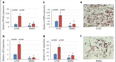
Next, we evaluated the adipogenic differentiation po-tential capacities of ATSCs and BMSCs. The cells were treated with AIM for several days, and then the mRNA expression levels of genes related to adipogen-esis were detected by qRT-PCR. Our results showed that the expression levels of adipogenesis-related marker genes such as peroxisome proliferator-activated receptor gamma (PPARγ), CCAAT/enhan-cer-binding protein alpha (CEBPα), adipocyte protein 2 (AP2), and lipoprotein lipase (LPL) were signifi-cantly increased in ATSCs compared with the BMSCs (Fig. 3a–d). After 21 days of AIM induction, the cells were fixed for Oil Red O staining. The result showed that BMSCs had lower adipogenic differentiation po-tential as compared with ATSCs (Fig. 3e, f ).
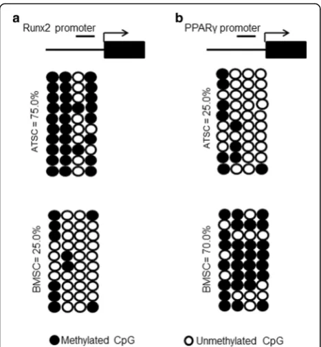
DNA methylation analysis of main transcription factors
Because epigenetic regulation is an important factor to control MSC differentiation and the methylation status of DNA is the most common epigenetic modification of the genome in mammalian cells [24], we asked whether DNA
methylation was involved in fate determination of ATSCs and BMSCs. Runx2 and PPARγare the main master tran-scriptional factors controlling osteogenesis and adipogene-sis, respectively. So, revealing the DNA methylation status of these two transcription factors may demonstrate their relationship with MSC fate determination. We calculated the percentage of methylated CpG loci (percent CpG methylation) in the total four CpG loci in Runx2 promoter and in four CpG loci in PPARγpromoter, respectively. We found that Runx2 promoter was hypermethylated whereas PPARγpromoter was hypomethylated in ATSCs (75% and 25% CpG methylation) (Fig. 4a, b). On the other hand, the methylation status of Runx2 was hypomethylated and PPARγ promoter was hypermethylated. These data sug-gest that DNA demethylation could be involved, at least partially, in the regulation of Runx2 and PPARγin ATSCs and BMSCs; the source of MSCs is a direct factor influen-cing fate determination of MSCs.
Compare chondrogenesis of ATSCs and BMSCs in vitro
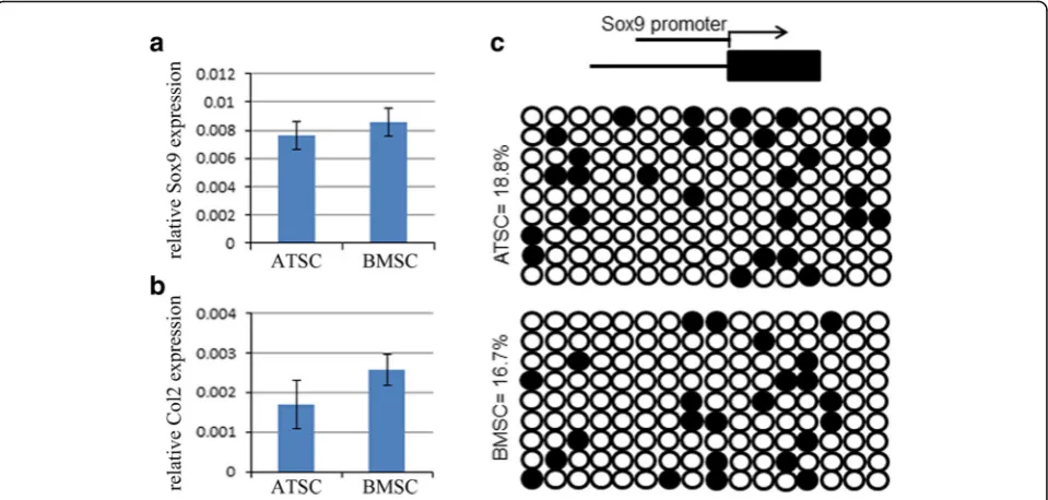
Next, we wanted to know whether there is any difference in chondrogenesis ability between ATSCs and BMSCs.
The cells were treated with CIM for 10 days, and then the expression levels of Sox9 and Collagen type II were evaluated by qRT-PCR. The results showed that both Sox9 and Collagen type II were slightly lower in ATSCs (Fig. 5a, b). Further bisulfite sequencing data showed that there was no significant difference in the methyla-tion status of CpG sites in the promoter of Sox9 be-tween ATSCs and BMSCs (Fig. 5c).
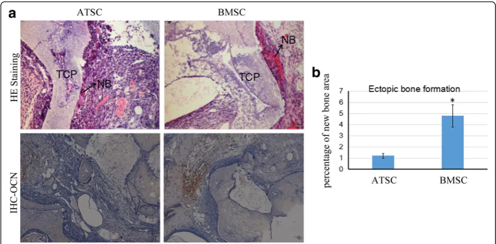
Ectopic bone formation of ATSCs and BMSCs in vivo
To further evaluate the advantages of BMSCs in osteo-genic differentiation in vivo, BMSCs and ATSCs were loaded onto sterilized Skelite® resorbable Si-TCP bone graft substitutes respectively and implanted subcutane-ously at the dorsal sides of nude mice. The transplants were harvested 8 weeks later and subjected to histo-logical examination with HE staining or immunohisto-chemical analysis to detect the distribution of osteoid and the expression of OCN. Our results showed that transplantation of BMSCs with Si-TCP resulted in more bone-like tissue formation and less loose fibrous tissue and adipose tissue formation around the scaffold com-pared to the ATSCs with Si-TCP in nude mice. The for-mation of bone-like tissue was confirmed by the presence of osteocalcin (Fig. 6a, b). These results indi-cated that BMSCs were superior to ATSCs in ectopic bone formation in vivo.
Bone regeneration in vivo using the calvarial defect model
To compare the effect of ATSCs and BMSCs on bone repair, the nude mice calvarial bone critical defect model was used. Then 50 μl of 2% hyaluronic acid hydrogel (5-mm-diameter cylinder) with 1 × 10⁵ hu-man ATSCs or BMSCs was immediately implanted into the defect cavity. Six weeks later, the samples were collected for X-ray and microCT reconstruction analysis. The result showed that more new bone tissue was observed in the BMSC group compared with the ATSC group (Fig. 7a, b). The ratio of bone volume/total volume in the BMSC group was signifi-cantly increased compared to that of the ATSC group (Fig. 7a, b).
Discussion
In the present study, we compared the differentiation capacities of ATSCs and BMSCs, and demonstrated that BMSCs possessed stronger osteogenic but lower adipo-genic differentiation potentials compared to ATSCs. There was no significant difference between their chon-drogenic differentiation potential. Interestingly, our re-sults provided important evidence that the DNA methylation status of the master transcription factors controlling MSC fate determination was responsible for
the regulation of MSC differentiation capacities. The epigenetic memory obtained from either bone marrow or adipose tissue favored their differentiation along osteoblastic or adipocytic lineage. This is of particular interest, since the finding provided a potential explan-ation to elucidate the mechanism which was responsible for the regulation of MSC differentiation capacities by the origin they derived.
MSCs have been isolated from the umbilical cord, um-bilical cord blood, bone marrow, adipose tissue, and many other adult tissues. They have been reported to share similar characteristics in vitro, such as plastic ad-herence, proliferation capacity, immunophenotype, and multilineage differentiation ability [25]. Lee et al. [26] re-ported that ATSCs were superior to BMSCs with respect to their maintenance of proliferating ability, but had similar morphology, phenotype, and microarray analysis of gene expression, and did not reveal differentially expressed osteogenic or adipogenic related genes between ATSCs and BMSCs. Our results have also provided evi-dence that ATSCs and BMSCs have similar morphology and cell surface markers, but their differentiation capacity was different. However, the intrinsic molecular mechanism supporting these observations has been lacking. We have partially addressed this question by exploring the methyla-tion status of promoters of main transcripmethyla-tion factors regu-lating trilineage differentiation potentials of MSCs.
DNA methylation is one of the most important epi-genetic regulations which possesses the power to control the differentiation of or maintain the self-renewal of MSCs [17]. Accumulating evidence has demonstrated that changes in the methylation states of the CpG islands in the promoter regions or the first exon are in-versely responsible for expression of the corresponding genes [27, 28]. The bivalent loci in MSCs are often re-duced in DNA methylation and can be further methyl-ated or activmethyl-ated, distinct from those in embryonic stem cells and differentiated cells [18]. A recent study demon-strated that the promoter regions of key genes in osteo-genic differentiation such as BMP2 and ALP are epigenetically locked in MSCs to prevent their expres-sion in nonosteogenic cells [29].
Sørensen et al. [30] reported that MSC differentiation did not affect lineage-specific promoter methylation states, arguing that these methylation patterns in differ-entiated cells are already established at the progenitor stage. But they did not compare the methylation status of the main transcription factors from different source-derived MSCs. We produced bisulfite PCR analysis to check the DNA methylation status of Runx2, PPARγ, and Sox9 from ATSCs and BMSCs. We demonstrated that BMSCs possessed stronger osteogenic and lower adipogenic differentiation potentials, which is completely different from ATSCs. The conclusion is that the
Fig. 5BMSCs showed slightly stronger chondrogenic differentiation potential.a, bTotal RNA extracted from ATSCs and BMSCs when cells were subjected to CIM for 10 days. Relative expression levels of Sox9 and Col2 checked by qRT-PCR.β-actin as an internal control. Data expressed as mean ± SD.p< 0.05.cDNA methylation status of Sox9 promoter in ATSCs and BMSCs using sodium bisulfite sequencing. Top panel indicates CpG di-nucleotide position of the Sox9 promoter region and numbers show positions of CpGs relative to the translation start site. Each PCR product was sub-cloned and subjected to nucleotide sequencing analysis. Ten representative sequenced clones depicted by filled (methylated) and open
differentiation potential of MSCs is highly influenced by their tissue of origin through epigenetic regulations such as DNA methylation of important transcription factors.
MSCs hold great promise for the treatment of a variety of difficult diseases such as myocardial infarction [31], neural diseases [32], and bone and cartilage defect [33, 34]. Bone marrow is the one of the major sources of MSCs, where
they represent only approximately 0.001–0.01% of the nu-cleated cells, which is much less abundant than hematopoietic stem cells (HSCs). Adipose tissue is the main alternative source of MSCs and is more abundant and easier to isolate. Which type of MSCs is more suitable for clinical application? Some studies indicate that the clinical applica-tion potential of ATSCs is more effective or as effective as
Fig. 7ATSCs and BMSCs enhanced calvarial bone repair in nude mice.aX-ray and microCT analysis 3D reconstruction of calvarial bone samples.
bMicroCT analysis showed the new bone volume in the BMSC group was significantly increased (*) compared to that of the control group. ATSC adipose tissue-derived MSC, BMSC bone marrow-derived MSC, CT computed tomography, MSC mesenchymal stem cell
that of BMSCs, while others conclude that BMSCs are su-perior to ATSCs [11–13]. In this study, we isolated bone marrow and adipose-derived MSCs from people aged older than 60 years. ATSCs and BMSCs from the same donors were evaluated. The results showed that BMSCs and ATSCs exhibited different trilineage differentiation potentials, al-though they expressed similar cell surface makers and had a similar phenotype. The bisulfite sequencing data further provided a mechanism basis to make this conclusion, which explicated why MSCs from bone marrow were better for bone regeneration. The differential ability in trilineage dif-ferentiation is determined by the origin of MSCs. Checking DNA methylation of the main transcription factors govern-ing MSC differentiation may be a predictive approach to distinguish different subpopulations of MSCs which may lead to better outcome for tissue regeneration. For example, MSCs with a lower DNA methylation rate in Runx2 pro-moter and a higher methylation rate in PPARγ promoter would result in better bone regeneration.
Conclusion
Taken together, our results demonstrated that the methy-lation status of the main transcription factors controlling MSC fate influenced their expression, which resulted in the different differentiation capacities of ATSCs and BMSCs. This study has provided strong evidence that BMSCs are superior to ATSCs in terms of osteogenic dif-ferentiation rather than adipogenic difdif-ferentiation, which is meaningful for the application of BMSCs in tissue en-gineering and regeneration of bone instead of ATSCs.
Additional file
Additional file 1:Table S1.Sequences of primers for real-time PCR analysis,Table S2.Sequences of primers for bisulfite sequencing analysis, andTable S3.Sequences of primers for CHIP-PCR analysis.
Abbreviations
AIM:Adipogenic induction medium; ALP: Alkaline phosphatase; AP2: Adipocyte protein 2; ATSC: Adipose tissue-derived MSC; BMSC: Bone marrow-derived MSC; CEBPα: CCAAT/enhancer-binding protein alpha; CIM: Chondrogenic induction medium; HSC: Hematopoietic stem cell; LPL: Lipoprotein lipase; MSC: Mesenchymal stem cell; OCN: Osteocalcin; OIM: Osteogenic induction medium; OPN: Osteopontin; PBS: Phosphate-buffered saline; PPARγ: Peroxisome proliferator-activated receptor gamma; Runx2: Runt-related transcription factor 2;α-MEM: Minimum Essential Media alpha
Funding
The work was partially supported by grants from Hong Kong Government Research Grant Council, General Research Fund (14119115, 14160917, 9054014 N_CityU102/15 and T12-402/17-N); National Natural Science Foun-dation of China (81371946, 81430049 and 81772322); Hong Kong Innovation Technology Commission Funds (ITS/UIM-305); Shenzhen City Science and Technology Bureau, China (JCYJ20150630165236960). This study was also supported in part by SMART program, Lui Che Woo Institute of Innovative Medicine, The Chinese University of Hong Kong and the research was made possible by resources donated by Lui Che Woo Foundation Limited.
Availability of data and materials
All of the supporting data can be downloaded.
Authors’contributions
BW (Guangzhou University of Traditional Chinese Medicine) and GL conceived and designed the study. LLX and YML carried out the experimentation and wrote the first draft of the manuscript. YXS and BW (Chinese University of Hong Kong) performed the animal experiments. YPX and WPL collected clinical samples and cultured BMSCs and ATSCs. QSW participated in data interpretation. HBW and WH participated in the design of the study and assisted in data interpretation. All authors read and approved the final manuscript.
Authors’information
Not applicable.
Ethics approval and consent to participate
Human ethics approval was obtained from the Joint CUHK-NTEC Clinical Research Ethics Committee of the Chinese University of Hong Kong (Reference No: CRE-2011.383). Animal surgery was carried out under the animal license issued by the Hong Kong SAR Government and the approval of the Animal Experimentation Ethics Committee of the Chinese University of Hong Kong (Ref No: 16-109-GRF). Human adipose tissue and bone marrow samples of three patients (females 60–65 years old) with total hip arthroplasty who gave informed consent were obtained from The Third Affiliated Hospital of Guangzhou University of Chinese Medicine (Guangzhou, China).
Consent for publication
All the authors give their consents for publication in the journal.
Competing interests
The authors declare that they have no competing interests.
Publisher’s note
Springer Nature remains neutral with regard to jurisdictional claims in published maps and institutional affiliations.
Author details
1Key Laboratory of Orthopaedics & Traumatology, The First Affiliated Hospital
of Guangzhou University of Chinese Medicine, The First Clinical Medical College, Guangzhou University of Chinese Medicine, Guangzhou, China.
2
Departments of Diagnostics of Traditional Chinese Medicine, Guangzhou University of Traditional Chinese Medicine, Guangzhou, Guangdong 510006, People’s Republic of China.3Department of Orthopaedics & Traumatology, Faculty of Medicine, The Chinese University of Hong Kong, Prince of Wales Hospital, Shatin, Hong Kong, Special Administrative Region of China.4Key Laboratory for Regenerative Medicine, Ministry of Education, School of Biomedical Sciences, Faculty of Medicine, The Chinese University of Hong Kong, Hong Kong, SAR, China.5Stem Cells and Regenerative Medicine
Laboratory, Lui Che Woo Institute of Innovative Medicine, Li Ka Shing Institute of Health Sciences, The Chinese University of Hong Kong, Prince of Wales Hospital, Shatin, Hong Kong, Special Administrative Region of China.
6The CUHK-ACC Space Medicine Centre on Health Maintenance of
Musculoskeletal System, The Chinese University of Hong Kong Shenzhen Research Institute, Shenzhen, People’s Republic of China.7Department of
Traumatology, The Third Affiliated Hospital of Guangzhou University of Traditional Chinese Medicine, Guangzhou, Guangdong 510240, People’s Republic of China.8Room 904, 9/F, Li Ka Shing Institute of Health Institute, Prince of Wales Hospital, The Chinese University of Hong Kong, Shatin, Hong Kong, Special Administrative Region of China.9Department of Traumatology, The First Affiliated Hospital of Guangzhou University of Traditional Chinese Medicine, Guangzhou, China.
Received: 20 February 2017 Revised: 9 August 2017 Accepted: 30 October 2017
References
1. Parekkadan B, Milwid JM. Mesenchymal stem cells as therapeutics. Annu Rev Biomed Eng. 2010;12:87–117.
and enriched environment after cortical stroke in rats: cell survival and functional recovery. Eur J Neurosci. 2009;29(3):562–74.
3. Li Y, Chen J, Chen XG, Wang L, Gautam SC, Xu YX, et al. Human marrow stromal cell therapy for stroke in rat: neurotrophins and functional recovery. Neurology. 2002;59(4):514–23.
4. Swanger SA, Neuhuber B, Himes BT, Bakshi A, Fischer I. Analysis of allogeneic and syngeneic bone marrow stromal cell graft survival in the spinal cord. Cell Transplant. 2005;14(10):775–86.
5. Chen J, Sanberg PR, Li Y, Wang L, Lu M, Willing AE, et al. Intravenous administration of human umbilical cord blood reduces behavioral deficits after stroke in rats. Stroke. 2001;32(11):2682–8.
6. Hoogduijn MJ, Dor FJMF. Mesenchymal stem cells: are we ready for clinical application in transplantation and tissue regeneration? Front Immunol. 2013;4:144.
7. Puissant B, Barreau C, Bourin P, Clavel C, Corre J, Bousquet C, et al. Immunomodulatory effect of human adipose tissue-derived adult stem cells: comparison with bone marrow mesenchymal stem cells. Br J Haematol. 2005;129(1):118–29.
8. Chung CS, Fujita N, Kawahara N, Yui S, Nam E, Nishimura R. A comparison of neurosphere differentiation potential of canine bone marrow-derived mesenchymal stem cells and adipose-derived mesenchymal stem cells. J Vet Med Sci. 2013;75(7):879–86.
9. Cooper GM, Durham EL, Cray Jr JJ, Bykowski MR, DeCesare GE, Smalley MA, et al. Direct comparison of progenitor cells derived from adipose, muscle, and bone marrow from wild-type or craniosynostotic rabbits. Plast Reconstr Surg. 2011;127(1):88–97.
10. Danisovic L, Varga I, Polak S, Ulicna M, Hlavackova L, Bohmer D, et al. Comparison of in vitro chondrogenic potential of human mesenchymal stem cells derived from bone marrow and adipose tissue. Gen Physiol Biophys. 2009;28(1):56–62.
11. Elman JS, Li M, Wang F, Gimble JM, Parekkadan B. A comparison of adipose and bone marrow-derived mesenchymal stromal cell secreted factors in the treatment of systemic inflammation. J Inflamm (Lond). 2014;11(1):1. 12. Rasmussen JG, Frobert O, Holst-Hansen C, Kastrup J, Baandrup U, Zachar V, et al.
Comparison of human adipose-derived stem cells and bone marrow-derived stem cells in a myocardial infarction model. Cell Transplant. 2014;23(2):195–206. 13. Huang JI, Kazmi N, Durbhakula MM, Hering TM, Yoo JU, Johnstone B.
Chondrogenic potential of progenitor cells derived from human bone marrow and adipose tissue: a patient-matched comparison. J Orthop Res. 2005;23(6):1383–9.
14. Hsiao ST, Asgari A, Lokmic Z, Sinclair R, Dusting GJ, Lim SY, et al. Comparative analysis of paracrine factor expression in human adult mesenchymal stem cells derived from bone marrow, adipose, and dermal tissue. Stem Cells Dev. 2012;21(12):2189–203.
15. Ahmadian Kia N, Bahrami AR, Ebrahimi M, Matin MM, Neshati Z, Almohaddesin MR, et al. Comparative analysis of chemokine receptor's expression in mesenchymal stem cells derived from human bone marrow and adipose tissue. J Mol Neurosci. 2011;44(3):178–85.
16. Rui Y, Xu L, Chen R, Zhang T, Lin S, Hou Y, et al. Epigenetic memory gained by priming with osteogenic induction medium improves osteogenesis and other properties of mesenchymal stem cells. Sci Rep. 2015;5:11056. 17. Boquest AC, Noer A, Collas P. Epigenetic programming of mesenchymal
stem cells from human adipose tissue. Stem Cell Rev. 2006;2(4):319–29. 18. Leu YW, Huang TH, Hsiao SH. Epigenetic reprogramming of mesenchymal
stem cells. Adv Exp Med Biol. 2013;754:195–211.
19. Hsiao SH, Lee KD, Hsu CC, Tseng MJ, Jin VX, Sun WS, et al. DNA methylation of the Trip10 promoter accelerates mesenchymal stem cell lineage determination. Biochem Biophys Res Commun. 2010;400(3):305–12. 20. Zuk PA, Zhu M, Mizuno H, Huang J, Futrell JW, Katz AJ, et al. Multilineage
cells from human adipose tissue: implications for cell-based therapies. Tissue Eng. 2001;7(2):211–28.
21. Haynesworth SE, Baber MA, Caplan AI. Cell surface antigens on human marrow-derived mesenchymal cells are detected by monoclonal antibodies. Bone. 1992;13(1):69–80.
22. Zinn RL, Pruitt K, Eguchi S, Baylin SB, Herman JG. hTERT is expressed in cancer cell lines despite promoter DNA methylation by preservation of unmethylated DNA and active chromatin around the transcription start site. Cancer Res. 2007;67(1):194–201.
23. Rui YF, Lui PP, Lee YW, Chan KM. Higher BMP receptor expression and BMP-2-induced osteogenic differentiation in tendon-derived stem cells
compared with bone-marrow-derived mesenchymal stem cells. Int Orthop. 2012;36(5):1099–107.
24. Poloni A, Maurizi G, Leoni P, Serrani F, Mancini S, Frontini A, et al. Human dedifferentiated adipocytes show similar properties to bone marrow-derived mesenchymal stem cells. Stem Cells. 2012;30(5):965–74. 25. Meirelles LD, Caplan AI, Nardi NB. In search of the in vivo identity of
mesenchymal stem cells. Stem Cells. 2008;26(9):2287–99.
26. Lee RH, Kim B, Choi I, Kim H, Choi HS, Suh K, et al. Characterization and expression analysis of mesenchymal stem cells from human bone marrow and adipose tissue. Cell Physiol Biochem. 2004;14(4-6):311–24.
27. Wagner JR, Busche S, Ge B, Kwan T, Pastinen T and Blanchette M. The relationship between DNA methylation, genetic and expression inter-individual variation in untransformed human fibroblasts. Genome Biology. 2014;15(2):R37.
28. Schultz MD, He YP, Whitaker JW, Hariharan M, Mukamel EA, Leung D, et al. Human body epigenome maps reveal noncanonical DNA methylation variation. Nature. 2015;523(7559):212–6.
29. Cho YD, Yoon WJ, Kim WJ, Woo KM, Baek JH, Lee G, et al. Epigenetic modifications and Canonical Wingless/int-1 Class (WNT) signaling enable trans-differentiation of nonosteogenic cells into osteoblasts. J Biol Chem. 2014;289(29):20120–8.
30. Sorensen AL, Timoskainen S, West FD, Vekterud K, Boquest AC, Ahrlund-Richter L, et al. Lineage-specific promoter DNA methylation patterns segregate adult progenitor cell types. Stem Cells Dev. 2010;19(8):1257–66. 31. Joggerst SJ, Hatzopoulos AK. Stem cell therapy for cardiac repair: benefits
and barriers. Expert Rev Mol Med. 2009;11:e20
32. Martino G, Franklin RJM, Baron-Van Evercooren A, Kerr DA, STEM SCMS. Stem cell transplantation in multiple sclerosis: current status and future prospects. Nat Rev Neurol. 2010;6(5):247–55.
33. Huang S, Xu LL, Zhang YF, Sun YX, Li G. Systemic and local administration of allogeneic bone marrow-derived mesenchymal stem cells promotes fracture healing in rats. Cell Transplant. 2015;24(12):2643–55. 34. Lin WP, Xu LL, Zwingenberger S, Gibon E, Goodman S, Gang L.
Mesenchymal stem cells homing to improve bone healing. J Orthop Translat. 2017;9:19–27.
• We accept pre-submission inquiries
• Our selector tool helps you to find the most relevant journal
• We provide round the clock customer support
• Convenient online submission
• Thorough peer review
• Inclusion in PubMed and all major indexing services
• Maximum visibility for your research
Submit your manuscript at www.biomedcentral.com/submit