University of Pennsylvania
ScholarlyCommons
Publicly Accessible Penn Dissertations
2017
Sleep And Activity Problems In Mouse Models Of
Neurodevelopmental Disorders
Christopher Angelakos
University of Pennsylvania, [email protected]
Follow this and additional works at:
https://repository.upenn.edu/edissertations
Part of the
Neuroscience and Neurobiology Commons
This paper is posted at ScholarlyCommons.https://repository.upenn.edu/edissertations/2748 For more information, please [email protected].
Recommended Citation
Angelakos, Christopher, "Sleep And Activity Problems In Mouse Models Of Neurodevelopmental Disorders" (2017).Publicly Accessible Penn Dissertations. 2748.
Sleep And Activity Problems In Mouse Models Of Neurodevelopmental
Disorders
Abstract
Adequate sleep is important for long-term health and day-to-day function. Compared to the general population, patients diagnosed with neurodevelopmental disorders have substantially higher prevalence of sleep, activity, and circadian problems, dramatically affecting their quality of life, and potentially exacerbating other adverse symptomologies. Despite this, the neurobiological underpinnings of sleep problems in
neurodevelopmental disorders remain unknown, and accurate rodent models capable of recapitulating human sleep and activity problems are lacking. In this dissertation, I investigate sleep, activity, and circadian rhythms in genetic mouse models of human neurodevelopmental disorders, with a focus on autism spectrum disorder (ASD). In Chapter 1, I review the importance of—and mechanisms contributing to—sleep/wake regulation, sleep problems in neurodevelopmental disorders, and the utility of rodent genetic models to address these problems. In Chapter 2, I investigate hyperactivity and male-specific sleep deficits found in the 16p11.2 del/+ chromosomal copy number variation mouse model of neurodevelopmental disorders (Angelakos et al., 2016). In Chapter 3, I highlight REM sleep reductions and altered electroencephalography (EEG) spectra in the SYGNAP1+/- mouse model of intellectual disability and ASD. In Chapter 4, I discuss home-cage hypoactivity observed in four different mouse models of ASD.
Degree Type
Dissertation
Degree Name
Doctor of Philosophy (PhD)
Graduate Group
Neuroscience
First Advisor
Ted Abel
Keywords
Autism, Circadian Rhythms, Mouse Models, Neurodevelopmental Disorders, Sex Differences, Sleep
Subject Categories
Neuroscience and Neurobiology
SLEEP AND ACTIVITY PROBLEMS IN MOUSE MODELS OF
NEURODEVELOPMENTAL DISORDERS
Christopher Caleb Angelakos
A DISSERTATION
in
Neuroscience
Presented to the Faculties of the University of Pennsylvania
in
Partial Fulfillment of the Requirements for the
Degree of Doctor of Philosophy
2017
Supervisor of Dissertation
_______________________
Ted Abel
Brush Family Professor of Biology
Graduate Group Chairperson
_______________________
Joshua Gold
Professor of Neuroscience
Dissertation Committee:
David Raizen, Associate Professor of Neurology (Committee Chair)
Max Kelz, David E. Longnecker Associate Professor of Anesthesiology and Critical Care
Claire Mitchell, Professor of Anatomy and Cell Biology
Mark Opp, Professor of Anesthesiology and Pain Medicine and Vice Chair for Basic
ii
ACKNOWLEDGMENT
I would like to thank my advisor, Dr. Ted Abel, for his mentorship and guidance
over the last five years. Ted has challenged me to ask pertinent questions and encouraged
me to be an independent thinker. He has been patient and understanding with me as I
made mistakes, and afforded me the time to troubleshoot problems. I have grown
substantially as a scientist, critical thinker, and writer thanks to Ted’s mentorship.
I would also like to thank my thesis committee—Dr. David Raizen, Dr. Max Kelz,
Dr. Claire Mitchell, Dr. Amita Sehgal, and Dr. Mark Opp. Their intellectual input has been
immensely helpful, and they are all tremendously supportive and compassionate people.
I could not have asked for a better committee. I would like to thank Amita for taking me
into her lab and providing me with a sense of community after Ted’s lab moved, and Mark
for serving as my external reviewer and providing me with a strong foundation in sleep
research when I was an undergraduate at the University of Michigan.
I would like to acknowledge all the members of the Abel lab, past and present, for
helping me along the way. In particular, I would like to recognize Jen, Robbert, Sarah,
Rolf, and Vince for their tutelage during my time in the lab. I would also like to thank the
many undergraduates and technicians who kept the lab running smoothly. I have made
many great relationships via the Abel lab, but Sarah and Vince deserve special recognition
for being tremendous friends, for helping me during the thesis-writing process, and for
always being there for me in lab and life in general.
Thank you to my friends in the Neuroscience Graduate Group for keeping graduate
iii
Peter, Andrew, Jen, and Preetika. Thank you all for your kindness, generosity,
hospitability, and friendship over the past five years. I would especially like to thank Greg
for being an amazing friend, co-worker, and for his scientific input and comments on this
dissertation and throughout graduate school.
I would like to acknowledge the National Defense Science and Engineering
Graduate Fellowship, which funded me for the last three years of my graduate study. I
would also like to thank the mice who gave their lives for these studies. I have worked very
hard to do efficient and purposeful studies, and I hope their sacrifices were not in vain.
Finally, and most importantly, I need to thank my family. Thank you for all your love
and support over the past 28 years. Mom and dad, thank you for always supporting me,
encouraging me, believing in me, providing for me, and pushing me to be better. To my
siblings—Matt, Kaily, and Taya—thank you for being lifelong friends and unwavering
supporters. Thank you to my grandparents, aunts, uncles, and cousins for supporting me
and indulging my passion for science. To my cousins and fellow neuroscientists, Ashley
and Aaron, thank you for helping me to get started in the field and for your scientific
discussions around the holidays. I could not have done any of this without my incredible
iv
ABSTRACT
SLEEP AND ACTIVITY PROBLEMS IN MOUSE MODELS OF
NEURODEVELOPMENTAL DISORDERS
Christopher Caleb Angelakos
Ted Abel
Adequate sleep is important for long-term health and day-to-day function. Compared to
the general population, patients diagnosed with neurodevelopmental disorders have
substantially higher prevalence of sleep, activity, and circadian problems, dramatically
affecting their quality of life, and potentially exacerbating other adverse symptomologies.
Despite this, the neurobiological underpinnings of sleep problems in neurodevelopmental
disorders remain unknown, and accurate rodent models capable of recapitulating human
sleep and activity problems are lacking. In this dissertation, I investigate sleep, activity,
and circadian rhythms in genetic mouse models of human neurodevelopmental
disorders, with a focus on autism spectrum disorder (ASD). In Chapter 1, I review the
importance of—and mechanisms contributing to—sleep/wake regulation, sleep problems
in neurodevelopmental disorders, and the utility of rodent genetic models to address
these problems. In Chapter 2, I investigate hyperactivity and male-specific sleep deficits
found in the 16p11.2 del/+ chromosomal copy number variation mouse model of
neurodevelopmental disorders (Angelakos et al., 2016). In Chapter 3, I highlight REM
sleep reductions and altered electroencephalography (EEG) spectra in the SYGNAP1
+/-mouse model of intellectual disability and ASD. In Chapter 4, I discuss home-cage
v
TABLE OF CONTENTS
ACKNOWLEDGMENT ... ii
ABSTRACT...iv
LIST OF TABLES ... viii
LIST OF FIGURES ...ix
CHAPTER 1: Mechanisms and functions of sleep and circadian rhythms ... Error! Bookmark not defined. 1.1 Mechanisms of sleep and circadian regulation ... 2
1.1.1 Circadian rhythms and regulation ... 2
1.1.2 Sleep homeostat (Process S) ... 4
1.1.3 Sleep-state switching ... 5
1.2 Functions of sleep and circadian rhythms, and consequences of their dysfunction ... 6
1.2.1 Consequence of inadequate sleep ... 7
1.2.2 Consequence of circadian dysfunction ... 9
1.3 Summary of the functional importance of sleep and circadian rhythms ....10
1.4 Sleep and circadian problems in autism spectrum disorders and related neurodevelopmental disorders ...11
1.5 Mouse genetic models for studying sleep and circadian rhythms ...13
Contributions ...15
CHAPTER 2: Hyperactivity and male-specific sleep deficits in the 16p11.2 deletion mouse model of autism...16
Introduction ...18
vi
Results ...26
Discussion ...30
Contributions ...35
Figure Legends ...36
Figures ...38
CHAPTER 3: REM sleep reductions, EEG spectra alterations, and altered homestatic sleep rebound in Syngap1+/1 mice ...42
Introduction ...43
Materials and Methods ...44
Results ...49
Discussion ...53
Contributions ...57
Figure Legends ...58
Figures ...61
CHAPTER 4: Home-cage hypoactivity in four mouse models of autism spectrum disorder ...69
Introduction ...71
Materials and Methods ...72
Results ...75
Discussion ...77
Contributions ...80
Figure Legends ...81
Figures ...83
vii
5.1 Conclusions ...88
5.2 Genetics of ASD ...89
5.3 Sex differences in ASD ...90
5.4 Targeted genetic manipulations of ASD-associated genes and CNVs ...91
5.5 Reversal of phenotypes and impact of development ...92
5.6 Summary and future directions ...93
Contributions ...95
APPENDIX: Circadian gene expression alterations and prolonged free-running period in CBPKix/Kix mice ...96
Introduction ...97
Materials and Methods ...99
Results ... 103
Discussion ... 107
Contributions ... 111
Figure Legends ... 112
Tables ... 115
Figures ... 136
viii
LIST OF TABLES
Table A.1. CBPKix/Kix gene expression experimental groups ... 115
Table A.2. Home-cage RNA-seq downregulated genes in CBPKix/Kix mice ... 116
Table A.3. Home-cage RNA-seq upregulated genes in CBPKix/Kix mice ... 120
Table A.4. Fear conditioned RNA-seq downregulated genes in CBPKix/Kix mice
... 122
ix
LIST OF FIGURES
Figure 2.1. 16p11.2 del/+ mice are hyperactive throughout the diurnal cycle ..38
Figure 2.2. Male 16p11.2 del/+ mice sleep less than wildtype littermates ...39
Figure 2.3. Male 16p11.2 del/+ mice spend a significantly higher proportion of
wake time in prolonged bouts of wakefulness ...40
Figure 2.4. Male 16p11.2 del/+ mice have increased alpha power during wake
...41
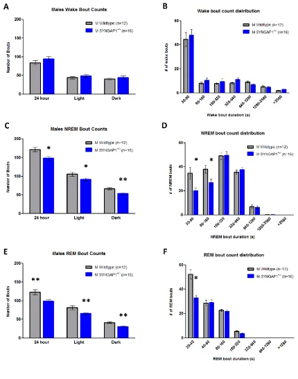
Figure 3.1. Syngap1+/- mice have REM sleep reductions in comparison to WT
...61
Figure 3.2. Syngap1+/- mice have decreased quantities of NREM and REM
bouts compared to WT ...62
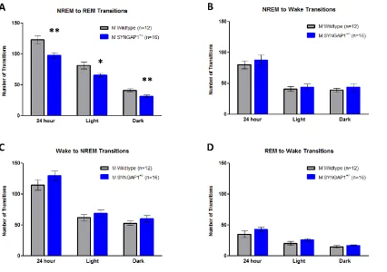
Figure 3.3. Syngap1+/- mice display less NREM to REM transitions than WT
littermates ...63
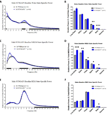
Figure 3.4. Syngap1+/- mice exhibit lower alpha and beta power during wake,
and elevated delta power during NREM sleep than WT mice ...64
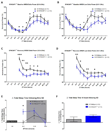
Figure 3.5. Syngap1+/- mice have altered homeostatic sleep drive during both
baseline sleep and following 6 hours of sleep deprivation ...65
Supplementary Figure 3.1. Syngap1+/- mice have normal mean durations of
x
Supplementary Figure 3.2 Syngap1+/- are hyperactive in the home-cage in
comparison to WT mice ...67
Supplementary Figure 3.3 Normal circadian rhythms in Syngap1+/- mice ...68
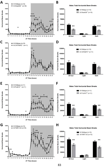
Figure 4.1. Male Shank3B-/-, CNTNAP2-/-, Pcdh10+/-, and Fmr1 KO mice are
hypoactive in the home-cage relative to sex-matched WT controls ...83
Figure 4.2. Female Shank3B-/- are hypoactive, but female CNTNAP2+/- and
Pcdh10+/- display no home-cage activity differences, relative to sex-matched WT
controls ...84
Supplementary Figure 4.1. Male Shank3B-/-, CNTNAP2-/-, Pcdh10+/-, and Fmr1 KO
mice display less home-cage rearing behavior than sex-matched WT controls ...85
Supplementary Figure 4.2 Female Shank3B-/- have reduced rearing behavior, but
female CNTNAP2+/- and Pcdh10+/- display no vertical activity differences, relative to
sex-matched WT controls ...86
Supplementary Figure 4.3 Normal circadian rhythms in Shank3B-/-, CNTNAP2-/-,
Pcdh10+/-, and Fmr1 KO mice ...87
Figure A.1. CBPKix/Kix gene expression experimental setup ... 136
Figure A.2. Altered expression of circadian regulatory genes in CBPKix/Kix mice
... 137
xi
Figure A.4. Lengthened circadian Tau in CBPKix/Kix mice relative to WT ... 139
1
CHAPTER 1: Mechanisms and functions of sleep and circadian rhythms
Abstract
Sleep is highly conserved across the animal kingdom. A diverse range of organisms
including nematodes, flies, fish, reptiles, birds, and mammals exhibit sleep or sleep-like
behavioral states. The fact that sleep is so evolutionarily conserved across phylogeny,
and humans spend nearly one-third of their lives in this vulnerable state, supports its
importance. Indeed, adequate sleep has been shown to be important for health,
metabolism, learning, cognition, mood, synaptic scaling, immune system function,
hormone release, motor performance, and more. However, in autism spectrum disorder
and related neurodevelopmental disorders, sleep is disturbed. In this chapter, I review
some important functions of sleep and mechanisms underlying normal sleep and circadian
regulation. I also summarize sleep issues highly prevalent in neurodevelopmental
disorders, with a focus on autism spectrum disorder (ASD). Finally, I address the utility
and need for genetic mouse models of neurodevelopmental disorders capable of
reproducing sleep, activity, and circadian problems found in human neurodevelopmental
2
1.1 Mechanisms of sleep and circadian regulation
Sleep is a perplexing and highly conserved behavioral phenomenon. It is a
reversible period of altered consciousness marked by behavioral quiescence, heightened
arousal threshold, and in humans and other animals with a sufficiently developed cortex—
altered brain firing patterns that can be measured by electroencephalography (Haas,
2003). Through polysomnography—the combination of electroencephalography (EEG)
(brain waves), electromyography (EMG) (skeletal muscle activity), and sometimes
electrooculography (EOG) (eye movements)—three broad sleep/wake stages can be
ascertained: wake, non-rapid eye movement sleep (NREM), and rapid eye movement
sleep (REM). Wake is characterized by high frequency, low amplitude waveforms in the
EEG with frequent muscle activity in the EMG. NREM sleep is characterized by low
frequency, high amplitude (synchronized) waveforms in the EEG and minimal muscle tone
in the EMG. For these reasons, NREM sleep is also referred to as slow wave sleep. REM
sleep is characterized by high frequency, low amplitude waveforms in the EEG, similar in
its desynchronized activity to wake, but with an increase of power in the theta frequency
(4-8Hz). Distinct from wake, however, REM sleep is also characterized by rapid eye
movements and a complete loss of muscle tone in the EMG (Dement & Kleitman, 1957).
Sleep and wakefulness is believed to be regulated by two distinct overarching modulators:
circadian rhythms (also known as “Process C”) and homeostatic drive (also known as
“process S”) (Borbély, 1982). These processes work in concert to wake us in the morning
and drive us toward sleep in the evening.
1.1.1 Circadian rhythms and regulation
3
al., 1999) that is entrained by environmental cues to the 24-hour rhythm of the rotating
earth (J C Dunlap, 1999). The endogenous biological clock is robust, tightly regulated, and
persists independent of the prior amount of time spent awake/asleep. Genomics studies
indicate that anywhere from 2% (Duffield et al., 2002) to 8-10% (Akhtar et al., 2002; Panda
et al., 2002; Storch et al., 2002) of the genome is circadian regulated, and approximately
40% of coding genes oscillate in at least one organ (Zhang et al., 2014). In mammals, the
“master” circadian clock is located within the suprachiasmatic nucleus (SCN) of the
hypothalamus (Moore & Eichler, 1972; Stephan & Zucker, 1972; Welsh et al., 1995). The
SCN receives direct environmental light cues via the retinohypothalamic tract, and in turn
signals circadian timing peripherally throughout the body via neural outputs, downstream
endocrine secretion, and by SCN-driven circadian regulation of body temperature
(Okamura, 2007; Dibner et al., 2010; Buhr & Takahashi, 2013). The molecular clock is
driven by a transcriptional-translational feedback loop of core clock molecules. The basic
helix-loop-helix-PAS-containing transcription factors CLOCK and BMAL1 heterodimerize
and bind to Enhancer box sequences in the promoters of the negative regulators mPER
and mCRY, driving their transcription. mPER and mCRY in turn feedback onto
CLOCK:BMAL1, inhibiting their transcription (Reppert & Weaver, 2002). In addition to this
molecular negative feedback loop, a separate feedback loop involving RAR-related
orphan receptor alpha (RORα) and Rev-Erbα maintains the circadian regulation of BMAL1
by activating and repressing BMAL1 transcription, respectively (Preitner, Damiola,
Luis-Lopez-Molina, et al., 2002). Numerous other transcriptional activators and repressors
have been identified that play supplementary but important roles in maintaining precise
circadian timing (for recent reviews, see: Golombek & Rosenstein, 2010; Buhr &
4
1.1.2 Sleep homeostat (Process S)
A second process known as the sleep homeostat (or Process S), is dependent
upon prior time spent awake or asleep (A A Borbély, 1982).The longer we stay awake,
the more intense is the drive to sleep. This is evident after sleep disruption, chronic sleep
restriction, or total sleep deprivation following an all-nighter (Klerman & Dijk, 2005; A. C.
Reynolds & Banks, 2010). Conversely, the need to sleep decreases as the prior duration
of sleep increases. Behavioral and physiological correlates following sleep deprivation
provide evidence for the existence of a sleep homeostat. Following sleep deprivation,
there is an increase of sleep time and slow wave activity (0.5-4.0Hz) relative to the
non-sleep restricted baseline day. Even without non-sleep deprivation, slow wave activity is
modulated as a function of prior wakefulness. Slow wave activity increases as a function
of time spent awake, and declines progressively over subsequent NREM bouts (Borbély
et al., 1981; Dijk et al., 1991; Achermann et al., 1993; Dijk et al., 1993; Campbell et al.,
2006). In addition to physiological correlates, there are also molecular homeostatic
correlates. Interstitial concentration of the molecule adenosine increases with
wakefulness, and decreases with sleep. Following sleep deprivation, adenosine tone
increases in the basal forebrain and cortex relative to baseline levels, and declines during
recovery sleep (Porkka-Heiskanen et al., 1997; Porkka-Heiskanen et al., 2000). In rats,
stimulation of the Gi-coupled adenosine A1 receptor increases NREM slow wave activity,
mimicking the effects observed following sleep deprivation (Ticho & Radulovacki, 1991;
Benington et al., 1995), while A1 receptor antagonism reduces recovery sleep and slow
wave activity (Gass et al., 2009). Caffeine, the world’s most widely consumed
5
antagonist (Smits et al., 1987; Fredholm, 1995). Numerous other molecules, including
glucocorticoids and peptide hormones likely influence the sleep homeostat; however, at
present time, adenosinergic contribution is the most extensively studied.
1.1.3 Sleep-state switching
In addition to circadian rhythms and homeostatic drive, which regulate sleep and
wakefulness broadly, myriad interconnected circuits and transmitter systems work in
interplay to regulate the transitions between wake, NREM sleep, and REM sleep
(reviewed in Fort et al., 2009; Saper et al., 2010). Briefly, and certainly not exclusively,
arousal networks include nuclei of the reticular formation of the brainstem (Moruzzi &
Magoun, 1949), cholinergic neurons of the pedunculopontine tegmental nuclei (PPT),
laterodorsal tegmental nuclei (LDT), and basal forebrain (el Mansari et al., 1989; Cape &
Jones, 2000; Lee et al., 2005a; Kaur et al., 2008; Boucetta & Jones, 2009; Hassani et al.,
2009a), noradrenergic projections from the locus coeruleus (Aston-Jones & Bloom, 1981),
serotonergic efferents from the dorsal and median raphe (Monti, 2011), dopaminergic
neurons in the ventral periaqueductal gray (Lu et al., 2006a), dorsal raphe (Cho et al.,
2017) and ventral tegmental area (Eban-Rothschild et al., 2016), histaminergic neurons
of the tuberomammillary nucleus (Steininger et al., 1999; Takahashi et al., 2006), and
orexinergic neurons of the lateral hypothalamus (Lin et al., 1999; Lee et al., 2005b;
Adamantidis et al., 2007; Carter et al., 2009). NREM sleep-promoting networks include a
subset of GABAergic neurons of the parafacial zone of the brainstem (Anaclet et al., 2012,
2014) and the ventrolateral preoptic nucleus (Nauta, 1946; McGinty & Sterman, 1968;
Sherin et al., 1996; Lu et al., 2000; Chung et al., 2017) and median preoptic nucleus of
6
project to and receive extensive inputs from many of the wake-promoting regions
mentioned above (Chou et al., 2002; Uschakov et al., 2007). Finally, REM promoting
circuitry includes subset of GABAergic neurons in the ventral medulla (Weber et al., 2015),
as well as the reciprocally interacting ON sublaterodorsal nucleus (SLD) and
REM-OFF GABAergic neurons of the ventrolateral periaqueductal gray and lateral pontine
tegmentum (vlPAG-LPT) (Sastre et al., 1996; Boissard et al., 2002; Lu et al., 2006b; Luppi
et al., 2006). Additional REM sleep-modulating networks exist in the cholinergic neurons
of the PPT and LDT (Webster & Jones, 1988; el Mansari et al., 1989), as well as
melanin-concentrating hormone-expressing neurons of the lateral and posterior hypothalamus
(Verret et al., 2003; Hassani, Lee, & Jones, 2009; Jego et al., 2013).Clearly, the regulation
of sleep and wake is an intricate and complicated process resulting from the delicate
interaction of dozens of brain regions and transmitter systems. Aberrant neural signaling
in a number of brain regions can lead to misregulation of sleep and circadian rhythms.
Consequences of altered sleep and circadian rhythms are discussed in the next section.
1.2 Functions of sleep and circadian rhythms, and consequences of their
dysfunction
The function of sleep is still debated. In truth, functions of sleep are likely
multi-faceted, situation-dependent, and different across development. Sleep is less
energetically demanding than wake, bringing about decreased body temperature, heart
rate, and oxygen consumption (Kleitman & Ramsaroop, 1948). Thus, sleep has been
proposed to be restorative, allowing for the clearance of detrimental oxygen free radicals
and β-amyloid that may accumulate during the metabolically taxing state of an active,
7
system function (Besedovsky et al., 2012), is affected and modulated by cytokines (Opp,
2005), and is believed to play an important role in recovery from illness (Toth et al., 1993;
Imeri & Opp, 2009). Sleep also plays a role in growth and development. Slow wave sleep
promotes release of growth hormone, which curtails with age (Takahashi et al., 1968; Van
Cauter et al., 2000). Adequate sleep in infants is related to cognitive performance and
language-learning (Gómez et al., 2006; Touchette et al., 2007; Beebe, 2011). Moreover,
sleep is crucial for the consolidation of both procedural and declarative memories
(Diekelmann & Born, 2010; Abel et al., 2013). During the subsequent sleep episode
following explorative learning, hippocampal place cells increase their firing rate and
“replay” the pattern of place cell activity experienced during exploration, suggesting a role
for sleep in spatial consolidation (Pavlides & Winson, 1989; Skaggs & McNaughton, 1996).
At the synaptic level, sleep is believed to modulate spine growth and connectivity, allowing
for the remembering of useful information and the forgetting of non-essential material
(Tononi & Cirelli, 2006; G. Yang et al., 2014). Still, the most striking evidence for the
function and importance of sleep and circadian rhythms comes from the severe
consequences resulting from their disruption.
1.2.1 Consequences of inadequate sleep
Insufficient sleep, a common problem in modern society, is concomitant with
numerous health and cognitive issues. Chronic sleep loss is associated with an increased
prevalence for obesity, diabetes, hypertension, and heart attacks (Taheri et al., 2004;
Spiegel et al., 2005; Mullington et al., 2009), as well as shorter lifespans (Kripke et al.,
2002; Patel et al., 2004; Tamakoshi et al., 2004). Insufficient sleep increases plasma
8
impacts insulin sensitivity, and modifies levels of leptin and ghrelin (Vgontzas et al., 1997;
Spiegel et al., 1999; Knutson et al., 2007). Psychologically, long-term sleep deficit is
positively correlated with depression, anxiety, and alcohol abuse (Pilcher & Huffcutt,
1996). Total sleep deprivation, partial sleep deprivation, and chronic sleep restriction all
impair performance on a number of neurocognitive tasks of executive function and working
memory. Moreover, performance generally worsens proportional to the amount of sleep
deprivation and the length of the cognitive task (Van Dongen et al., 2003; Durmer &
Dinges, 2005). Following learning, restricted sleep and total sleep deprivation impairs
memory consolidation and subsequent recall (Walker, 2008; Diekelmann & Born, 2010;
Abel et al., 2013). Clearly, the effects of insufficient sleep in humans are substantial and
widespread.
Work from our lab, in mice, has uncovered cellular and molecular consequences
of acute sleep deprivation, especially as it relates to the consolidation of
hippocampus-dependent memories. Six hours of sleep deprivation following learning impairs spatial
object recognition and contextual fear memory 24 hours later (Graves et al., 2003; Halassa
et al., 2009). Sleep deprivation decreases levels of cAMP—a second messenger
important for protein synthesis and long-term memory formation—by increasing levels of
phosphodiesterase isoform 4A5 (PDE4A45), an enzyme that breaks down cAMP (Vecsey
et al., 2009). During sleep deprivation, pharmacogenetic augmentation of cAMP or
blockade of hippocampal PDE4A5 function prevents spatial memory impairments (Vecsey
et al., 2009; Havekes et al., 2014). Moreover, acute sleep deprivation attenuates
translational mechanisms by interfering with the mammalian target of rapamycin (mTOR)
pathway, preventing normal memory consolidation (Tudor et al., 2016). At the cellular
9
and density in hippocampus region CA1, which is normalized by recovery sleep (Havekes
et al., 2016). Further, 6 hours of sleep deprivation impairs certain forms of long-term
potentiation in the hippocampus, the presumptive cellular correlate for learning (Vecsey
et al., 2009). Thus, even one night of acute sleep deprivation has molecular consequences
and is capable of altering neural structure and connectivity.
1.2.2 Consequences of circadian dysfunction
Proper alignment between endogenous circadian clocks and exogenous
environmental stimuli are important for health and function. It is estimated that 15% of all
metabolites are under circadian control (Dallmann et al., 2012), and metabolic diseases
are associated with circadian disturbances (reviewed in: Delezie & Challet, 2011).
Circadian disruption caused by shift work is associated with obesity, heightened levels of
triglycerides, type 2 diabetes, and an increased risk for cancer and cardiovascular disease
(Bøggild & Knutsson, 1999; Davis et al., 2001; Karlsson et al., 2001; Schernhammer et
al., 2001, 2003; Pan, et al., 2011). In addition to long-term health detriments, shift work is
also associated with fatigue resulting in significantly more work accidents (Smith et al.,
1994) and increased work errors due to sleepiness (Åkerstedt, 1988).
Important circadian influences on metabolism and health have also been
demonstrated in cyanobacteria, Drosophila, and rodents. Strains of cyanobacteria whose
internal oscillator closely match the external environment out-compete strains of
cyanobacteria that do not oscillate in tune with the environment (Woelfle et al., 2004).
Drosophila with impaired circadian clock function have decreased reproductive success
(Beaver et al., 2002), altered carbohydrate and lipid homeostasis (Seay & Thummel,
10
gene mutants are hyperphagic, have altered glucose metabolism, and become obese
(Turek et al., 2005). Chronically “jet lagged” wildtype mice who undergo an 8 hour phase
advance at the beginning of each week develop leptin resistance and become obese
(Kettner et al., 2015). Moreover, mice receiving a chronic jet lag protocol of four
consecutive weekly 6-hour phase advances have impaired immune system function,
hypothermia, and a 4-fold increased mortality rate following LPS injection
(Castanon-Cervantes et al., 2010). Additionally, tau-shortened hamster mutants kept on a normal
12h:12h light:dark cycle develop cardiomyopathies, renal disease, and die prematurely.
Importantly, if the light:dark cycle is adjusted to match the endogenous circadian rhythm
of these hamster mutants with short endogenous rhythms, no detriments were observed
in these organs (Martino et al., 2008).
In humans, there are five currently recognized circadian disorders: delayed sleep
phase disorder, advanced sleep phase disorder, irregular sleep-wake disorder,
non-24-hour sleep-wake disorder, and shift work sleep disorder. Circadian disorders are comorbid
with ADHD and depression (Bunney, 2000; Baird et al., 2012). Much of the problems
resulting from circadian disorders are societal in nature, as afflicted individuals may have
trouble going to school or holding jobs requiring a standard 9-5 schedule without
constantly suffering from sleepiness, fatigue, headaches, and cognitive impairments
(Okawa and Uchiyama, 2007).
1.3 Summary of the functional importance of sleep and circadian rhythms
In summary, adequate sleep and precisely timed circadian rhythmicity is important
11
hormone release, motor performance, and more. Sleep and circadian problems result in
profound behavioral, cellular, and molecular consequences, affecting quality of life and
long-term health. Much of what we know about the circuitry and molecular mechanisms
underlying normal and deficient sleep and circadian rhythmicity comes from studies in
rodent models. These factors motivated my dissertation work—to investigate sleep,
activity, and circadian rhythms in mouse genetic models of neurodevelopmental
disorders—a patient population with profoundly enhanced sleep issues relative to the
general population. The long-term goal of these studies is to identify accurate genetic
models capable of recapitulating some of the diverse sleep and activity issues that cause
substantial burden to the majority of individuals diagnosed with a neurodevelopmental
disorder.
1.4 Sleep and circadian problems in autism spectrum disorders and related
neurodevelopmental disorders
Autism spectrum disorder (ASD) is a multifaceted disorder of neural development,
affecting an estimated 1 in 68 children born today. ASD is strongly sex-biased, with males
4.5 times more likely than females to receive an ASD diagnosis (Christensen et al., 2016),
even with the same genetic insult (Robinson et al., 2013; Jacquemont et al., 2014).
Patients diagnosed with ASD typically exhibit social deficits, communication problems,
and repetitive behaviors. In addition to these “core” ASD symptomologies, it is now
recognized that sleep problems are a hallmark of ASD, as well as other
neurodevelopmental disorders (for reviews, see: Richdale & Schreck, 2009; Glickman,
12
The prevalence of sleep problems in neurodevelopmental disorders are estimated
to be as high as 80%, representing a 2-3 fold increase over the typically developing
population (Robinson-Shelton & Malow, 2016; Souders et al., 2017). The most common
sleep problem in ASD is insomnia, which is 10x more likely in children with ASD than in
children without a neurodevelopmental disorder diagnosis (Sivertsen et al., 2012). Other
sleep problems prevalent in ASDs include difficulties falling asleep, difficulties staying
asleep (increased night awakenings), and early morning rising (Limoges et al., 2005;
Malow et al., 2006; Miano et al., 2007; Goldman et al., 2009; Richdale & Schreck, 2009;
Souders et al., 2009; Picchioni et al., 2014). ASD patients have been reported to have
decreased REM sleep time and reduced rapid-eye movements during REM sleep
(Limoges et al., 2005; Buckley et al., 2010). On top of the direct consequences of altered
sleep in ASD, sleep problems are correlated with the severity of other negative ASD
symptomologies, and have been estimated to account for 22-32% of the variance in
behavioral problems in ASD (Park et al., 2012; Mazurek & Sohl, 2016).
Insomnia and early morning wakings in ASD suggest circadian abnormalities.
Melatonin is a circadian-regulated hormone produced by the pineal gland that regulates
timing of sleep and wakefulness. Indeed, numerous studies have reported altered
melatonin synthesis, concentration, and circadian profile in ASD patients relative to
typically developing controls (Ritvo et al., 1993; Nir et al., 1995; Kulman et al., 2000;
Tordjman et al., 2005; Bourgeron, 2007; Melke et al., 2008; Mulder et al., 2010).Nocturnal
melatonin has helped mitigate insomnia in some ASD patients (Rossignol & Frye, 2011;
Cortesi et al., 2012; Cuomo et al., 2017), but may result in earlier morning awakenings
(Gringras et al., 2012). Further, meta-analyses of randomized, double-blinded, and
13
improvements in night-time awakenings in ASD patients (Rossignol & Frye, 2011; Cuomo
et al., 2017).
1.5 Mouse genetic models for studying sleep and circadian rhythms
The benefits of using genetically-modified mice to study sleep and circadian
rhythms are multiple. Mice have brains with similar structure, circuitry, and regional
functionality as humans. Unlike simpler model organisms such as C. elegans and
Drosophila, polysomnography can be performed in rodents, they exhibit REM sleep, and
they demonstrate rebound slow wave activity and sleep time following sleep deprivation
(Franken et al., 1991). Moreover, experimental protocols for studying sleep and circadian
rhythms in mice are well-defined. Most importantly, among animals amenable to powerful
genetic manipulations, mice are the closest phylogenetic relative to humans.
Despite the fact that dozens of genetic mouse models of ASD have been
generated and the high prevalence of sleep problems in human patients, at the present
time, only two studies have quantified sleep and circadian rhythms in any rodent genetic
model of ASD (Thomas et al., 2016, 2017). In the following chapters, I will discuss my
dissertation research outlining sleep, activity, and circadian alterations in multiple rodent
genetic models of neurodevelopmental disorders. In Chapter 2, I describe male-specific
sleep deficits in the 16p11.2 del/+ mouse model of neurodevelopmental disorders. In
Chapter 3, I show altered REM sleep and cortical EEG spectra in the Syngap1+/- mouse
model of intellectual disability and ASD. In Chapter 4, I present an unexpected, but shared
home-cage hypoactivity phenotype across four different mouse models of ASD. Utilizing
14
and molecular etiologies of the sleep, activity, and circadian alterations described herein,
and 2) test potential treatment strategies—with the ultimate goal of improving quality of
15
Author Contributions: This chapter was written by Christopher Angelakos with input and
16
CHAPTER 2: Hyperactivity and male-specific sleep deficits in the 16p11.2
deletion mouse model of autism
Abstract
Sleep disturbances and hyperactivity are prevalent in several neurodevelopmental
disorders, including autism spectrum disorders (ASDs) and attention deficit-hyperactivity
disorder (ADHD). Evidence from genome-wide association studies indicates that
chromosomal copy number variations (CNVs) are associated with increased prevalence
of these neurodevelopmental disorders. In particular, CNVs in chromosomal region
16p11.2 profoundly increase the risk for ASD and ADHD, disorders that are more common
in males than females. We hypothesized that mice hemizygous for the 16p11.2 deletion
(16p11.2 del/+) would exhibit sex-specific sleep and activity alterations. To test this
hypothesis, we recorded activity patterns using infrared beam breaks in the home-cage of
adult male and female 16p11.2 del/+and wildtype (WT) littermates. In comparison to
controls, we found that both male and female 16p11.2 del/+mice exhibited robust
home-cage hyperactivity. In additional experiments, sleep was assessed by polysomnography
over a 24-hour period. 16p11.2 del/+male, but not female mice, exhibited significantly
more time awake and significantly less time in non-rapid-eye-movement (NREM) sleep
during the 24-hour period than wildtype littermates. Analysis of bouts of sleep and
wakefulness revealed that 16p11.2 del/+males, but not females, spent a significantly
greater proportion of wake time in long bouts of consolidated wakefulness (greater than
42 minutes in duration) compared to genetic controls. These changes in hyperactivity,
wake time, and wake time distribution in the males resemble sleep disturbances observed
17
be a useful genetic model for studying sleep and activity problems in human
18
Introduction
Sleep and activity problems are extremely prevalent in autism spectrum disorders
(ASDs) and attention-deficit/hyperactivity disorder (ADHD), yet they remain poorly
understood. Disrupted sleep affects up to 80% of ASD patients and 55% of children with
ADHD, compared to only 7-30% in the control population (Ivanenko & Johnson, 2008;
Goldman et al., 2011; Cohen et al., 2014; Kirov & Brand, 2014). The most commonly
reported sleep issues in these neurodevelopmental disorders include insomnia, delayed
sleep onset, and increased night awakening (Ivanenko & Johnson, 2008; Ming & Walters,
2009; A. M. Reynolds & Malow, 2011). These subjective observations have been
supported by objective polysomnography and actigraphy sleep recordings (Miano et al.,
2007; Cortese et al., 2009; Goldman et al., 2009; Souders et al., 2009). In addition, sleep
problems are positively correlated with the severity of core ASD symptomology including
communication deficits, withdrawal, and repetitive and stereotyped behavior (Cortesi et
al., 2010; Park et al., 2012). Despite this, few well-controlled studies have addressed the
neurobiological underpinnings of sleep problems in these disorders. The high prevalence
of sleep problems in ASDs and ADHD and the impact of disrupted sleep on symptomology
highlight the utility of identifying an appropriate genetic model with which to test potential
treatments and elucidate mechanisms underlying these disorders.
Increasing evidence from genome-wide association studies suggests that
chromosomal copy number variations (CNVs) are significantly enhanced in many
neurodevelopmental disorders (Sebat et al., 2007; Grayton et al., 2012). In particular,
hemideletion in chromosomal region 16p11.2 profoundly increases the risk for several
19
high comorbidity between these disorders (Hanson et al., 2014). 16p11.2 hemideletion is
associated with an estimated 0.6% of all ASD diagnoses (Weiss et al., 2008), and 16p11.2
hemideletion patients display cognitive deficits and other ASD symptomology even if they
do not meet the criteria for ASD diagnosis (Stefansson et al., 2014). Unlike many factors
believed to contribute to neurodevelopmental disorders, the 16p11.2 chromosomal region
is highly conserved in the syntenic 7qF3 region in the mouse, and thus copy number
variation in this region can be accurately modeled. To date, three different mouse lines of
16p11.2 hemideletion have been created, varying in the size of the deletion as well as
genetic background of the mice (Horev et al., 2011; Portmann et al., 2014; Arbogast et al.,
2016). Indeed, 16p11.2 hemideletion (16p11.2 del/+) mice have deficits in brain structure,
cognition, and communication (Horev et al., 2011; Portmann et al., 2014; Pucilowska et
al., 2015; Brunner et al., 2015; Yang et al., 2015a; Yang et al., 2015b; Arbogast et al.,
2016).
ASDs and ADHD also show significant sex bias risk, but the mechanisms
contributing to this remain unknown. Males are 4 times more likely than females to be
diagnosed with ASD (Werling & Geschwind, 2013) and 3 times more likely to be diagnosed
with ADHD (Schneider & Eisenberg, 2006). For this reason, developing genetic models
that also demonstrate this risk bias is important for construct validity. Previous studies
utilizing the 16p11.2 del/+ mouse model have not directly compared males and females
(Brunner et al., 2015; Pucilowska et al., 2015; Yang et al., 2015a; Yang et al., 2015b;
Arbogast et al., 2016). In this study, we assessed home-cage activity using infrared beam
breaks and sleep/wake behavior using polysomnography in both male and female adult
20
both male and female 16p11.2 del/+ mice compared to sex-matched wildtype littermates.
In addition, we found male-specific sleep/wake decrements in total sleep time and
distribution of wakefulness. In 16p11.2 del/+ males, but not females, the proportion of
wake time distributed in long bouts of continuous wakefulness was significantly greater
than in sex-matched WT littermates. When compared to sex-matched controls, these
male-specific sleep and activity alterations parallel deficits seen in human ASD and ADHD
patients. Additionally, while no systematic analysis of sleep has been performed in a
population of human 16p11.2 hemideletion patients, sleep disturbances have been
reported in two 16p11.2 hemideletion and two 16p11.2 duplication patients (Fernandez et
al., 2009; Tabet et al., 2012). To our knowledge, the present study is the first study
showing male-specific sleep deficits in a rodent genetic model of neurodevelopmental
disorders. These findings suggest that 16p11.2 del/+ mice are an appropriate genetic
model for investigating treatment strategies and potential mechanisms underlying sleep
problems, hyperactivity, and sex differences found commonly in human ASD and ADHD
patients.
Materials and Methods
Animals
16p11.2 del/+ male mice on a mixed C57BL/6J and 129S1/SvImJ background
purchased from The Jackson Laboratory (Stock #013128) were bred with females on a
mixed C57BL/6J and 129S1/SvImJ background (Stock #101043). All available pups were
used for experiments, no litters were culled, and all cages were fitted with Nestlets
21
remained group housed with sex-matched littermates (4-5 mice/cage) until
experimentation. Adult (2.5-4.5 month old) male and female 16p11.2 del/+ and WT
littermate offspring were used for all experiments in accordance with age ranges utilized
in previous mouse sleep experiments (Vassalli et al., 2013; Wimmer et al., 2013; Mang et
al., 2016) and guidelines for assessment of behavior in adult mice (Crawley, 2007). The
age distribution was consistent between the 4 experimental groups (mean ± SEM; Male
WT: 117.3 ± 3.9 days, Male 16p11.2 del/+: 115.7 ± 3.9 days, Female WT: 118.2 ± 4.6
days, Female 16p11.2 del/+: 117.5 ± 4.1 days). Separate cohorts of mice were used for
all behavioral experiments. Animals were provided food and water ad libitum and
maintained on a 12 hour light / 12 hour dark cycle with light onset at 7:00 am. All animal
care and experiments were approved by the Institutional Animal Care and Use Committee
of the University of Pennsylvania and conducted in accordance with the National Institutes
of Health guidelines.
Activity Monitoring
Activity was monitored using an infrared beam-break system (Opto M3, Columbus
Instruments, Columbus, OH), which provided a scaffold of high-resolution infrared lights
and detectors. The beams were spaced 0.5 inches apart and beam breaks were sampled
every 10 seconds. Two infrared grids at 0.75 inches and 2.75 inches from the cage floor
measured horizontal and vertical (rearing) activity, respectively. Mice were housed
individually in noise-attenuating chambers (22′′ × 16′′ × 19′′, Med Associates, St. Albans,
VT) equipped with individual lights (250 lux) and fans. Cages were placed within the beam
break system and covered with a lid to contain the mouse and to reduce brightness (80
22
acclimate to the lighting and social isolation of the activity chambers for 1 week before
experimentation. Following 1 week of acclimation, activity data was collected for 1 week
under 12-hour light / 12-hour dark conditions. Beam break counts were binned into 1 hour
bins and averaged over the 1 week of data collection. Lighting conditions were then
switched to 24-hour constant darkness for 2 weeks and counts of beam breaks were
compiled every 1 minute. Circadian period (Tau) was calculated from Day 2 to Day 14 of
constant darkness using ClockLab software (Actimetrics). Because of the circadian
manipulations of activity experiments, and to ensure consistent age ranges between sleep
and activity experiments, separate cohorts of mice were used for activity monitoring and
polysomnography sleep experiments.
Polysomnography
Animals were surgically implanted with electroencephalography (EEG) and
electromyography (EMG) electrodes under isoflurane anesthesia as described previously
(Wimmer et al., 2013). Briefly, electrodes consisted of Teflon-coated wires (Cooner wires,
Chatsworth, CA) soldered to gold socket contacts (Plastics One, Roanoke, VA) and
pushed into a six-pin plastic plug (MS363 plug, Plastics One). Electrodes were held in
place with miniature screws (J.I. Morris Co, Southbridge, MA) and dental cement (Ketac,
3M, St Paul, MN). Mice were allowed to recover from surgery for a minimum of 2 weeks.
During the second week of recovery, animals were connected to amplifiers using
lightweight cables (363, Plastics One) attached to a rotating commutator (SLC6, Plastics
One). Mice were allowed to acclimate to the cables and to the noise-attenuating faraday
recording chambers (38” x 39” x 33”, Med Associates, Georgia, VT) for one week before
23
mm from bregma) electrodes referenced to an electrode over the cerebellum (-1.5 mm
from lambda). Cerebellar reference was chosen based upon a lack of signal disruption
from either neck muscles or other brain areas, and has been previously validated by our
lab (Wimmer et al., 2013) and others (McShane et al., 2010; Vassalli et al., 2013).
EEG/EMG signals were sampled at 256 Hertz (Hz) and filtered at 0.3-30 Hz and
1-100 Hz, respectively, with 12A5 amplifiers (Astro-Med, West Warwick, RI). Data
acquisition and manual visual scoring was performed using SleepSign software (Kissei
Comtec Inc, Japan). EEG/EMG data was collected for a total of 24 consecutive
undisturbed hours beginning at 7:00am (onset of the light phase). EEG/EMG data was
analyzed in 4 second epochs as wake, non-rapid eye movement (NREM) sleep, or rapid
eye movement (REM) sleep by a trained experimenter blind to experimental conditions.
Wake was classified by increases in higher frequency waves (>10 Hz), decreases in
amplitude of the EEG, and high movement in the EMG. NREM was classified by increases
in delta power (0.5-4 Hz) and amplitude of the EEG, along with low amplitude activity in
the EMG. REM was determined by high EEG activity in the theta range (4-8 Hz) and very
low activity in the EMG. One 16p11.2 del/+ male was excluded from all analyses because
its total wake time over the 24-hour period was 236 minutes less than the group average,
>2.5 standard deviations below the group mean. For EEG spectral analysis, fast Fourier
transform (FFT; Hanning window; 0.5-20 Hz, 0.25 Hz resolution) was performed across
the 24-hour period on artifact-free epochs, and wake, NREM, and REM were normalized
and expressed as a percentage of the total spectral power. For a subset of the animals
(15 males and 5 females), spectral analysis was not appropriate due to technical error
24 Bout Distributions
Bouts of wake, NREM, and REM were classified as reported previously (Watson
et al., 2015). Briefly, wake and NREM bouts were identified as 32 seconds (8 epochs) or
greater of consecutive wake or NREM, respectively, while bouts of REM were identified
as 20 seconds (5 epochs) or greater of consecutive REM. Bouts of NREM and REM were
considered broken once interrupted by 8 epochs of any other state, while bouts of wake
were terminated by a single epoch of any other state. Bouts of wake, NREM, and REM
were then sorted into duration bins following a log2 pattern (30-80 s, 84-160 s, 164-320 s,
324-640 s. 644-1280 s, 1284-2560 s, and >2560 s) in accordance with previously
published analysis (Mochizuki et al., 2004; Kantor et al., 2013). One WT male was
excluded from all analyses due to a bout of >7 hours of consecutive wakefulness, >2.5
standard deviations above the group average.
Elevated Zero-maze
Mice were individually placed on a 5.5 cm wide circular track with an external
diameter measuring 45 cm, raised 40 cm above the floor (San Diego Instruments, San
Diego, CA). The track had two open and two enclosed segments of equal dimensions.
Mice were gently placed in the center of a closed segment to begin a 5 minute trial. Time
spent in open arms, risk assessment (Karlsson et al., 2005), and total distance traveled
were assessed using an automated MATLAB (Mathworks) based image analysis
software as described previously (Patel et al., 2014).
25
Spontaneous activity was assessed in an open field arena (14” x 14”, San Diego
Instruments, San Diego, CA) fitted with photocells to detect motion. Mice were gently
placed individually into the center of the open field for a 10 minute trial. Total ambulation,
center activity, periphery activity, and rearing behavior were recorded. Each trial was
digitally recorded and analyzed with image analysis software (Mathworks). Separate
cohorts of mice were used for elevated zero-maze and open field tasks.
Statistics
All statistical analyses were performed using SPSS for Windows (V. 22.0). For
beam break activity, Mixed Design ANOVAs were used with genotype (WT or 16p11.2
del/+) and sex (male or female) as the between-subjects factors and time as the
within-subjects factor. Two-way ANOVAs were used to compare wake, NREM and REM sleep
times averaged over 24 hours, and to analyze elevated zero-maze and open field data.
For 4x6-hour binned sleep state analysis, sleep state bout distribution experiments, and
FFT spectral analysis, Mixed Design ANOVAs were used with genotype (WT or 16p11.2
del/+) as the between-subjects factor and time (or in the case of FFT analysis, frequency
bin) as the within-subjects factor. Post hoc multiple comparisons were performed using
Bonferroni’s adjustment for multiple comparisons. In instances where the assumption of
sphericity was violated, Greenhouse-Geisser corrected F values are given. Mann-Whitney
U-tests were performed on normalized FFT data binned into low delta, delta, theta, alpha,
and beta frequency bins. Multivariate analysis of variance (MANOVA) was used on the
proportion of time spent in each sleep state during the light phase and the dark phase,
with alpha corrected for multiple ANOVAs and set at α = 0.05/2, followed by post hoc
26
Results
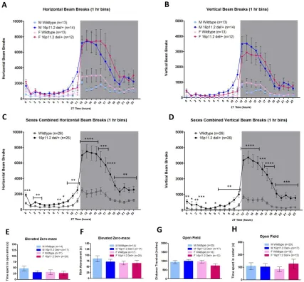
Home-cage hyperactivity in 16p11.2 del/+ mice
Male and female 16p11.2 del/+ mice and WT littermates were acclimated and
assessed for home-cage activity across the diurnal cycle by breaks of infrared beams in
the horizontal axis (Fig. 2.1a) as well as the vertical axis (Fig. 2.1b) to measure rearing
behavior. Mixed Design ANOVAs revealed that 16p11.2 del/+ mice had significantly more
activity than WT mice in both the horizontal (main effect of genotype; F(1,48) = 27.808, p
< 0.001, Fig. 2.1c) and vertical axis (main effect of genotype; F(1,48) = 34.714, p < 0.001
Fig. 2.1d). There was no effect of sex (F(1,48) = 0.814, p = 0.371) nor a sex*genotype
interaction (F(1,48) = 0.083, p = 0.774). To test whether the hyperactive behavior
observed in 16p11.2 del/+ mice may be related to stress/anxiety, we performed elevated
zero-maze on a separate cohort of 16p11.2 del/+ and WT mice. There were no significant
differences in time spent in the open arm (Two-way ANOVA, p = 0.279, Fig. 2.1e) or risk
assessment behavior (Karlsson et al., 2005) (p = 0.54, Fig. 2.1f), demonstrating that
16p11.2 del/+ mice display normal anxiety-like behavior. There were also no differences
between 16p11.2 del/+ mice and WT mice in distance traveled (Two-way ANOVA, p =
0.404, Fig. 2.1g) or time spent in the center (p = 0.501, Fig. 2.1h) in a 10 minute novel
open field task.
Because the hyperactivity observed in 16p11.2 del/+ males and females was most
pronounced during the dark (active) phase, and circadian problems such as increased
latency to sleep and early night awakenings are commonly reported in ASD patients
27
circadian rhythms. Male and female 16p11.2 del/+ mice were entrained to constant
darkness for 2 weeks and activity was quantified via infrared beam breaks. There were no
effects of genotype on circadian period (tau) in male (WT: 23.70 ± 0.04 hrs, 16p11.2 del/+:
23.76 ± 0.05 hrs; p = 0.36) or female (WT: 23.57 ± 0.05 hrs, 16p11.2 del/+: 23.64 ± 0.02
hrs; p = 0.22) mice. This indicates that 16p11.2 del/+ mice have normal circadian rhythms
and also that the hyperactivity observed in these mice is intrinsic and not a product of
environmental light cues.
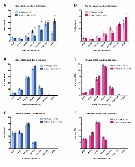
Male-specific sleep decrements in 16p11.2 del/+ mice
Because 16p11.2 del/+ male and female mice exhibited robust hyperactivity, we
assessed sleep and wake in 16p11.2 del/+male and female mice using polysomnography
recordings to distinguish between activity and sleep. EEG/EMG was recorded and
assessed across a 24-hour period. Female mice were awake significantly more than male
mice across the 24-hour day (main effect of sex; F(1,51) = 6.318, p = 0.015) consistent
with previously published results showing sex differences in sleep time and architecture
between male and female mice (Koehl et al., 2006; Paul et al., 2006). There was no main
effect of genotype (F(1,51) = 2.628, p = 0.111) nor sex*genotype interaction (F(1,51) =
2.110, p = 0.152). Because of the inherent sleep differences between male and female
mice and the main effect of sex within our dataset, male and female 16p11.2 del/+ mice
were analyzed separately and compared against sex-matched WT littermates in order to
focus on the biologically relevant comparisons. Male 16p11.2 del/+ mice exhibited more
wake across the 24-hour day compared to sex-matched WT mice (Student’s t-test, p =
0.008; Fig. 2.2a). There was a significant difference in 16p11.2 del/+ male wake time
28
2.2a). Post hoc analysis revealed that 16p11.2 del/+ males have significantly more wake
time during the light phase (F(1,28) = 5.322, p = 0.029), but not during the dark (active)
phase (F(1,28) = 2.780, p = 0.107) than WT males. Concordant with increased total wake
time, 16p11.2 del/+ male mice have less NREM sleep time than WT mice across the
24-hour period (Student’s t-test, p = 0.013; Fig. 2.2b). In contrast to differences in total wake
and NREM time, there was no difference in REM time in 16p11.2 del/+ males relative to
WT mice (Student’s t-test, p = 0.94; Fig. 2.2c). Sleep data was next analyzed in 6 hour
time bins as previously published (Franken et al., 1999). Mixed Design ANOVA revealed
that the decreased NREM time observed in 16p11.2 del/+ males (main effect of genotype;
F(1,28) = 7.019, p = 0.013; Fig. 2.2d) was due primarily to the final 6 hours of the light
phase (Student’s t-test, t(28) = 9.055, p = 0.005; Fig 2.2d). In contrast to the males,
16p11.2 del/+ females exhibited no differences in wake (Student’s t-test, p = 0.92; Fig.
2.2e), NREM (Student’s t-test, p = 0.53; Fig. 2.2f), or REM (Student’s t-test, p = 0.067;
Fig. 2.2g) time compared to WT females across the 24-hour period. Female 16p11.2 del/+
mice also exhibited no differences in wake, NREM, or REM in the light or dark phases
(wake: F(2,22) = 0.414, p = 0.67, Fig 2.2e; NREM: F(2,22) = 1.633, p = 0.218, Fig 2.2f;
REM: F(2,22) = 3.121, p = 0.064, Fig 2.2g). Analyzing female 16p11.2 del/+ NREM sleep
in 6 hour time bins revealed no significant main effect (F(3,69) = 0.397, p = 0.535; Fig. 2h)
nor a significant genotype*time interaction (F(3,69) = 1.272, p = 0.29; Fig. 2.2h). This data
suggests that 16p11.2 del/+ males, but not females, have deficits in either sleep initiation
or sleep maintenance.
29
Because 16p11.2 del/+ male mice have decreased sleep and increased
wakefulness, we binned and analyzed the distribution of wake, NREM, and REM bout
durations as described previously (Mochizuki et al., 2004; Kantor et al., 2013) to elucidate
the factor(s) contributing most strongly to this phenotype. We found that 16p11.2 del/+
male mice have an altered distribution of wake bout length duration (genotype*bout time
interaction; F(6,168) = 6.599, p = 0.001; Fig. 2.3a). Post hoc comparisons indicated that
16p11.2 del/+ males spent a higher proportion of their wake time in prolonged bouts of
continuous wakefulness (>42 consecutive minutes: t(28) = 11.340, p = 0.002) and less
time in bouts of shorter duration (160-320 seconds: t(28) = 5.356, p = 0.028; 320-640
seconds: t(28) = 10.248, p = 0.003; 640-1280 seconds: t(28) = 6.480, p = 0.017). By
contrast, females exhibited no differences in wake bout length distribution (F(6,138) =
1.576, p = 0.22; Fig. 2.3d). There were no differences in NREM bout time distribution in
16p11.2 del/+ males (F(6,168) = 0.500, p = 0.59; Fig 2.3b) or females (F(6,138) = 1.924,
p = 0.16; Fig. 2.3e). Likewise, there were no differences in REM bout length distribution in
either males (F(6,168) = 0.785, p = 0.48; Fig. 2.3c) or females (F(6,138) = 1.816, p = 0.16;
Fig. 2.3f) Together, this data indicates that 16p11.2 del/+ males, but not females, sleep
less than WT mice due to deficits in sleep initiation, rather than sleep maintenance.
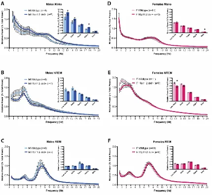
16p11.2 del/+ males have reduced alpha power during wake
Next, we performed fast Fourier transform (FFT) to analyze the EEG power spectra
of 16p11.2 del/+ males and females during wake, NREM, and REM. There was no main
effect of genotype for any sleep state for either sex (Males wake: F(1,13) = 2.649, p =
0.13, Fig. 2.4a; NREM: F(1,13) = 3.851, p = 0.07, Fig. 2.4b; REM: F(1,13) = 0.760, p =
30
1.680, p = 0.21, Fig. 2.4e; REM: F(1,18) = 0.744, p = 0.40, Fig. 2.4f). However, binning
the data into low delta (0.5-1.5 Hz), delta (0.5-4.0 Hz), theta (4.0-8.0 Hz), alpha (8.0-12.0
Hz), and beta (12.0-20.0 Hz) frequency bands revealed that 16p11.2 del/+ males have
significantly increased alpha power during wake relative to WT littermates (Mann-Whitney
U = 8, p = 0.021; Fig. 2.4a, inset), suggesting increased arousal and vigilance during quiet
wake in 16p11.2 del/+ males (Cantero et al., 2002). Increased arousal during quiet wake,
in concert with increases in total wake time and prolonged bouts of continuous
wakefulness, suggests that 16p11.2 del+/ males may possibly have deficits initiating
wake-to-sleep transitions. In contrast, there were no differences in alpha power during
wake between 16p11.2 del/+ females and WT littermates (Mann-Whitney U = 31, p = 0.18,
Fig. 2.4d, inset). 16p11.2 del/+ females, however, had significantly increased beta power
during wake (Mann-Whitney U = 22, p = 0.038, Fig. 2.4d, inset). There were no differences
in any other frequency bands apart from a decrease in low delta power during NREM in
16p11.2 del/+ females in comparison to WT littermates (Mann-Whitney U = 16, p = 0.010,
Fig. 2.4e, inset), which may indicate differences in sleep homeostasis.
Discussion
We investigated home-cage activity and sleep patterns in one mouse model of
human 16p11.2 chromosomal hemideletion. We report robust and reliable home-cage
hyperactivity across the light-dark cycle that is present in both males and females. These
findings expand upon previous reports of hyperactivity in 16p11.2 del/+ mice (Horev et al.,
2011; Brunner et al., 2015; Arbogast et al., 2016) by comparing males and females,
assessing activity in the home-cage, and quantifying activity over a week of consecutive
31
hemideletion differing in deletion size and genetic background, which should be
considered when comparing findings between studies. In addition, we report the first
male-specific sleep decrements in a rodent model of ASD. Decreased total sleep time and
prolonged bouts off wakefulness, suggesting difficulties in initiating wake-to-sleep
transition, recapitulate common sleep and activity problems reported in human ASD
patients (Miano et al., 2007; Krakowiak et al., 2008; Reynolds & Malow, 2011; Baker &
Richdale, 2015). The sleep/wake deficits in 16p11.2 del/+ males, but not females relative
to sex-matched controls support theories of female protectiveness from ASD given the
same genetic insult (Robinson et al., 2013; Werling & Geschwind, 2013; Jacquemont et
al., 2014).
Additionally, compared to sex-matched controls, the sleep/wake differences
observed only in male 16p11.2 del/+ mice are contrasted by activity differences in both
16p11.2 del/+ males and females. This distinction suggests disparate neurobiological
mechanisms underlying the two alterations. The etiology of sleep and activity deficits in
ASDs remains unknown, but numerous lines of evidence strongly support a relationship
between imbalanced excitatory/inhibitory signaling, sleep, and hyperactivity. Although
both ADHD and ASD are associated with imbalanced excitatory/inhibitory signaling, the
neurochemical and neuroanatomical mechanisms mediating these disorders are likely
distinct. The sex differences in sleep/wake, but not activity in 16p11.2 del/+ mice therefore
suggest that the 16p11.2 del/+ mouse model may be a useful rodent genetic model for
investigating neurobiological mechanisms mediating sex differences in
neurodevelopmental disorders.