in a recreational athlete:
a case report
Chris deGraauw,
DC*
* Sports Sciences Resident, Canadian Memorial Chiropractic College, 1900 Bayview Avenue, Toronto, Ontario M4G 3E6. © JCCA 1999.
A 23-year-old recreational male athlete presented with intermittent pain of three weeks duration, localized to the left ankle. Pain was aggravated by walking, although his symptoms had not affected the patient’s jogging activity which was performed three times per week. Past history revealed an inversion sprain of the left ankle, sustained fifteen months previously. Examination showed mild swelling anterior to the ankle mortise joint while other tests including range of motion, strength and motion palpation of specific joints of the ankle were noted to be unremarkable. Radiographic findings revealed a defect in the medial aspect of the talus. An orthopaedic referral was made for further evaluation. Tomography revealed a Grade III osteochondral lesion of the talus.
It was determined that follow-up views be taken in three months to demonstrate if the lesion was progressing or healing. Within the three month period, activity modifications and modalities for pain control were indicated. Surgery was considered a reasonable option should conservative measures fail.
The present case illustrates an osteochondral lesion of the talus, a condition which has not previously been reported in the chiropractic literature. A review of the pertinent orthopaedic literature has indicated an average delay of three years in diagnosing the existence of this lesion.
Although considered rare, the diagnostic frequency of the condition appears to be on the rise due to increased awareness and the use of bone and CT scans. The osteochondral lesion of the talus deserves particular consideration by practitioners working with athletes due to its higher incidence within this group. This diagnosis should be considered in patients presenting with chronic
Un homme de 23 ans, faisant du sport durant ses heures de loisir, vient consulter pour une douleur intermittente ressentie à la cheville gauche depuis trois semaines. La marche accentue la douleur, mais les symptômes n’empêchent pas le patient de faire de la course trois fois par semaine. Les antécédents du patient révèlent qu’il a subi, 15 mois auparavant, une entorse en varus de la cheville gauche. L’examen physique permet d’observer une légère tuméfaction antérieure à l’articulation à mortaise de la cheville, mais les autres examens, dont l’amplitude des mouvements, la force et la palpation en mouvement de certaines articulations de la cheville, s’avèrent négatifs. Les radiographies montrent une anomalie sur la face interne de l’astragale et le patient est dirigé vers un orthopédiste pour une évaluation plus poussée. La tomographie laisse voir la présence d’une lésion ostéo-cartilagineuse de niveau ill sur l’astragale.
Il est décidé que des radiographies de contrôle seront prises dans trois mois afin de vérifier si la lésion
progresse ou régresse. Entre-temps, il y a modification des activités et diverses mesures sont prises pour soulager la douleur. La chirurgie est présentée comme une solution de rechange acceptable en cas d’échec du traitement conservateur.
Le présent cas fait état d’une lésion
ostéo-cartilagineuse de l’astragale, affection qui n’a jamais été signalée dans la documentation scientifique en chiropratique. Un examen de la documentation médicale sur le sujet révèle qu’il s’écoule en moyenne un délai de trois ans avant que le diagnostic de lésion ostéo-cartilagineuse ne soit posé.
Introduction
In today’s society there is an increasing awareness in health and the importance of exercise. Emerging attitudes reflect the knowledge that exercise increases cardiovascu-lar fitness and reduces the risk of cardiovascucardiovascu-lar disease. Studies have shown that increased overall fitness can lead to higher energy levels, a more positive mood and in-creased creativity, leading to a better quality of life.1
The boom in the number of people participating regu-larly in recreational sport comes with an increase in sports injuries. Many people who exercise regularly have experi-enced the aggravation of having injuries interfere with their activity levels. Worse are the instances where partici-pants allow injuries to go unattended or are managed improperly.
The ankle is one of the most commonly injured joints in sports. One condition associated with the ankle joint that has not previously been reported in the chiropractic litera-ture, but has been extensively cited in the orthopaedic lit-erature, is an osteochondral lesion of the talus.2–6 Although
this lesion is relatively rare (0.9% of all fractures),2 its
incidence may be on the rise and is noted to be higher in the athletic population. It is a diagnosis often overlooked and deserves greater consideration from practitioners working with athletes. This may prevent delays in receiving appro-priate treatment.
Chiropractors may play an important role in managing many athletic injuries. Chiropractors are well trained in the diagnosis, treatment and rehabilitation of these injuries. The challenge is often making the correct diagnosis. Once the diagnosis is made, a proper plan of management can be devised.
The purpose of the following is to heighten the aware-ness of this condition and demonstrate its clinical presen-tation. The discussion is intended to provide a brief historical perspective on the osteochondral lesion itself and review its diagnosis and management, both from con-servative and surgical perspectives.
Case report
A 23-year-old male presented with a primary complaint of intermittent pain in the left ankle. The pain was insidious in onset, three weeks prior to presentation. The pain was described as dull and aching, localized to the anterior ankle mortise joint and extended to the lateral malleolus. The pain was aggravated by walking. The patient also reported weakness resulting in altered gait including a shorter stride on the affected side, “walking with the foot inverted” and a slight limp due to pain.
The pain did not prevent participation in any of his regu-lar activities. He had not received any previous treatment for this condition and was not taking any medication.
ankle pain particularly when a history of an inversion sprain exists.
The purpose of this report is to increase awareness of this condition, and review diagnosis and management strategies.
(JCCA 1999; 43(1):15–21)
K E YW O R D S: osteochondral lesion, talus, osteochondritis
dissecans, diagnosis, chiropractic, athletic injuries, ankle.
plus grande sensibilité des médecins à l’égard de l’affection et du recours aux scintigraphies osseuses et à la tomodensitométrie. Les praticiens travaillant auprès des athlètes devraient prêter une attention particulière à la lésion ostéo-cartilagineuse de l’astragale étant donné la fréquence accrue de cette maladie dans ce groupe de population. Le diagnostic devrait être envisagé dans les cas de douleur chronique à la cheville, surtout lorsqu’il y a des antécédents d’entorse en varus.
Le but du présent article est d’éveiller l’attention des chiropraticiens à l’égard de cette affection et de passer en revue les stratégies diagnostiques et thérapeutiques.
(JACC 1999; 43(1):15–21)
M O T S C L É S : lésion ostéo-cartilagineuse, astragale,
Past history indicated that he had sustained an inversion sprain to the left ankle fifteen months earlier. The injury resulted in significant pain and swelling, however no treatment was received and the patient felt there were no sequelae.
The patient was an active individual who enjoyed play-ing volleyball, basketball and runnplay-ing. He had initiated a running program three weeks prior to the onset of the com-plaint. The program consisted of approximately three runs per week, each of 20-30 minutes duration.
Inspection revealed mild swelling of the left ankle, lo-calized anterior to the talus with no other visible abnor-malities. Physical examination consisted of ranges of motion, neurological, and orthopaedic assessments and specific joint play tests of the ankle. The ranges of motion of the ankle were full and pain free. The neurological exam assessed reflexes, sensation and motor strength of the lower limbs bilaterally. The reflexes were 2+, sensation to light touch was determined to be normal and muscle tests were graded as 5/5. Functional muscle testing including heel raises and hopping on one leg revealed mild discom-fort but there was no weakness or signs of early fatigue. The muscle girth of the left thigh and gastroc soleus com-plex showed a slight difference of 1 cm less as compared to the right side. Orthopaedic testing and motion palpation of the forefoot and ankle were unremarkable.
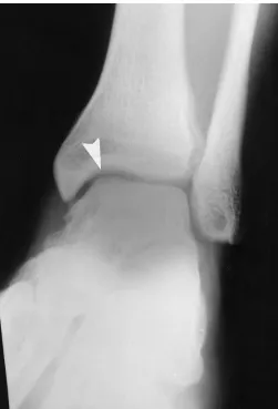
Radiographic examination
The radiograph shown in Figure 1 revealed a bony defect without a distinct outline at the medial aspect of the left talar dome, possibly associated with a bony fragment and soft tissue swelling. This finding was highly suggestive of an osteochondral lesion of the talus.
Referral/advanced imaging
An orthopaedic referral was initiated and tomograms were taken to further evaluate the lesion. The plain film x-ray shown in Figure 2 clearly delineates the lesion to be 12 mm by 6 mm in size, localized to the medial aspect of the left talus with a non-displaced fragment. This indicated a Grade III lesion by the Berndt and Harty classification system.7
Management
Activity modification and modalities for pain control were indicated for this condition. Activity modification for three
months was recommended with follow-up views to be taken to ascertain healing or progression. The goal of the activity modification was to prevent undue stress occur-ring in the ankle joint. Therefore, previous activities such as running, volleyball, basketball and ball hockey were eliminated for three months. Suggested alternatives to maintain fitness capacity included cycling, swimming and water running. These alternatives were emphasized to assure patient compliance. Modalities including inter ferential current and microcurrent were used for pain con-trol as required.
Discussion
Early literature indicates that osteochondral lesions of the talus account for approximately 4% of all cases of osteo-chondritis.2 However more recent literature suggests an
even higher incidence.3 One data collecting centre
ob-served a sevenfold increase in the diagnostic frequency of osteochondral lesions of the talus between the years 1981 to 1986 and 1987 to 1992.3 They concluded that such a
dramatic increase may be due to better imaging techniques (bone scan and CT), as well as increased awareness of the lesion.3 The increased awareness seems to be the key as
this diagnosis is often overlooked. Various studies report delays in diagnosis from the onset of symptoms to be be-tween 6.5 months and 3 years.3,4
To better understand the osteochondral lesion, it is nec-essary to examine how the terminology associated with
this condition has evolved. This is important in order to eliminate the confusion surrounding its definition and the liberal use of various terms for this condition.
In 1870 Paget8 described a pathological process that he
termed “quiet necrosis”, which resulted in the creation of loose, necrotic osteochondral fragments from articular surfaces. This report was followed in 1888 when Konig9
first coined the term “osteochondritis dessicans”. Konig theorized that inflammation of the bone and cartilage, fol-lowed by spontaneous necrosis led to a fragment dissect-ing away, hence the term. Such early descriptions were generally cases involving the knee, which accounts for approximately 75% of all osteochondritis cases.10 In 1932
Rendu reported an intra-articular fracture of the talus that appeared similar to osteochondritis dissecans. This report led to the publication by Roden5 in 1953 of 55
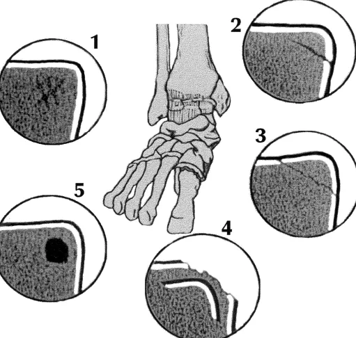
Figure 3 Loomer’s revision of the Berndt and Harty Classification System I. Compression, II. Partially fractured but undisplaced, III. Completely fractured but undisplaced, IV. Displaced fracture, V. Radiolucent (fibrous defect).
dritis dissecans-like lesions of the talus.
In 1959, Berndt and Harty7 demonstrated that both
me-dial and lateral lesions of the talus, previously considered to be osteochondritis dessicans, were actually osteochon-dral fractures caused by trauma with viable bone rather than the necrotic bone found with osteochondritis dessicans.7 Simultaneously, it was shown that there was no
associated inflammatory component to osteochondritis dessicans.11 Therefore the name was incorrect, and there
was strong evidence for a traumatic etiology.
The lack of understanding of the specific pathological process describing this condition has led to a plethora of terms, including: osteochondrosis dissecans, osteo-chondral fractures, transosteo-chondral fractures, osteonecro-sis, subchondral cysts, osteochondral lesion and of course osteochondritis dissecans which has persisted to present.3,4,6,12,13,14,15
Due to the high frequency of traumatic origin, osteo-chondral or transosteo-chondral fracture seems appropriate but this does not explain the non-traumatic presentation or rare familial forms which involve genetic factors such as iden-tical lesions in ideniden-tical twins.16 Therefore, the most
ap-propriate term may be an osteochondral lesion.
Berndt and Harty developed a classification system for osteochondral lesions which divides such fractures into four different stages. This system has recently been ex-panded to include a fifth stage introduced by Loomer et al.3
All stages are shown and described in Figure 3.3,7
Berndt and Harty were also able to produce two distinc-tive lesions in cadavers, anterolaterally and a postero-medially. The anterolateral lesion was produced by forceful inversion and strong dorsiflexion. The posterome-dial lesion was caused by inversion, plantar flexion and lateral rotation of the tibia.7 The classic inversion sprain of
the ankle, one of the more common running injuries seems to be a frequent mechanism of injury.
Clinically, the most common history given is a previous injury, likely an ankle sprain, often followed by an asymp-tomatic period which then becomes painful. The pain usu-ally increases proportionately with exercise and the patient will often exhibit swelling. Physical examination is usu-ally inconclusive.
Other signs and symptoms include catching, locking, giving way, reduction in dorsiflexion and painful anterior drawer but these findings are rare.3
Following a thorough history and physical examination,
the next step for a patient presenting with chronic ankle pain should be a radiographic assessment. The lesion will typically be visualized on the talar dome with routine ankle views (AP, lateral and internal oblique). Thompson and Loomer, however, suggests a technique for better identifi-cation of the posteromedial lesion. It consists of an AP view of the ankle with maximum plantar flexion taken at a focal film distance of 1 metre using 70 kVP.2
If radiographs are normal and the chronic ankle pain cannot be explained by any other ankle or foot condition, a bone scan is indicated. Bone scans have been shown to have 99% sensitivity in detecting osteochondral lesions.17
A positive bone scan with uptake in or near the talus should be followed up with a CT scan or MRI for definitive diag-nosis and localization.
In a study with 92 patients Loomer et al. (1993),3
re-ported that a CT scan gave the correct diagnosis in 98% of cases.3 The radiolucent lesion (Grade V) depicted in
Fig-ure 3 accounted for an unexpected 77% of the lesions, which is surprising since this majority had gone unrecog-nized until the grade V lesion was introduced by Loomer and colleagues in 1993.3 The grade V lesion was mainly
diagnosed through bone scans and CT scans following normal x-rays.
Treatment options include conservative or surgical management. Conservative management may consist of the use of a short leg non-weight bearing cast for approxi-mately six weeks, coupled with the administration of non-steroidal antiinflammatory drugs (NSAIDS). Alter-natively, activity modification may be recommended since there has been very little success in the management of this condition, with or without prolonged immobilization. This approach may be co-managed with electrotherapy, exer-cises, stretching and protected weight bearing.3,5,6,18,20
Historically surgical arthrotomy has been the treatment of choice. Arthroscopic procedures, however are now accessible for specific lesions. The procedure usually involves curettage and drilling, and when present, re-moval of the fragment. If the fragment is large enough, fixation of the fragment to the site of separation may be possible.3,5,6,18,20
Surgical procedures are generally prescribed for a grade IV lesion (a displaced fragment; see figure 3), particularly if symptomatic. A grade V lesion which may progress to any other stage should undergo initial conservative treat-ment but if problematic, has had good surgical results.3
It has been shown that postponing the operation does not adversely affect the ultimate outcome of surgery and some lesions have been shown to heal without sur-gery.19,20 Therefore a trial of conservative management is
appropriate.
The final points to consider are the patient’s goals and attitudes. Each individual should take part in the decision to have surgery based on the knowledge that the lesion left alone, will likely not lead to osteoarthritis and that the lesion may heal over a time frame extending from months to years.20 Therefore the patient’s athletic inclination,
oc-cupation and eagerness to return to full activity, may deter-mine their choice for early surgical management.
Conclusions
The diagnosis of an osteochondral lesion in this patient’s case was timely. He was given the opportunity to make the choice between surgical and conservative management without undue delay in the correct diagnosis and pro-longed mismanagement. The onus is on the primary care practitioner to be aware of this condition and it’s presenta-tion in order to decrease the morbidity surrounding the osteochondral lesion. At this point, the diagnostic hall-mark is a history of trauma followed by chronic pain often with normal x-rays. Such points should be remembered to avoid excessive delays in diagnosis.
References
1 Steinberg H. Exercise increasing creativity independent of mood. Br J Sports Med 1997; 31:240–245.
2 Thompson JP, Loomer RL. Osteochondral lesions of the talus in a sports medicine clinic. Am J Sports Med 1984; 12(6):460–463.
3 Loomer R, Fisher C, Loyd-Smith R, Sisler J, Cooney T. Osteochondral lesions of the talus. Am J Sports Med 1993; 21(1):13–19.
4 Hagmeyer RHM, Van der Wurff P. Transchondral fractures of the talus on an inversion injury of the ankle: a frequently overlooked diagnosis. J Ortho Sports Phys Ther 1987; Jan:362–367.
5 Canale ST, Belding RH. Osteochondral lesions of the talus. J Bone Joint Surg (Br) 1980; 62–A:97–102. 6 Schenck RC, Goodnight JM. Osteochondritis dissecans.
current concepts review. J Bone Joint Surg (Br) 1996; 78–A:439–456.
7 Paget J. On the production of some of the loose bodies in joints. St. Bartholomew’s Hosp Rep 1870; 6:1–4. 8 Konig F. Uber freie Lorper in den Gelenken. Deutsche
Zeitschr. Chir 1888; 27:90–109. In- Schenck RC, Goodnight JM. Osteochondritis Dissecans. Current Concepts Review. J Bone Joint Surg (Br) 1996; 78–A:439–456.
9 Clanton TO, DeLee JC. Osteochondritis dissecans. History, pathophysiology, and current treatment concepts. Clin Ortho 1982; 167:50–64.
10 Berndt AL, Harty M. Transcondylar fractures
(osteochondritis dissecans) of the talus. J Bone and Joint Surg 1959; 41–A:988–1020.
11 Nagura S. The so-called osteochondritis dissecans of Konig. Clin Orthop 1960; 18:100–121.
12 Pappas AM. Osteochondrosis dissecans. Clin Orthop 1981; 158:59–69.
13 O’Donoghue DH. Chondral and osteochondral fractures. J Trauma 1966; 6:469–481.
14 Paul GR. Ankle Injuries. Ankle Injuries. New York, Churchill Livingstone, 1983:113–130.
15 Pfeiffer WH, Gross ML, Seeger LL. Osteochondritis dissecans of the patella. Clin Orthop 1991; 271:207–211. 16 Woods K, Harris I. Osteochondritis dissecans of the talus
in identical twins. Orthop 1995; 77B:331.
17 Sisler J. Radiologic imaging of the post traumatic chronic tarsal pain. Clin J Sports Med 1989; 1:67–70.
18 Anderson AF, Lipscomb AB, Coulam C. Antegrade curettement, bone grafting and pinning of osteochondritis dissecans in the skeletally mature knee. Am J Sports Med 1990; 18:254–261.
19 Alexander AH, Lichtman DM. Surgical treatment of transchondral talar-dome fractures (osteochondritis dissecans). J Bone Joint Surg (Br) 1980; 62–A:646–652. 20 McCullough CJ, Venugopal V. Osteochondritis dissecans