Diagnosis and rehabilitation of gastrocnemius
muscle tear: a case report
Virginia Nsitem,
BSc (Hon), DC, FRCCSS(C), MEd** Total Health & Family Care Centre
1090 Dundas Street East, Suite, L-105, Mississauga, Ontario, L4Y 2B8 [email protected] Tel: 905-275-4993; Fax: 905-275-5046 ©JCCA 2013
Objective: This case study presents the epidemiology,
etiology, diagnostic criteria, and therapeutic interventions for a common clinical condition – gastrocnemius injury.
Clinical Features: A 44-year old male presented with acute calf pain with a palpable defect, loss of range of motion, and loss of strength after sustaining a soft tissue injury to the lower leg. The differential diagnosis of tear of the medial head of the gastrocnemius was confirmed by physical examination and diagnostic ultrasound imaging.
Intervention and Outcome: The patient was treated over a 6 week period. Initially, rehabilitation was approached using the PRICE principles for symptomatic relief, followed by stretching, strengthening,
proprioception, and conditioning exercises. At 9-month follow-up post injury, there was no residual impairment in the gastrocnemius muscle function.
Summary: This case demonstrates the importance of epidemiology, clinical assessment, and the use of diagnostic ultrasound and MRI imaging in the diagnosis of a tear of the medial head of the gastrocnemius muscle. With an accurate diagnosis and comprehension
Objectif : cette étude de cas présente l’épidémiologie,
l’étiologie, les critères diagnostiques et les interventions thérapeutiques pour une affection clinique courante – la blessure du muscle gastrocnémien (muscles jumeaux).
Caractéristiques cliniques : un patient de 44 ans
a présenté des douleurs aiguës au mollet avec une anomalie palpable, une perte d’amplitude de mouvement et une perte de tonus après avoir subi une blessure aux tissus mous de la jambe inférieure. Le diagnostic différentiel de la déchirure du chef médial des muscles jumeaux a été confirmé par un examen physique et une échographie diagnostique.
Intervention et résultat : le patient a été traité pendant une période de 6 semaines. Au début, la réadaptation a été abordée selon les principes PRICE du soulagement symptomatique, suivi par de l’étirement, d’exercices de musculation, de la kinesthésie et d’exercices de mise en forme. Au suivi, neuf mois après la blessure, on n’a révélé aucune détérioration résiduelle dans la fonction des muscles jumeaux.
Résumé : ce cas démontre l’importance de
Introduction
Muscle injuries in the calf are a relatively common clin-ical condition1-6, and are also termed “tennis leg” in
gen-eral because of the prevalence in that sport3,7. However,
middle-aged or older patients, usually over the age of 40, often present with lower leg muscle injuries following strenuous exercise or sometimes innocuous activity.5
There is a consensus to classify myotendinous strains as first degree (stretch injury), second degree (partial tear), and third degree (complete rupture).5,8 This type of
classi-fication takes into consideration the physical findings and pathological correlation as described above, and the dis-abilities that is, absent, mild, or complete loss of muscle function.6,7 The term strain does not accurately reflect the
structural characteristics of injuries of muscles; rather it is more of a biomechanical description of the mechan-ism of injury9, and as such, the term tear should be used
as it more accurately describes the structural injuries of muscle fibres8. Mueller-Wohlfahrt et al.8 discusses the use
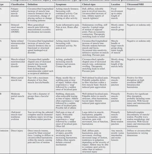
of a classification system that describes four types of in-direct (acute) muscle injuries, and recommends the use of the term tear to describe the injuries of muscle fibres and bundles (Table 1).
A tear to the gastrocnemius muscle is, more often than not, implicated in lower leg trauma and is considered at high risk of injury because of its position spanning across two joints: the knee and ankle, and because of the high density of type-two fast twitch muscle fibres.2,3,6 The
gas-trocnemius muscle functions to plantar flex the foot at the ankle joint and flex the leg at the knee joint in the non-weight-bearing state.7 Although studies documenting
the injury rates of calf muscle tears are limited7, a 5-year
study of European soccer players revealed that 12% of the muscle injuries sustained were injuries to the
gastrocn-emius muscle, and the gastrocngastrocn-emius was categorized as one of the top five muscles injured5.
The diagnosis of the gastrocnemius tear is often clin-ical. Sudden onset of pain, tenderness localized to the musculotendinous junction of the medial head of the gas-trocnemius, and a palpable defect in the medial belly of the gastrocnemius just above the musculotendinous junc-tion are pathognomonic for a gastrocnemius tear.4,10
Muscle tears located in the calf region are often as-sociated with other pathologies such as thrombophleb-itis, soleus tear, Achilles tendon rupture, and posterior compartment syndrome, making it more difficult for the practitioner to formulate a correct diagnosis, despite per-forming an accurate clinical examination.1 Researchers
concur that conservative management is effective in the treatment of gastrocnemius tears.1,4,5,7,10
This paper will draw attention to the diagnostic and therapeutic procedures associated with gastrocnemius tears. A case will be presented which emphasizes the use of advanced imaging to reach an accurate diagnosis which aides in the process of developing an appropriate course of treatment.
Case
A 44-year old male presented with a chief complaint of right posterior calf pain, and provided consent to the use of specifics related to his clinical information. He reported the onset of symptoms a few days prior during a dancing session. He stated that he experienced a sud-den and sharp sensation at the back of his calf as he ex-tended the leg backwards while planting the heel on the ground. He stated “I thought someone kicked the back of my leg…but when I turned around, there was no one there”. The pain was located along the medial aspect of
of classification of muscle injuries, management of gastrocnemius tears is straightforward.
(JCCA 2013;57(4):327-333)
k e y w o r d s: injury, muscle, gastrocnemius, diagnosis
lésions musculaires, la gestion des déchirures des muscles jumeaux est simplifiée.
(JCCA 2013;57(4):327-333)
m o t s c l é s : blessure, muscle, muscles jumeaux,
Comprehensive muscle injury classification: type-specific definitions and clinical presentations
Type Classification Definition Symptoms Clinical signs Location Ultrasound/MRI
1A
Fatigue-induced muscle disorder
Circumscribed longitudinal increase of muscle tone (muscle firmness) due to overexertion, change of playing surface or change in training patterns
Aching muscle firmness. Increasing with continued activity. Can provoke pain at rest. During or after activity
Dull, diffuse, tolerable pain in involved muscles, circumscribed increase of tone. Athlete reports of ‘muscle tightness’
Focal involvement up to entire length of muscle Negative 1B Delayed-onset muscle soreness (DOMS) More generalised muscle pain following unaccustomed, eccentric deceleration movements.
Acute inflammative pain. Pain at rest. Hours after activity
Oedematous swelling, stiff muscles. Limited range of motion of adjacent joints. Pain on isometric contraction. Therapeutic stretching leads to relief
Mostly entire muscle or muscle group
Negative or oedema only
2A Spine-related
neuromuscular muscle disorder
Circumscribed longitudinal increase of muscle tone (muscle firmness) due to functional or structural spinal lumbopelvical disorder.
Aching muscle firmness. Increasing with continued activity. No pain at rest
Circumscribed longitudinal increase of muscle tone. Discrete oedema between muscle and fascia. Occasional skin sensitivity, defensive reaction on muscle stretching. Pressure pain
Muscle bundle or larger muscle group along entire length of muscle
Negative or oedema only
2B Muscle-related
neuromuscular muscle disorder
Circumscribed (spindle-shaped) area of increased muscle tone (muscle firmness). May result from dysfunctional neuromuscular control such as reciprocal inhibition
Aching, gradually increasing muscle firmness and tension. Cramp-like pain
Circumscribed (spindle-shaped) area of increased muscle tone, oedematous swelling. Therapeutic stretching leads to relief. Pressure pain
Mostly along the entire length of the muscle belly
Negative or oedema only
3A Minor partial
muscle tear Tear with a maximum diameter of less than muscle fascicle/bundle.
Sharp, needle-like or stabbing pain at time of injury. Athlete often experiences a ‘snap’ followed by a sudden onset of localised pain
Well-defined localised pain. Probably palpable defect in fibre structure within a firm muscle band. Stretch-induced pain aggravation
Primarily muscle-tendon junction
Positive for fibre disruption on high resolution MRI*. Intramuscular haematoma 3B Moderate partial muscle tear
Tear with a diameter of greater than a fascicle/ bundle
Stabbing, sharp pain, often noticeable tearing at time of injury. Athlete often experiences a ‘snap’ followed by a sudden onset of localised pain. Possible fall of athlete
Well-defined localised pain. Palpable defect in muscle structure, often haematoma, fascial injury Stretch-induced pain aggravation
Primarily muscle-tendon junction
Positive for significant fibre disruption, probably including some retraction. With fascial injury and intermuscular haematoma
4 (Sub)total
muscle tear/ tendinous avulsion
Tear involving the subtotal/ complete muscle diameter/ tendinous injury involving the bone-tendon junction
Dull pain at time of injury. Noticeable tearing. Athlete experiences a ‘snap’ followed by a sudden onset of localised pain. Often fall
Large defect in muscle, haematoma, palpable gap, haematoma, muscle retraction, pain with movement, loss of function, haematoma Primarily muscle-tendon junction or Bone-tendon junction Subtotal/complete discontinuity of muscle/ tendon. Possible wavy tendon morphology and retraction. With fascial injury and intermuscular haematoma
Contusion Direct injury Direct muscle trauma, caused by blunt external force. Leading to diffuse or circumscribed haematoma within the muscle causing pain and loss of motion
Dull pain at time of injury, possibly increasing due to increasing haematoma. Athlete often reports definite external mechanism
Dull, diffuse pain, haematoma, pain on movement, swelling, decreased range of motion, tenderness to palpation depending on the severity of impact. Athlete may be able to continue sport activity rather than in indirect structural injury Any muscle, mostly vastus intermedius and rectus femoris
Diffuse or circumscribed haematoma in varying dimensions
* Recommendations for (high-resolution) MRI: high field strength (minimum 1.5 or 3 T), high spatial resolution (use of surface coils), limited field of view (according to clinical examination/ultrasound), use of skin marker at centre of injury location and multiplanar slice orientation.
Table 1:
the posterior calf and extended upwards toward the knee and distally towards the ankle. He described the pain as tight and throbbing. The pain was aggravated with gen-eral ankle movements.
Examination revealed an antalgic gait, favouring the right leg. He was unable to balance on the right leg un-assisted. Inspection revealed moderate soft tissue swell-ing of the right calf with discoloration and bruisswell-ing ex-tending to the posterior aspect of the foot. Measurement of the calf was 41 cm on the right and 38.5 cm on the left, measured 10 cm below the patella. A visible defect of the medial gastrocnemius muscle was evident and was palp-able at this juncture. In addition, a mass was palpated at the posterior calf, likely the rupture of the gastrocnemius muscle. Palpation revealed tenderness along the entire medial gastrocnemius muscle, particularly at the mus-culotendinous junction.
The Thompson Squeeze Test was negative for an Achil-les tendon rupture, as it was painful but produced plantar flexion. Active and passive ankle dorsiflexion produced moderate pain. Resisted plantar flexion of the ankle also reproduced the symptoms. There was mild pain with ac-tive knee flexion. There was difficulty performing a single leg calf raise with the affected leg.
The patient was referred for a venous doppler of the right lower leg that normal compressibility, phasic flow and augmentation in the deep veins of the lower extremity from the common femoral vein to the popliteal conflu-ence below the knee. A diagnostic ultrasound study of the right calf revealed an abnormality at the medial aspect of the gastrocnemius muscle, described as “a complex cystic structure at the medial aspect of the gastrocnemius muscle…adjacent to the plantaris tendon/muscle”. There was concern of a soft tissue hematoma with a partial thickness tear of either the medial gastrocnemius muscle or the plantaris tendon/muscle. Based on the epidemiol-ogy, mechanism of injury, clinical findings, and diagnos-tic ultrasound findings, the patient was diagnosed with a type 3 tear of the medial gastrocnemius muscle.
Bleakley11 elaborates on the widely accepted approach
in the treatment of soft tissue injuries as protection, rest, ice, compression, and elevation, commonly shortened by the acronym ‘PRICE’. Campbell4 suggests that rest, ice,
compression, and elevation, along with the use of protec-tion may be required for symptomatic relief of gastrocn-emius tears. The components of the PRICE principle were
applied during the first phase of therapy (week 1-2) to minimize pain and discomfort.
At the onset of treatment, the patient was advised to limit activities. The use of a compression sleeve for the calf was recommended to decrease the hemorrhaging. He was directed to apply ice to the area with 10 minutes on, 10 minutes off and then repeat for symptomatic relief. He was educated on the proper technique of elevating the limb slightly above the level of the heart to reduce the swelling. The patient was comfortable with weight-bear-ing and declined the use of crutches and/or walkweight-bear-ing boot to assist his mobility.
range of motion in the affected leg. Manual muscle test-ing showed MRC grade five strength in all major muscle groups of lower limbs, including hip, knee, and foot mus-cles. There were no signs of giveaway weakness from pain inhibition noted in the muscle testing of the affected limb.
At 9-month follow-up post injury, there was no resid-ual impairment in the gastrocnemius muscle function, however, palpation and inspection revealed a slight de-fect of the medial wall of the gastrocnemius tendon. The strengthening and stretching exercise routine was con-tinued to reduce the risk of re-injury.
Discussion
The tear of the gastrocnemius muscle is sometimes termed “tennis leg”, due to its frequent occurrence in younger athletes involved in the sport.1,2 This injury,
how-ever, is not limited to the athlete, and is commonly seen in middle-aged or older patients, usually over the age of 401,2,4 participating in physically demanding activities
de-spite suboptimal physical presentation10. In this category,
the muscle may become predisposed to injury as a result of certain factors such as physiologic changes associated with muscle aging and a general loss of flexibility.4 The
mature athlete may experience gastrocnemius muscle tears while performing maneuvers that require sudden and swift changes in direction leading to overstretching of the muscle.2
The mechanism of a gastrocnemius tear is related to the extension of the knee with simultaneous forced dor-siflexion of the ankle.2 In an effort to contract, the forces
of the eccentric movement on the already lengthened gastrocnemius muscle lead to injury at the myotendinous junction.12
In the pathogenesis of this injury, studies have associ-ated the tearing of the medial head of the gastrocnemius muscle at the musculotendinous junction.1 Several factors
have been documented to contribute to the susceptibility of a muscle to tear including the composition of type II fibres (fast contracting), extension across two joints, ec-centric action, and fusiform stretch.4,5,6,9 The
gastrocn-emius muscle injury is caused by the combination of its “biarthrodial architecture” and “rapid forceful contraction of type two muscle fibers”.7
In an effort to identify the nature of the injury, a thor-ough interview may reveal that the patient is able to
clear-ly recall the single major traumatic event at the source of the pain.6 In addition, there is a significant decrease in the
level of function of the patient immediately following the specific moment of injury. With a muscle tear or rupture, the patient is likely to have difficulty continuing with the sport or action.13 The patient may make it know that there
was sudden calf pain with a concomitant audible “pop”.4
The patient may also report the feeling of being physic-ally struck in the lower calf.4
Observation of the patient typically shows an antal-gic gait, bruising in the calf area, visible ecchymosis and significant swelling.4,10 The physical examination aids in
clinical diagnosis and may reveal a palpable defect in the medial belly of the gastrocnemius just above the mus-culotendinous junction.2 There is usually muscle
weak-ness with plantar flexion in the affected leg.12
Research has established that it is more common to find the involvement of the medial head of the gastrocnemius muscle in calf muscle tear injuries.1,2,5,10 However, injuries
to other soft tissue elements of the lower leg can lead to a differential diagnosis of tear to the plantaris tendon, so-leus tendon, or peroneus longus.2,4,12,13 The lateral head of
the gastrocnemius has also been found to be involved in calf muscle tear injuries, although, rarely.4 Clinical
pres-entation may also suggest Achilles tendon strain or deep vein thrombosis or thrombophlebitis.4,9,10,14 Likewise, the
findings can often implicate a ruptured Baker’s cyst as the source of the pain.1
The differential diagnoses can present a challenging obstacle for the practitioner and further imaging is often required.15 Radiological examinations can prove
invalu-able in order to confirm the diagnosis and prepare an ap-propriate course of therapy.10 The practitioner will find
plain x-rays of no benefit as muscle tears in the calf do not affect the bones.4,13
Diagnostic ultrasound imaging can be considered the modality of choice to confirm or exclude grastrocnemius tear, to determine the extent of soft tissue injury, and to evaluate possible hematomas.1 Diagnostic ultrasound
technique can differentiate partial tears from complete tears of the muscle rupture, and detect the size of the defect.4 When there is a question of possible deep vein
thrombosis, Doppler ultrasound investigation is very use-ful for diagnostic clarification.4 Diagnostic ultrasound
hypoechogen-icity indicative of intramuscular fluid collection.9 Some
studies have concluded that the presence of a hematoma at the musculotendinous junction suggests that the tear is located at the medial head of the gastrocnemius muscle as opposed to the plantaris tendon which is an avascular structure.5
Magnetic resonance imaging (MRI) is most often used to delineate muscle injuries and allows differentia-tion between gastrocnemius and other soft tissue injur-ies, improving treatment management.4 MRI imaging
often reveals intramuscular or musculotendinous junction hyperintensity, indicative of edema and hemorrhage and hematoma at the musculotendinous junction is pathog-nomic.9
When considering the use of imaging, cost and avail-ability are limiting factors that may determine the selec-tion of certain diagnostic imaging modalities. There are advantages and disadvantages of diagnostic ultrasound and MRI investigations12, but both can be used to confirm
the tear, localize the injured muscle and determine extent of injury7. However, affordability and accessibility make
diagnostic ultrasound the modality of choice.5,16
The literature supports the conservative treatment of gastrocnemius tears1,4,5,7,10,13,17 with healing occurring
any-where from 3-6 weeks2 with comprehensive rehabilitation.
Factors such as non-compliance to treatment and the pres-ence of widespread bruising for days preceding treatment have been demonstrated to delay recovery.13 Henning et
al.18 emphasized that with respect to the natural history of
the gastrocnemius tear, there are no clear patterns. While research has demonstrated that rehabilitation can facili-tate return to function, the threat of re-injury remains. In addition, the patient may experience long term symptoms of pain and limited function based on the severity of the injury and success of therapy.7
Initial intervention includes limitation of activities and the use of crutches or a cane to assist with mobil-ity, and this can be useful for the first 1-2 weeks. This can help control hemorrhaging and pain.7 In addition to
protection of the lower leg, researchers recommend rest, ice, compression, and elevation.4,5,7,10,13 Neoprene sleeves
are useful for early compressive treatment to decrease in the amount of hemorrhage following the injury amount and facilitate early ambulation.10,19 However, studies have
shown that the application of heat and soft tissue tech-niques such as massage are contraindicated in the initial
phase of therapy as these therapeutic interventions may increase the risk of hemorrhage.7
In the sub-acute phase, rehabilitation should consist of passive and active stretching program, soft tissue tech-niques, and proprioception training.7 The use of
modal-ities such as low level laser therapy, therapeutic ultra-sound, and electrical stimulation are appropriate as part of the treatment plan.7 In addition, friction massage may
help decrease the formation of adhesions.7 As range of
motion improves, the patient may progress from isomet-ric and isotonic exercises, to dynamic training exercises.7
The general conditioning exercises, closed-chain exer-cises, and sport-specific exercises helped the patient gain strength and agility.7
Orchard et al.20 concluded that there is a lack of
agree-ment in the current research regarding guidelines for return-to-sport following muscle tears. An appropriate benchmark for return to pre-traumatic activity level is the ability to ambulate without pain.10
Medical management is required if a large hematoma is present and requires drainage or there is the develop-ment of myositis ossificans that complicates the clinical presentation.7,13 Surgical intervention, such as a
fasciot-omy, is considered when there is an associated acute com-partment syndrome.8,13,21 In fact, some studies have shown
the inability to perform a single heel rise is an indicator for surgical intervention.13
Conclusion
Calf muscle tear injury, also termed “tennis leg”, is a rela-tively common clinical condition involving damage to the medial head of the gastrocnemius muscle. Understanding the epidemiology and obtaining a comprehensive clin-ical history can aid in the diagnosis. The physclin-ical exam, including observation, palpation, orthopedic testing, and gait analysis, allows the practitioner to localize the area of injury and asses the severity of soft tissue damage. Both diagnostic ultrasound and MRI imaging allow the practi-tioner to rule out other pathologies and provide useful in-formation to direct therapeutic management. MRI imaging is considered the gold standard in suspected gastrocnemius tears due to the better-quality soft tissue contrast and spa-tial resolution, in addition to greater reproducibility.5 The
With an accurate diagnosis and comprehension of classification of muscle tear injuries, management of gastrocnemius tears is straightforward. Applying the principles of protection, rest, ice, compression, and eleva-tion at the onset of injury is critical. Rehabilitaeleva-tion in the subacute phase facilitates the healing process and timely return to pre-accident activities.
The possibility of an isolated tear of the medial gas-trocnemius tendon should be considered in a patient pre-senting with posterior lower leg pain and a palpable de-fect in the posterior aspect of the calf.
References
1. Flecca D, et al. US evaluation and diagnosis of rupture of the medial head of the gastrocnemius (tennis leg). J Ultrasound. 2007; 10: 194–198.
2. Delgado GJ, et al. Tennis Leg: Clinical US study of 71 patients and anatomic investigation of four cadavers with MR Imaging and US. Radiology. 2002; 224: 112-9. 3. Goddard A. The ‘Runner’s Point’: an extra point for the
treatment and prevention of lower leg injuries in athletes. J Chinese Medicine. 2011;97: 25-28.
4. Campbell J. Posterior calf injury. Foot Ankle Clin N Am. 2009; 14(4):761–771.
5. Armfield D, et al. Sports-related muscle injury in the lower extremity. Clin Sports Med. 2006; 25: 803–842.
6. Bencardino J, et al. Traumatic musculotendinous injuries of the knee: diagnosis with MR Imaging. Radiographics. 2000; 20: S103-20.
7. Dixon J. Gastrocnemius vs. soleus strain: how to differentiate and deal with calf muscle injuries. Curr Rev Musculoskelet Med. 2009; 2: 74–77.
8. Mueller-Wohlfahrt H, et al. Terminology and classification of muscle injuries in sport: The Munich consensus statement. Br J Sports Med. 2013; 47: 342–350. 9. Hayashi D, et al. Traumatic injuries of thigh and calf
muscles in athletes: role and clinical relevance of MR imaging and ultrasound. Insights Imaging. 2012; 3: 591– 601.
10. Kwak H, et al. Diagnosis and follow-up US evaluation of ruptures of the medial head of the gastrocnemius (“Tennis Leg”). Korean J Radiol. 2006; 7: 193-198.
11. Bleakley CM. Current concepts in the use of PRICE for soft tissue injury management. Physiotherapy Ireland. 2009; 30: 19-20.
12. Watura C, et al. Isolated partial tear and partial avulsion of the medial head of gastrocnemius tendon presenting as posterior medial knee pain. BMJ Case Rep. 2010.
13. Swords M, Dietzel D. Muscles, Strains and Contusions In: Johnson D, Pedowitz R. editors. Practical Orthopaedic Sports Medicine & Arthroscopy. 1st edition. Lippincott Williams & Wilkins. 2007. p. 843-847.
14. Anouchi Y, et al. Posterior compartment syndrome of the calf resulting from misdiagnosis of a rupture of the medial head of the gastrocnemius. J Trauma. 1987; 27: 678-80. 15. Anton E. Tennis Leg: a look from the geriatric side. JAGS.
2005; 53: 356-357.
16. Jeshil R, Shah J, et al. Pictorial essay: Ultrasonography in ‘tennis leg’. Indian J Radiol Imaging. 2010; 20: 269–273. 17. Spina A. The plantaris muscle: anatomy, injury, imaging, and treatment. J Can Chiropr Assoc. 2007; 51: 158–165. 18. Henning PT, Finoff JT. Gastrocnemius Tear (Tennis Leg)
In: Musculoskeletal, Sports, and Occupational Medicine. Buschbacher, R. editor. MD Demos Medical Publishing 2011. p. 88-89.
19. Kwak H. Ruptures of the medial head of the gastrocnemius (“tennis leg”): clinical outcome and compression effect. Clin Imaging. 2006; 30: 48-53.
20. Orchard J, et al. Return to play following muscle strains. Clinical J Sport Medicine. 2005; 15: 436-441.