The Mechanical Basis of Memory - the MeshCODE Theory
Benjamin T. Goult
School of Biosciences, University of Kent, Canterbury, Kent, CT2 7NJ, UK;
Abstract
The MeshCODE framework outlined here represents a unifying theory of data storage in
animals, providing read/write storage of both dynamic and persistent information in a binary
format. Mechanosensitive proteins, that contain force-dependent switches, can store
information persistently which can be written/updated using small changes in mechanical
force. These mechanosensitive proteins, such as talin, scaffold each and every synapse
creating a meshwork of switches that forms a code, a MeshCODE. Synaptic transmission and
action potential spike trains would operate the cytoskeletal machinery to write and update the
synaptic MeshCODEs, propagating this coding throughout the brain and to the entire
organism. Based on established biophysical principles, a mechanical basis for memory
provides a physical location for data storage in the brain. Furthermore, the conversion and
storage of sensory and temporal inputs into a binary format identifies an addressable
read/write memory system supporting the view of the mind as an organic supercomputer.
Keywords: Memory, talin, mechanobiology, information-processing, MeshCODE, brain, neuroscience, integrin, learning, cytoskeleton, REM sleep.
Introduction, the computer
I would like to propose a unifying theory of rewritable data storage in animals. This theory is
based around the realisation that mechanosensitive proteins, that contain force-dependent
binary switches, can store information persistently in a binary format, with the information
stored in each molecule able to be written/updated using small changes in mechanical force.
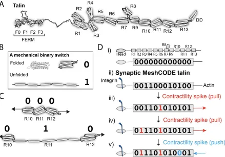
The protein talin, contains 13 of these switches (Goult et al., 2018; Wang et al., 2019; Yao et
al., 2016) and it is my assertion that talin is the memory molecule of animals. These
mechanosensitive proteins scaffold each and every synapse and were previously considered
to be mainly structural. However, this scaffold contains a meshwork of binary switches that I
propose form a code, a MeshCODE (Fig 1). The identification of such a network of switches
and the machinery that controls them leads to a new hypothesis for the way the brain might
The MeshCODE array of mechanical switches, would be operated by the cytoskeletal
machinery, with synaptic signalling triggering the cytoskeleton to push and pull on these
switches to constantly alter and update the coding in each neuron. Together, this mechanical
layer would integrate with the chemical and ionic signalling layers to provide the mechanism
for information-processing and storage. Electrochemical signalling between neurons would
coordinate a network of 100s of trillions of mechanically operated switches, each able to store
one bit of data, with action potential spike trains serving as the means to enter new information
into this calculation. This mechanical coding would be running continuously in every neuron,
extending into every cell in the organism amounting to a machine code coordinating the
animal.
Figure 1. The mechanical cell. Cartoon of a neuron showing the binary coding that results from the 100s of mechanical switches built into each synapse. The cytoskeleton is represented as a mechanical machine that operates these switches in response to neuronal activity, altering and updating the coding.
Whilst this essay focuses on the mechanical switches of talin as the minimal unit of memory,
there are other mechanosensitive proteins that are also present in the periphery of the
computer, the MeshCODE framework only requires that there are mechanical binary switches
present in each synapse that can be controlled by synaptic transmission.
The concept of the mechanical computer described herein provides a new hypothesis for how
the brain might be performing computation, identifying an addressable read/write memory
mechanism. Such a memory would facilitate both storage of data in an indexed, hierarchical
structure and provide a basis for how information-processing machinery can call this data as
required. This novel concept for biochemical data storage and organic computation, has
similarities with computers, old and new. These similarities span from the earliest mechanical
computing machines, through to solid state disks (SSD) and the complex subroutines that
enable complex computation. Remarkably, humankinds efforts to produce optimal
computation in silico has led to architectures that bear a striking similarity to what nature has
already arrived at in vivo.
Charles Babbage and the first mechanical computer.
The original concept of a digital programmable computer is attributed to Charles Babbage,
whose “Analytical Engine” mechanical computer was first described in 1837 (reviewed in
(Babbage et al., 1973)). These early visions of how a computer could be used to automate
calculations were prescient, defining a pipeline of events, starting with taking an input, running
it through a central processing unit, holding values in memory, and performing calculations
before generating outputs as a result of the calculation. This pipeline represents the same
core architecture of modern day computers.
Early computers were built using mechanical components, complex machines of levers and
gears that were used to perform calculations, by turning gears and incrementing counters and
ultimately output displays. The inputs initiate the calculation and the levers and gears crunch
the numbers, and push and pull until the calculation is complete and the output returned. The
programs that the Analytical Engine uses take the form of punched cards, where the pattern
of holes on the card run different parts of the machine. By changing the pattern of holes on
the card, a different program will run to give a different calculation and obtain a different output.
The output in many of these devices was a display of numbers, where gears turning
incremented the display.
The cell as an organic calculating machine
There are considerable similarities between a mechanical computer and a cell. Each cell
contains a series of levers, pulleys and gears in the form of its cytoskeleton. The cytoskeleton
microtubules and intermediate filaments. These filaments can assemble and disassemble
rapidly and robustly in response to cellular signals, and the interplay between them is complex.
The cytoskeleton can be used to generate forces, with motor proteins pushing and pulling on
actin and microtubule filaments, exerting forces on to specific targets. These filaments can
also serve as railroads to transport cargos to precise locations in the cell. There are hundreds
of cytoskeletal regulators that control these networks with the precise linkages, filaments,
adapters, etc. determined by the programme that cell is running.
Almost all cells in our bodies rely on cell adhesion molecules, that adhere the cell to adjacent
cells and/or the surrounding meshwork of proteins called the extracellular matrix (ECM). The
cytoskeleton is wired up to the cells adhesions, to the nucleus and to all the organelles in a
highly ordered (but dynamic) manner. Once a cell is established in its environment, the
cytoskeleton is maintained under tension but is not constantly generating large forces or
battling against itself. This homeostasis is achieved when all the forces and tensional
restraints are balanced in the system, at which point it is said to be under tensional integrity
or “tensegrity” (Fuller, 1961; Ingber, 1997). In a muscle, the actin filaments and myosin motors
are highly organised to enable the generation of forces and motion. In a similar fashion the
cytoskeleton in the brain would be ordered in a functional way that connects all the synaptic
adhesions together via mechanical linkages.
Adhesions as information-processing centres.
The adhesions to the ECM, mediated by the integrin family of ECM receptors, serve as
sensitive mechanosensors, able to feel the surrounding environment and instruct the cell how
to function (Iskratsch et al., 2014). A seminal study by Engler et al. (Engler et al., 2006)
demonstrated the powerful computing capabilities of integrin adhesions. Based solely on the
physicality of their environment a stem cell is able to reprogram into different cell lineages.
This remarkable ability of a cell to interrogate the environment and adjust accordingly hints at
the complexity of signalling through adhesions and demonstrates the role of adhesions as
information-processing centres. Via these adhesions both physical and geometric constraints
are sensed and used to induce modular gene expression patterns. These physical cues are
detected by exquisitely sensitive mechanosensitive proteins, that detect changes in
mechanical stiffness and alter the signalling of the cell.
Talin
The major linkage between the integrin-ECM connections and the cytoskeleton is the protein
talin (Calderwood et al., 2013; Klapholz and Brown, 2017). In each adhesive structure sits
cytoskeleton creating a complex array of talin molecules. Talin is perfectly positioned to
respond to changes in forces, both from outside and inside the cell, and has emerged as a
master mechanosensor in that it can sense these forces and convert them into biological
signals. Each talin molecule contains 13 force-dependent binary switches (Yao et al., 2016)
(Fig 2A). These talin domains, can be reversibly switched between two thermodynamically
stable states, “folded” and “unfolded”, using mechanical force (Fig 2B). The conformational
state of each switch determines what signalling molecules are recruited, providing different
instructions to the cell as a function of force (Goult et al., 2018). As the environment changes,
such as when a cell migrates, these switches detect these changes enabling the cell to
respond appropriately. Most current models envisage talin as a rope in a “Tug-of-War”
between extracellular forces, and those generated by the cells force-generating machinery
(Fig 2C).
Talin, the data molecule of life.
Every animal known to humankind has the same 13 switches in talin, and the high
conservation of switch pattern suggests a role that is explicitly dependent on the order of the
string of switches. The best way to visualise such a role is to consider the scenario where the
extracellular environment is built in such a way that it presents a mechanically stable,
predictable environment. Instead of a tug-of-war scenario, these cells would be pulling on talin
molecules attached to a surface. In this scenario, the talin switches are no longer required to
sense the extracellular environment as that is, to all intents and purposes, constant. Instead,
the cell can use its force-generating machinery to operate these mechanical switches and in
doing so write data into the adhesions (Fig 2D). This means that, in all animal cells, there is
an array of talin switches, wired up to an extensive cytoskeletal machinery, that provides the
capacity to store huge amounts of data, and to form persistent signalling complexes that
control cell behaviour as a function of mechanical force.
This has profound implications for data storage in animals, as it means that the cell can
repurpose the talin switches for use as data-storage systems. Adhesions in controlled, stable
environments can adopt the role of data-storage devices that store information in a manner
controlled by the system. I would like to propose that this complex meshwork of switches
operates as a code, a MeshCODE. To simplify the nomenclature of the switches in this view
of talin it is easier to regard the “folded” and “unfolded” states as “0” and “1” respectively to
better reflect that it is data that is being stored in the talin molecule.
Exquisite calculation in a single cell.
As a result, the cell is a mechanical computer with all of the architecture necessary for
computation. The cytoskeleton serves as the levers and gears that perform the calculation,
and the MeshCODE adhesions provide a multifunctional system that can be used to perform
calculations on, and serve as a memory storing the results in the conformations of the
switches. An input signal, be it an extracellular signal activating a receptor, a change in
physicality, or excitation by a chemical or electrical signal, perturbs the balanced state of the
cell, switching the computer on, triggering changes in the cytoskeleton as it seeks to return to
homeostasis. These changes in architecture and contractility, push and pull on the adhesions,
and result in reproducible alterations in the binary switches. When the calculation is complete,
homeostasis is restored. At this end point, the conformations of the switches in the adhesions
are altered and the array of 0s and 1s reflect the outcome of the calculation.
The appearance of talin at the dawn of multicellularity allowed cells to store information
as a memory, these switches also coordinate the cells signalling and provide a way to control
the reading of the genome from the periphery of the cell. Cells utilise this switch box in many
diverse ways to control phenotype and cell behaviour.
Organic calculation in the brain.
The brain is a colossal cell signalling machine with a trillion cells all communicating with each
other, leveraging the organic calculating power of each cell. Synapses are the perfect system
for optimised cell signalling between connected cells, and there are ~100 trillion synapses in
the brain (Pakkenberg et al., 2003) transmitting signals between neurons, to give rise to brain
activity. Synaptic transmission is one of the best studied biological events, and the details of
the transmission across the active zone, and the interactions in the pre-, and post-synaptic
density are well characterised (Asok et al., 2019; Mayford et al., 2012). Each synapse is
scaffolded by adhesions located around the edge of the synapse (Dityatev et al., 2014; Lilja
and Ivaska, 2018; Park and Goda, 2016), these adhesions are mediated by integrins binding
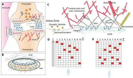
to ECM components such as laminin and fibronectin (Levy et al., 2014) (Fig 3A-C). The brain
is soft, which provides the perfect, protective environment for each neuron to tightly control its
own mechanical environment independently, building its own ECM nest around each synapse,
with the surroundings serving to dampen external forces. This isolates the neuron from
external mechanical forces, enabling mechanical computation to occur with meticulous
precision. Therefore, in the MeshCODE framework these scaffolds provide the capacity to
write data into the synapses themselves in the extensive arrays of binary switches located in
both the pre-, and postsynaptic side of each synapse. The capacity to store information in
every synapse, with the potential to orchestrate the flow of information through that synapse,
Figure 3. The MeshCODE. A) Schematic diagram of a tripartite synaptic junction. The integrin adhesion complexes and MeshCODEs in the presynaptic, postsynaptic and astrocyte are shown. The synapse is encapsulated by a specialised ECM that protects the mechanical environment of the MeshCODEs. Integrin (blue) and talin (grey) in one MeshCODE are shown wired up to the actin cytoskeleton (red). B) The MeshCODE crown around the synaptic cleft that scaffolds the synapse. C) Cartoon of a synaptic adhesion showing the MeshCODE intricately wired up to the cytoskeleton. D) A schematic of an array of ten talin molecules with the 13 switches arranged vertically. White = 0, Red = 1. E) Perturbations to the system, alter the pattern of 1s and 0s written in the MeshCODE and its resultant output in a defined way.
The ability for re-writable, long-term storage of information in the conformational patterning of
the MeshCODEs, means data storage in the brain would be encoded in a binary format written
into each and every synapse (Fig 3D). Each input signal would alter the activity of positive and
negative regulators of the cytoskeleton within that neuron, setting the machinery into action
and initiating a new calculation which results in altered MeshCODE patterns in that synapse
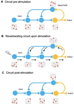
and other synapses in the neuron (Fig 3E). As this neuron would be communicating with other
neurons, part of this return to homeostasis would involve signals being emitted through
specific synapses, switching on the calculation in adjacent cells (Fig 4). The calculation would
trigger, and control the rate of, synaptic release meaning that synapses can be switched on
and off transiently on demand. In each of these synaptic transmissions the binary coding of
the MeshCODEs across that synapse would be altered. As a result, the re-balancing and
return to a metastable end state would need to occur across entire circuits. The pattern of 1s
and 0s in each synapse across the whole network of neurons would be inextricably linked, all
Figure 4. MeshCODE updating. Cartoon of a simplified neural circuit comprised of an input neuron, inputting into a 3 neuron circuit (blue) wired up to an output neuron (orange). A schematic of a MeshCODE is shown for each neuron (N.B. for clarity just a 6x6 array is shown). White = 0, Red = 1. A) At rest the MeshCODEs through the circuit are in a specific pattern of 1s and 0s. B)
Stimuli input into the system, perturbing the equilibrium state across the whole circuit. The calculation occurs as the cytoskeletal alterations cause changes to the coding of the system. The relevant output from the circuit is transmitted. C) Following the input, and the resulting calculation, the MeshCODEs in that circuit are altered in a way that encodes information.
Machine Code
Every MeshCODE of every synapse of every neuron of every circuit would contain information
representing the current state of the organism. The coding would therefore represent a type
of machine code that the organism is using. Like most computer machine code, in the
MeshCODE framework, the brain would be using a binary format. Every synapse in the brain
would be written with the code, which would be constantly changing in response to the signals
from the system. Some circuits would be stable, and others would be rapidly changing.
Furthermore, MeshCODEs are found at all places where cells engage the ECM, so having a
communication, providing an instruction set for every cell to work in synchrony. Like computer
machine code, without the necessary parsers to correctly decode the symbols, the code is
unreadable to an outsider, and would look like just pulses of electrical activity, triggering
alterations to vast strings of 1s and 0s. However, once the language of the code is understood
this information might be decipherable and might reveal an unimaginable level of
communication.
Mathematical representation – overcoming the binding problem
Stimuli acting on sensory receptors result in action potential spike trains that transmit external
information to the brain. Since every information sender and receiver in the organism is
controlled and running the same operating system, action potential spike trains would serve
as the vehicle to enable the transfer of information encoded in a way that allows the updating
of the coding of the sender and receiver. A machine code provides a mechanism for the brain
to integrate all sensory inputs and outputs into a single coherent whole, which can be
processed and compiled into a mathematical representation of the animals entire life. For
example, sensory input from vision does not just function in isolation, but inputs into the
calculation that contains every other sensory input contextualised in the entire learned
experience. All of these different cues are processed and form part of a unified cohesive
experience, and a mathematical representation would provide an explanation for how all this
information is bound together.
MeshCODEs provide a framework for organic calculation in the brain and the potential for
every synapse to contain the coding for the current best information available to the organism
for immediate usage. As a result, the reason the brain can process and react so quickly despite
electrochemical transmission being slow (eight orders of magnitude slower than in a
computer) would be because each neuron and motor synapse etc. is primed with information
that is being constantly updated. The calculation is performed predominantly in the brain but
Box 1: What is the memory capacity of the human brain in the MeshCODE framework?
Talin has 13 binary switches, but for simplicity of numbers let us assume that each talin contains 8 switches, or 8 bits of information = 1 byte per talin.
If we assume 100 talins per synapse (likely to be more), then each synapse can store 100 bytes of information.
If we assume 100 trillion synapses in the brain, then the MeshCODE data storage would be 100 bytes per 100 trillion synapses = 10,000 trillion bytes = 10 petabytes of global brain memory capacity*.
How much data can MeshCODE arrays in the cortex hold?
If we consider the cortex as the location of long-term memory information, then what is the storage capacity of this region? If each pyramidal cell has 10,000 synapses with 100 bytes/synapse, then each pyramidal cell can store ~1 MB. If 100 pyramidal cells form a minicolumn, then each minicolumn can contain 100 MB. There are ~100 million minicolumns meaning a potential storage capacity of 100 terabytes in the cortex.
*The memory capacity has potential to be hugely more than this as all cells in the body (and astrocytes and glia etc. in the brain) also have MeshCODE capacity. It is possible that each cell gets encoded with updated memory information as part of the brains computation.
Implications of a physical location for data storage.
A major requirement of any computational device is the capability for long-term memory
storage and retrieval (Gallistel and King, 2009). An array of MeshCODE binary switches would
provide a mechanism for physical storage of data. Information might be written into such an
array in a similar fashion to how data is stored on a solid state disk (SSD) in computers. In this
section I discuss how simply considering a physical location for data storage necessitates
consideration of the practicalities of storing terabytes of data (Box 1), not least how data might
be allocated and stored in the brain in a way that allows rapid recall.
Hypothesis: The brain cortex is an array of memory modules.
Memory associations and long-term memory are thought to be stored in the cerebral cortex
(Kandel et al., 2014). The architecture of the cortex is remarkably logical, made of distinct
sub-regions that control different processes (Zingg et al., 2014). The arrangement of neurons in
each sub-region of the cortex is highly ordered, arranged into 1-2 million cortical columns
(Mountcastle, 1997; Peters and Sethares, 1996) (Fig 5). Each cortical column has a logical
structure, containing many pyramidal neurons each with >10,000 synapses (Spruston, 2008).
Approximately ~100 pyramidal neurons arrange into minicolumns, which wire up to form the
together in highly ordered arrays, that might form memory modules. These memory modules
are connected to the hippocampus allowing information to travel back and forth between the
cortex and the hippocampus. If each MeshCODE in each synapse in each column can be
written to specifically, then this would represent a huge disk. This layered arrangement and
its intricate connectivity patterns has striking similarities to the architectural layout of an SSD.
In Fig 5 the basic architecture of a Flash SSD unit and a cortical column are drawn side by
side.
Figure5.Architectural similarities of memory storage in silico and in vivo.A) A high-capacity SSD storage device is incredibly complicated, but its general architecture is arranged into a logical repeating structure of Pages, arranged into Blocks that are arranged into memory modules (NAND Flash). These hierarchical structures are linked to channels that connect each memory module to a memory controller that controls the data coming in and out of the drive and directs current to the relevant pages and transistors for data read/write. B) The cortex shares a similar logical architecture. Three cortical columns are shown that are equivalent to the SSD Channels. These columns contain layers (I-VI) that are comprised of minicolumns (the memory modules equivalent to the NAND Flash), that are comprised of circuits of pyramidal neurons (the Blocks) that contain tens of thousands of MeshCODEs in the synapses and dendritic spines (the Pages). Each column is linked with read and write channels back to the hippocampus. Adapted with permission from (Peters and Sethares, 1996).
Synaptic connections that have the highest transmission might define the major connections
neurons into precisely mapped circuits, that allow memory allocation to each synapse in the
module from a central processing unit via some form of memory allocation circuitry. As with
an SSD information is read directly back from the stored information, in the case of the cortex
this would be achieved via synaptic transmissions passing through the column of memory
cells after being called by the information-processing units.
Hypothesis: Synapses are activated and deactivated transiently in response to signals down
the major circuit connections.
The presence of switches in the synapses raises the possibility that certain MeshCODE
patterns might transiently activate, or deactivate, transmission through specific synapses. It is
possible that these switches provide the basis for synaptic tagging (Frey and Morris, 1997;
Martin et al., 1997). As each neuron can have >10000 synapses, it might be that these can
be turned on and off with high specificity leading to precise stimulation of different synapses
and neuronal circuits directing the signals to specific synapses for data storage. MeshCODE
operations that allosterically enhance or dampen synaptic signalling would ensure that data is
directed to the right memory modules. Activation of one synapse in a neuron would lead to
alterations in other synaptic MeshCODEs in that neuron, mediated mechanically via the
cytoskeleton. These changes would dynamically alter both the stored information and the
transmission through that circuit.
Data mapping – the hippocampus as a memory controller?
A computer requires a digital interface circuit called a memory controller, which is positioned
between the computer and the memory, to manage the flow of data going back and forth
between the computer and the memory. The need for memory storage in the brain to be highly
ordered necessitates that the allocation of signal to each neuron in the network must also be
controlled via some form of memory allocation circuitry in the brain (this would seem to be a
requirement whatever the method of memory storage). To enable data to be written to such a
huge memory array, a detailed map of the memory architecture would be required to direct
the flow of traffic through the memory circuits, to mechanically write data in specific synapses.
Every neuron and synapse would be addressable by targeted communications. This process
would require the brain equivalent of a file system architecture to ensure that new data was
allocated to “free blocks” for writing but also that the data was stored in a logical, indexed way
so that it could be retrieved.
The hippocampus plays a major role in learning and memory (O’Keefe and Dostrovsky, 1971)
and has been identified as a key area of the brain for the processing of short-term memory
(reviewed in (Preston and Eichenbaum, 2013)). Studies have shown that initially memories
might be stored in the hippocampus in some representative form and that these
representations are later transferred into the cerebral cortex where the memory is stored as a
long-lasting engram in neocortical networks. This pipeline of data flow; collected by an input
source, processed and stored transiently in the hippocampus before being written to long-term
storage would suggest that the hippocampus might contain the equivalent of a file system that
maps out the data locations in the cortex and assigns information to specific cortical columns
for long-term storage. In doing so, data would be given a specific address in memory, indexed
and organised such that it is able to be retrieved and modified as and when required.
Therefore, the MeshCODE framework outlined here defines an addressable read/write
memory mechanism.
This transfer of data would be written by electrochemical signals going via these main
connections out into the cortical memory blocks, allocated seemingly at random to an outside
observer, but explicitly allocated via a memory controller to specific regions in the brain where
that data is stored. This data would be recorded by altering the pattern of 0s to 1s in specific
MeshCODEs within that memory module. Strikingly, during this writing process almost no
change would be visible in any of the synapses per se using current technology except for
alterations in firing patterns, even though major changes in data encoded in the synaptic
MeshCODEs would be occurring. However, if seen at the molecular level this process would
involve a whir of activity, with many changes in protein conformation as the data was written,
transferred, transmitted etc. across many synapses in the circuit.
How are memories retrieved so quickly?
The brain can recall data incredibly fast, in less than a second, and this speed of access
suggests that memory must be highly ordered. Neurons fire, and a lot of synaptic transmission
occurs to achieve this feat, but it is a non-trivial task to store such vast amounts of information
in a way that it is readily retrievable on demand. One way that huge amounts of data can be
organised to enable such rapid retrieval is to use a database. A database is an organized
collection of data, arranged in a hierarchical storage structure, that can be accessed, managed
and updated. By indexing the data, you can store it all, and only do data lookups when
required, by going directly to the address of that data, this is not only much faster than holding
everything in local memory, it also requires a smaller allocation of random access memory
when doing data lookups. Without a database and hierarchical storage structures all data is
given equal status, which means all data needs to be stored locally in case it is called. This
near infinite number of possibilities makes the requirement for a hierarchical storage structure
might also serve the role of a data management system. New memories would be processed
and indexed prior to being written in specific locations in the cortical MeshCODEs.
Hypothesis: A role for sleep in data management.
Studies have shown that neuronal activity during sleep plays a role in the transfer of memory
representations from the hippocampus to the cortex (Lee and Wilson, 2002; Marshall et al.,
2006). Following a period of being awake, the brain has received a lot of information, some
useful, some less so, and this needs to be processed and made coherent with the existing
data. The physical nature of memory outlined here suggests that the reason for sleep might
be the requirement to process huge amounts of data, integrate it with existing data, allocate it
to, and physically write it into specific memory modules in the cortex for storage. Furthermore,
as data is added, withdrawn and modified, overtime indexes become fragmented which
adversely affects performance. In a computer database it is necessary to run index
maintenance regularly and rebuild/reorganise indexes that require it. Rebuilding a database
is an intensive process that involves unloading information out of the database, before
reloading it back in a uniform, ordered fashion, optimising the filling space and re-indexing.
With such memory management in computers these processes are usually scheduled to be
performed overnight when the data is not being used. The process of the normal sleep cycle,
cycling between deep sleep and rapid eye movement (REM) sleep, might be the brain
performing data management.
Deep sleep
The initial deep sleep that occurs first in the normal sleep cycle might be required so that the
brain can complete the calculations from that day. Sleep, shuts down nearly all sensory inputs,
maintaining only sufficient awareness for self-preservation. During periods of deep sleep,
where brain activity is reduced, the lack of inputs might allow the system-level changes in
cytoskeleton and MeshCODE data storage to return to homeostasis which ensures the data
is correctly written and the newly formed memories are stabilised.
REM sleep might be the brain actively writing data to the cortex
Following this period of deep sleep, the sleep patterns change, and REM sleep begins. Here
the brain is very dynamic, with a lot of brain activity. It could be that REM is where the brain
can now work on this data, processing it and integrating it with the existing data. And this
requires lots of brain activity, stimulating the newly acquired data locations, moving old data
around and writing new data to specific regions of the brain. During the REM process a lot of
electrical activity provides a signature that something is happening, but little change in the
protein level, there would be a storm of activity as the coding is altered, the cytoskeletal
machinery mediating communications mechanically through each neuron, talin switches
switching back and forth, strings of information being used to signal to other neurons to write
other strings of information. The individual talin molecules themselves would likely not move
far, but they would help to drive the flow of information, altering the conformations and switch
patterns in other synapses and in other neurons. The overall effect would be that data, the
binary patterning of 0s and 1s, would be moved about, processed etc.
Following this reshuffling of the data, the brain goes back into a period of deeper sleep,
perhaps to ensure that the new data patterns are established, and to allow the system time
for further information-processing. Following memory stabilisation, another round of REM
sleep occurs, allowing further data writing and reordering. This cycling between sleep states
ideally occurs 4-5 times a night.
The result of this data management would see the transfer of newly encoded memories from
the hippocampus to the cortex where they are consolidated and stabilised as long lasting
MeshCODE patterns, that are the physical location of engrams. The next day the electrical
fingerprint of that memory association is seen to be different as the total number of neurons
involved would be reduced as the memory was consolidated. The process would see each
memory given a specific physical address, allowing it to be indexed so that it can be called
whenever needed. A result of sleep deprivation would be that the brains memory would soon
get fragmented with deleterious effects on recall and performance.
A recent study showing that sleep-associated activity patterns can also erase memories from
the hippocampus (Draguhn, 2018; Norimoto et al., 2018) suggests a volatility to hippocampal
data storage. The transient memory storage of the hippocampus sounds like Random Access
Memory (RAM) in computers where data is volatile and only retained whilst powered on. After
the data is successfully written to the cortex, the hippocampal MeshCODEs can be reset ready
for the next day.
Conclusion
The MeshCODE theory presented here provides an original concept for the molecular basis
of memory storage. I propose that memory is biochemical in nature, written in the form of
different protein conformations in each of the trillions of synapses. This concept is based on
the discovery of a complex network of mechanical switches (Goult et al., 2018; Yao et al.,
switches can be operated by the force generation machinery of the cells cytoskeleton leading
to a new view of the brain as a mechanical computer. The identification of an addressable
read/write memory mechanism clearly points to a way that the brain might carry information
forward in time and perform computation. Data written in symbolic form would provide a basis
for how the brain might function as an input/output system where its computation and data
processing systems are founded on physical and mathematical principles. The implications
for our understanding of information-processing in the brain should transform our view of
organic computation.
Action potential spike trains are well established as the organisms way of sending information
over long distances, similar to how electrical pulses carry information in electronic systems,
yet quite how these voltage spikes travelling down axons carry information has not been fully
understood. In this framework these spikes would transfer information by triggering precise
responses which alter the coding of the receiver cell. Diverse input signals including visual,
auditory, olfactory, temporal cues, self-movement (idiothetic) etc., are converted into spike
trains and the precise patterns of spikes trigger exact changes to the neurons, such that the
information they carry would be integrated into the organisms binary coding. It is possible to
imagine a complete mathematical representation of the world encoded in the MeshCODE
framework connecting all the inputs and outputs of the animal. This complex mechanical
coding amounts to a machine code that is constantly running in all animals. From an initial
state at birth, the life experiences and environmental conditions of the animal would be written
into the code, creating a constantly updating, mathematical representation of the animals
unique life. It is possible that consciousness is simply an emergent property arising from the
interconnectedness of electrical signals connecting all these MeshCODEs, forming a complete
mathematical representation of the world, that gives rise to precise electrical signals that
coordinate an entire biochemical organism in the context of the world.
Hypoxia and the death of the brain
The brain is the most energy intensive organ in the human body, requiring constant supply of
oxygen and nutrients. Loss of this supply is catastrophic, as ~6 minutes after the heart stops
there is irreversible brain damage. As a living machine, the mechanical coding of the brain
exists at a balance between opposing forces and factors. These forces are generated by the
cells force-generating machinery that requires energy to function. As these motors fail, the
synergy between the mechanical coding of all of the neurons will begin to be rapidly
scrambled. The loss of contractility will scramble the data stored in the MeshCODE switches,
irreversibly corrupting the data, as the synchronisation of coding across the brain is lost. As a
is re-established, the brain cannot recover this information and functioning once it is lost
beyond a certain point.
Implications for neurological disease
For MeshCODE storage to work correctly, it would require each switch to unfold and refold
with high fidelity. Having the protein conformations encoding memory located around the edge
of the synapse means it would be susceptible to getting clogged up, and proteins sticking
non-specifically to the mesh would disrupt the pattern of 1s and 0s. Any disturbance of
folding/refolding would corrupt the coding and scramble the information stored, leading to
memory loss. Abnormal accumulation of amyloid-β and tau protein is linked to the memory loss and cognitive decline seen in Alzheimer’s disease (Brion, 1998) and it is possible that
part of this effect is that these tangles interfere with the MeshCODE. Loss of synaptic integrity
in ageing would also result in loss of stored information. The MeshCODE framework should
therefore provide a novel therapeutic axis for a number of synaptopathies (Grant, 2012), and
neurodegenerative diseases such as Alzheimer’s (Dourlen et al., 2019) and dementia.
Future implications
As a final comment, physical storage of memory would have significant potential future
implications, not least that it might make the stuff of science fiction possible. If memory and
consciousness are biochemical in nature, it is possible that one day we will decipher this
MeshCODE, how it stores and computes information to form a mathematical representation
of the world. In doing so we may understand the computations of the human mind, which might
even allow the transfer of the human mind from neural networks onto silicon chips running the
human Operating System. A biochemical basis of memory storage also raises the possibility
of being able to read the memory of not only the living but also the dead. Short term memory
might be accessible only transiently after death, however, if long term MeshCODEs are “write protected” it might be possible to read the long-term memory for the duration of the integrity
of the brain.
Acknowledgements:
I thank Nick Brown, Martin Schwartz, Jie Yan, Peter Gunning, Dave Critchley and Ian Goult
for stimulating discussions, and Katy Goult, Martin Warren, Mike Geeves, Neil Kad and Jen
Hiscock for critical reading of the manuscript. BTG is funded by BBSRC grant BBS007245/1
References:
Asok, A., Leroy, F., Rayman, J.B., and Kandel, E.R. (2019). Molecular Mechanisms of the Memory Trace. Trends Neurosci. 42, 14–22.
Babbage, C., Merrifield, C.W., Babbage, H.P., Ludgate, P.E., Quevedo, L.T., and Couffignal, L. (1973). Analytical Engines. In The Origins of Digital Computers, (Berlin, Heidelberg: Springer Berlin Heidelberg), pp. 7–123.
Brion, J.P. (1998). Neurofibrillary tangles and Alzheimer’s disease. Eur. Neurol. 40, 130–140.
Calderwood, D. a, Campbell, I.D., and Critchley, D.R. (2013). Talins and kindlins: partners in integrin-mediated adhesion. Nat. Rev. Mol. Cell Biol. 14, 503–517.
Dityatev, A., Wehrle-Haller, B., and Pitkänen, A. (2014). Preface. In Progress in Brain Research, pp. xiii–xvii.
Dourlen, P., Kilinc, D., Malmanche, N., Chapuis, J., and Lambert, J.-C. (2019). The new genetic landscape of Alzheimer’s disease: from amyloid cascade to genetically driven synaptic failure hypothesis? Acta Neuropathol. 138, 221–236.
Draguhn, A. (2018). Making room for new memories. Science (80-. ). 359, 1461–1462.
Engler, A.J., Sen, S., Sweeney, H.L., and Discher, D.E. (2006). Matrix Elasticity Directs Stem Cell Lineage Specification. Cell 126, 677–689.
Frey, U., and Morris, R.G.M. (1997). Synaptic tagging and long-term potentiation. Nature 385, 533–536.
Fuller, B. (1961). Tensegrity. Portf. Art News Annu. 4, 112–127.
Gallistel, C.R., and King, A.P. (2009). Memory and the Computational Brain (Oxford, UK: Wiley-Blackwell).
Goult, B.T., Zacharchenko, T., Bate, N., Tsang, R., Hey, F., Gingras, A.R., Elliott, P.R., Roberts, G.C.K.K., Ballestrem, C., Critchley, D.R., et al. (2013). RIAM and vinculin binding to talin are mutually exclusive and regulate adhesion assembly and turnover. J. Biol. Chem. 288, 8238–8249.
Goult, B.T., Yan, J., and Schwartz, M.A. (2018). Talin as a mechanosensitive signaling hub. J. Cell Biol. 217, 3776–3784.
Grant, S.G.N. (2012). Synaptopathies: diseases of the synaptome. Curr. Opin. Neurobiol. 22, 522–529.
Ingber, D.E. (1997). TENSEGRITY: THE ARCHITECTURAL BASIS OF CELLULAR MECHANOTRANSDUCTION. Annu. Rev. Physiol. 59, 575–599.
Iskratsch, T., Wolfenson, H., and Sheetz, M.P. (2014). Appreciating force and shape — the rise of mechanotransduction in cell biology. Nat. Rev. Mol. Cell Biol. 15, 825–833.
Kandel, E.R., Dudai, Y., and Mayford, M.R. (2014). The Molecular and Systems Biology of Memory. Cell 157, 163–186.
130, 2435–2446.
Lee, A.K., and Wilson, M.A. (2002). Memory of Sequential Experience in the Hippocampus during Slow Wave Sleep. Neuron 36, 1183–1194.
Levy, A.D., Omar, M.H., and Koleske, A.J. (2014). Extracellular matrix control of dendritic spine and synapse structure and plasticity in adulthood. Front. Neuroanat. 8.
Lilja, J., and Ivaska, J. (2018). Integrin activity in neuronal connectivity. J. Cell Sci. 131, jcs212803.
Marshall, L., Helgadóttir, H., Mölle, M., and Born, J. (2006). Boosting slow oscillations during sleep potentiates memory. Nature 444, 610–613.
Martin, K.C., Casadio, A., Zhu, H., Yaping, E., Rose, J.C., Chen, M., Bailey, C.H., and Kandel, E.R. (1997). Synapse-specific, long-term facilitation of aplysia sensory to motor synapses: A function for local protein synthesis in memory storage. Cell 91, 927–938.
Mayford, M., Siegelbaum, S.A., and Kandel, E.R. (2012). Synapses and Memory Storage. Cold Spring Harb. Perspect. Biol. 4, a005751–a005751.
Mountcastle, V. (1997). The columnar organization of the neocortex. Brain 120, 701–722.
Norimoto, H., Makino, K., Gao, M., Shikano, Y., Okamoto, K., Ishikawa, T., Sasaki, T., Hioki, H., Fujisawa, S., and Ikegaya, Y. (2018). Hippocampal ripples down-regulate synapses. Science (80-. ). 359, 1524–1527.
O’Keefe, J., and Dostrovsky, J. (1971). The hippocampus as a spatial map. Preliminary evidence from unit activity in the freely-moving rat. Brain Res. 34, 171–175.
Pakkenberg, B., Pelvig, D., Marner, L., Bundgaard, M.J., Gundersen, H.J.G., Nyengaard, J.R., and Regeur, L. (2003). Aging and the human neocortex. In Experimental Gerontology, pp. 95– 99.
Park, Y.K., and Goda, Y. (2016). Integrins in synapse regulation. Nat. Rev. Neurosci. 17, 745–
756.
Peters, A., and Sethares, C. (1996). Myelinated axons and the pyramidal cell modules in monkey primary visual cortex. J. Comp. Neurol. 365, 232–255.
Preston, A.R., and Eichenbaum, H. (2013). Interplay of Hippocampus and Prefrontal Cortex in Memory. Curr. Biol. 23, R764–R773.
Spruston, N. (2008). Pyramidal neurons: Dendritic structure and synaptic integration. Nat. Rev. Neurosci. 9, 206–221.
Wang, Y., Yan, J., and Goult, B.T. (2019). Force-Dependent Binding Constants. Biochemistry
58, 4696–4709.
Yao, M., Goult, B.T., Klapholz, B., Hu, X., Toseland, C.P., Guo, Y., Cong, P., Sheetz, M.P., and Yan, J. (2016). The mechanical response of talin. Nat. Commun. 7, 11966.