Open Access
Research
Micro-morphologic changes around biophysically-stimulated
titanium implants in ovariectomized rats
Kivanc Akca
1,2, Ebru Sarac
3, Ugur Baysal
4, Mete Fanuscu
5, Ting-Ling Chang
6and Murat Cehreli*
7Address: 1Research Scholar, Division of Restorative Dentistry, The Jane and Jerry Weintraub Center for Reconstructive Biotechnology, UCLAc School of Dentistry, USA, 2Associate Professor, Department of Prosthodontics, Faculty of Dentistry, Hacettepe University, Ankara, Turkey, 3Private Practice, Obstetrics and Gynecology, Ankara, Turkey, 4Associate Professor, Department of Electrical and Electronics Engineering, Hacettepe University, Ankara, Turkey, 5Associate Clinical Professor, Division of Restorative Dentistry, The Jane and Jerry Weintraub Center for Reconstructive Biotechnology, UCLA School of Dentistry, USA, 6Associate Clinicial Professor, Division of Advanced Prosthodontics, The Jane and Jerry Weintraub Center for Reconstructive Biotechnology, UCLA School of Dentistry, USA and 7Associate Professor of Prosthodontics, CosmORAL Oral and Dental Health Polyclinics, Ankara, Turkey
Email: Kivanc Akca - [email protected]; Ebru Sarac - [email protected]; Ugur Baysal - [email protected]; Mete Fanuscu - [email protected]; Ting-Ling Chang - [email protected]; Murat Cehreli* - [email protected] * Corresponding author
Abstract
Background: Osteoporosis may present a risk factor in achievement of osseointegration because of its impact on bone remodeling properties of skeletal phsiology. The purpose of this study was to evaluate micro-morphological changes in bone around titanium implants exposed to mechanical and electrical-energy in osteoporotic rats.
Methods: Fifteen 12-week old sprague-dowley rats were ovariectomized to develop osteoporosis. After 8 weeks of healing period, two titanium implants were bilaterally placed in the proximal metaphyses of tibia. The animals were randomly divided into a control group and biophysically-stimulated two test groups with five animals in each group. In the first test group, a pulsed electromagnetic field (PEMF) stimulation was administrated at a 0.2 mT 4 h/day, whereas the second group received low-magnitude high-frequency mechanical vibration (MECHVIB) at 50 Hz 14 min/day. Following completion of two week treatment period, all animals were sacrificed. Bone sites including implants were sectioned, removed en bloc and analyzed using a microCT unit. Relative bone volume and bone micro-structural parameters were evaluated for 144 µm wide peri-implant volume of interest (VOI).
Results: Mean relative bone volume in the peri-implant VOI around implants PEMF and MECHVIB was significantly higher than of those in control (P < .05). Differences in trabecularthickness and -separation around implants in all groups were similar (P > .05) while the difference in trabecular-number among test and control groups was significant in all VOIs (P < .05).
Conclusion: Biophysical stimulation remarkably enhances bone volume around titanium implants placed in osteoporotic rats. Low-magnitude high-frequency MECHVIB is more effective than PEMF on bone healing in terms of relative bone volume.
Published: 16 July 2007
Head & Face Medicine 2007, 3:28 doi:10.1186/1746-160X-3-28
Received: 11 December 2006 Accepted: 16 July 2007
This article is available from: http://www.head-face-med.com/content/3/1/28
© 2007 Akca et al; licensee BioMed Central Ltd.
Background
Implant designs and treatment protocols are continuously evolving to promote osseointegration and the clinical suc-cess of oral implants. While many oral implant systems have been designed thus far to ameliorate biologic host response, biomechanical needs, which essentially domi-nate functioning has not been profoundly recognized [1]. To date, some methods of applying biophysical stimuli to implants such as electrical stimulation [2-4], pulsed elec-tromagnetic fields (PEMF) [5-7], and mechanical vibra-tion (MECHVIB) [8] have been tested for promovibra-tion of fracture healing and tissue differentiation at the bone-implant interface [9,10]. It is unfortunate that these exper-imental approaches have not been used to derive any ther-apeutic instrument coupled with an application schedule for oral/orthopedic implants so far.
The application of static and pulsed magnetic fields have been demonstrated to promote bone formation at sites of injury, such as fracture [11]. In the context of implants, magnetic fields under 10 mT have been shown to increase cell attachment and proliferation on titanium implant surfaces [12]. A study in the rat femur also showed that application of PEMF increased bone contact ratios of implants and that the applied dose had a critical role on bone apposition [7]. Under serum-free conditions, mechanical stimulation by intermittent hydrostatic com-pression has been demonstrated to increase sulfate con-tent mineralization of calcifying cartilage of fetal long bone rudiments [13]. The application of MECHVIB have shown that daily 1 Hz/100-sec regimen led to 28% bone ingrowth increase and at 20 Hz, the amount of ingrowth increased to 69% [8]. In addition, another study demon-strated that the intraosseous stability of implants sub-jected to direct 3 Hz mechanical vibration with a force of 5 N for 1800 cycli for 6 weeks were improved [14] Indeed, application of low-amplitude, high frequency mechanical stimuli seems very attractive from a therapeutic point of view to promote osseointegration [15], as the anabolic effects of low-magnitude mechanical signals have already been demonstrated on bone [16]. Further the risk of soft and hard tissue damage will be avoided at such low ampli-tudes, and the noninvasive and nonpharmacologic nature of the technique could improve the well-being of the patients during treatment. Nevertheless, current limita-tions of these approaches, i.e., long application sessions for PEMF and possible direct mechanical stimulation of implants via intraoral mechanical-shaking devices do not virtually seem feasible.
Osteoporosis leads to decrease in bone density and bone-implant contact ratio of bone-implants. A study has shown that transmitted MECHVIB could increase density of load-bearing bones in osteoporotic women [17]. In search of ways to promote histodynamics of tissue differentiation
and biomechanical potential of oral implants in oste-oporotic women, it was hypothesized that using transmit-ted MECHVIB could facilitate application of biophysical stimuli to the critical area [17,18] and its user-friendly nature could potentially be used for therapeutic applica-tion. In addition, it was assumed that the outcome of MECHVIB could be superior to PEMF in terms of bone response. The purpose of this study was, therefore, to compare micro-morphologic changes in bone around implants subjected to PEMF and transmitted (indirect) low-magnitude high-frequency MECHVIB in osteoporotic rats.
Materials and methods
Animals, care and ovariectomy
The experiments were undertaken in 15 locally-bred 12-week old Sprague-Dowley female rats. The animals were cared for according to the policies and principles estab-lished by the Animal Welfare Act and the NIH Guide for Care and Use of Laboratory Animals (publication # 86– 23). The surgical and experimental protocols for the mals were approved by the ethical committee of the ani-mal research facility of Hacettepe University (2004/45-9). During the entire test period, the animals were kept in rooms illuminated from 07:00 to 19:00 hours (12 h light/ 12 h dark cycle), maintained at 21–23°C, and had full access to low-calcium (0.1 %) powdered diet, prepared according to AIN-93M prescription, and water ad libitum. All surgical procedures were performed under general anesthesia using a mixture of ketamin (Ketalar, Parke-Davis; 50 mg/kg i.m.) and xylazine (Rompun, Bayer; 15 mg/kg i.m.). Following surgery, each animal was kept in a 25°C incubator until it regained consciousness.
1. Sample and addition of 2-Amino-2-methyl-1-propa-nol: 1.12 mol/L, pH 10.44 (30°C);magnesium acetate: 2.49 mmol/L; zinc sulfate: 0.50 mmol/L; N-(2-hydroxye-thyl)-ethylenediamine triacetic acid: 2.49 mmol/L
2. Addition of p-Nitrophenyl phosphate: 99.5 mmol/L; pH 8.50 (25°C); preservatives.
In the presence of magnesium and zinc ions, p-nitrophe-nyl phosphate is cleaved by phosphates into phosphate and p nitrophenol. The p nitrophenol is released propor-tional to the ALP activity and is measured photometri-cally.
Implants and surgery
A total of 30 cylindrical implants (Ø 1 mm × 5 mm) were obtained from a commercially-pure titanium rod (99.6%; Goodfellow Cambridge Ltd., Huntingdon, England). The implants were washed in ultrasonic deionized water, then further in trichloroethylene (99.5 %) and ethanol (70%), and sterilized before tests [22]. During surgery, both cor-tices of the tibia were perforated with low rotational speed under constant saline cooling with a surgical drill having a diameter smaller than the implant's diameter. The rationale behind this approach was to achieve good pri-mary stability of the cylindrical implants. Two implants were placed bilaterally placed in the proximal metaphyses of tibia. The flaps were closed with resorbable sutures (Vicryls 3-0, Ethicon GmbH, Norderstadt, Germany) and left to heal for 1 week.
Test groups and application of biophysical stimuli
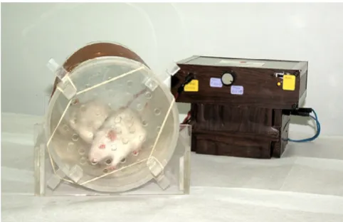
Upon placement of the implants, the animals were ran-domly divided into three groups. Group 1 served as con-trol. In Group 2, PEMF stimulation was administrated at 0.2 mT 4 h/day for the implants [7] (Fig. 1). The custom-made PEMF delivery device was fabricated at the Depart-ment of Electrical and Electronics Engineering of Hacet-tepe University and tested for accuracy using hall effect gauss/tesla meter (Sypris F.W. Bell Model 5080, Florida, USA) having 1% accuracy in measurement range. In Group 3, low-magnitude high-frequency MECHVIB at 5
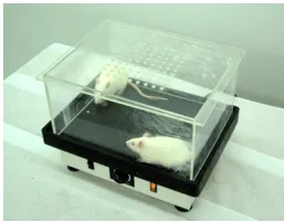
N/50 Hz 14 min/day was applied to the implants, while each animal was set on a mechanical vibrating plate (Vibratore Shaker 6, Carlo Degiorgi, Milano, Italy) with plexiglass borders to keep the test animal within the test zone during the therapeutic stimulation period (Fig. 2). The mechanical vibrating plate provides a barely percepti-ble stimulus, which does not alter animal behavior. This application allowed a ground-based whole body applica-tion through the hindfeets of the animal contacting the vibrating plate [17,18]. In addition, this technique allows the animal to move freely on the vibrating plate [18]. After 14 days, all animals were sacrificed and the tibia of each animal was removed en bloc and kept in physiologic saline maintained at 21–23°C.
Micro-morphologic evaluation of bone around implants
Each specimen was subjected to micro-tomographic scan-ning [23] using desktop MicroCT (µCT40, ScancoMedical, Bassersdorf, Switzerland) with a resolution of 16 × 16 × 16
µm3. The specimen were scanned with up to 320
trans-verse slices, where each slice consisted of 1024 × 1024 pix-els and followed by off-line reconstruction. Prior to micro-morphologic analyses, transverse slices cervically and apically resting in cortical bone were discarded. Three longitudinal volume of interest (VOI) each with 48 µ
m-thick (3 voxels × 16 µm resolution) were nominally
defined, and consecutively numbered 1 to 3 starting from the implant surface (Fig. 3). The resulting images were then segmented by using different thresholds for bone and implant [24]. The specific thresholds for titanium and bone were determined by superimposing segmented over original grayscale images.
The relative bone volume (BV/TV: %) and micro-morpho-metric bone parameters including trabecular thickness (Tb.Th: mm), trabecular separation (Tb.Sp: mm) and
p-nitrophenyl phosphate+H O2 ALP→phosphate+p nitrophenol
Fabricated custom-made device to deliver PEMF stimulation on osteoporotic rats
Figure 1
Fabricated custom-made device to deliver PEMF stimulation on osteoporotic rats.
Table 1: ALP levels (U/L) before and after ovariectomy in randomly selected 5 animals.
Animal BO AO
#1 72 285
#2 76 305
#3 87 298
#4 78 312
#5 85 264
trabecular number (Tb.N: 1/mm) were separately calcu-lated for each VOI. Morphometric indices, which can be directly determined from the binarized VOI, are the bone volume density (BV/TV), which is a measure of the vol-ume of the bone trabeculae relative to the total volvol-ume of the VOI, and the trabecular number (Tb.N). From these directly determined indices, other indices are derived such as the trabecular thickness (Tb.Th), the trabecular separa-tion (Tb.Sp) [25]. The correlasepara-tion between structural parameters obtained by micro-CT and conventional histo-morphometry has been investigated to determine which micro-CT parameter is most closely related to oste-oporotic fracture [26]. It has been ascertained that BV/TV, 3D-Tb.Th and 3D-Tb.N were correlated with those values on conventional histomorphometry. Therefore, trabecu-lar bone parameters gained from micro-CT scanning in this study seems to provide valuable information.
Statistical analysis
The data of each test group were compared with one-way analysis of variance (ANOVA) at a confidence level set at 95% and further with Post Hoc Tests (LSD) at 95% to determine different groups.
Results
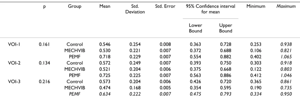
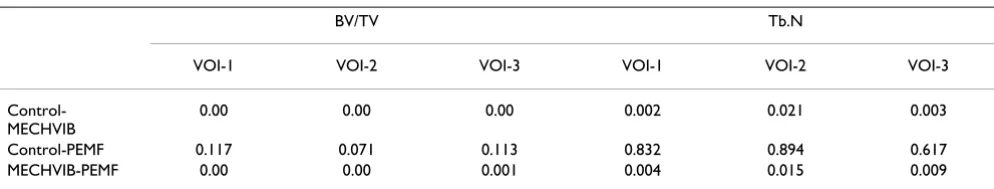
3-D microCT view of three VOIs around control, PEMF-stimulated, and MechV-stimulated implants are presented in Fig 4. Descriptive statistics of BV/TV, Tb.Th and Tb.Sp, and Tb.N are presented in Tables 2, 3, 4, 5 and Post Hoc Test (LSD) comparisons between groups are shown in Table 6. ANOVA of BV/TV revealed a significant difference among test and control groups (p = 0.000) in vicinities VOI-1, VOI-2 and VOI-3. The peri-implant relative bone volume around MECHVIB-stimulated implants were higher than control and PEMF-stimulated implants (p = 0.000), whereas similar values were obtained for the latter two groups (P > .05) (Table 2). ANOVA of morphologic evaluations revealed that Tb.Th and Tb.Sp around implants in all groups were similar (P > .05) (Table 3 and 4, respectively). The difference in Tb.N among test and control groups was significant in all VOIs (P < .05). Tb.N around MECHVIB-stimulated implants was higher than control and PEMF-stimulated implants (P < .05), and sim-ilar around control and PEMF-stimulated implants (P > .05) in all VOIs (Table 5).
Table 2: ANOVA of peri-implant relative bone volume (BV/TV: %) around implants of test and control groups (n = 15).
p Group Mean Std.
Deviation
Std. Error 95% Confidence interval for mean
Minimum Maximum
Lower Bound
Upper Bound
VOI-1 0.000 Control 4.0 0.002 0.0007 0.002 0.006
MECHVIB 15.5 0.005 0.001 0.113 0.196 0.083 0.225
PEMF 7.0 0.002 0.0007 0.005 0.009 0.049 0.123
VOI-2 0.000 Control 22.4 0.008 0.002 0.162 0.286 0.121 0.370
MECHVIB 61.4 0.216 0.006 0.459 0.769 0.358 0.950
PEMF 34.3 0.007 0.002 0.287 0.339 0.220 0.509
VOI-3 0.000 Control 17.0 0.005 0.001 0.128 0.211 0.084 0.265
MECHVIB 47.8 0.20 0.006 0.334 0.621 0.246 0.793 PEMF 26.5 0.008 0.002 0.207 0.322 0.180 0.447
Plexiglass bordered vibrating plate used to administer mechanical low-magnitude high-frequency to osteoporotic rats
Figure 2
Plexiglass bordered vibrating plate used to administer mechanical low-magnitude high-frequency to osteoporotic rats.
3a and 3b Figure 3
Discussion
The present study was designed to gain insight into micro-morphologic changes in bone around biophysically-stim-ulated implants in an animal study. In the present study, the rationale behind using ovariectomized rats was to explore the effect of MECHVIB and PEMF stimulation on bone micromorphology around titanium implants placed in metabolically compromised bone condition in terms of the possible worst case in bone to undertake the study [27]. Unlike previous studies [8] low-magnitude high-fre-quency MECHVIB was delivered using a "transmitted" approach, as this could facilitate administration of mechanical signals in a user-friendly nature and improve patients comfort. Indeed, the results of the present study show that low-magnitude high-frequency MECHVIB could transmit through bone [17], reach the implant, and improve osteogenic response in the vicinity of implants. The increased peri-implant relative bone volume (BV/TV) around MECHVIB-stimulated implants clearly shows that a transmitted application of 5 N/50 Hz for 14 min/day for 2 weeks improves trabecular bone, which may essentially
refer to improvement of implant anchorage and stability. Moreover, it is well-known that mechanical signals have a pronounced influence on the development and differenti-ation of mesenchymal tissues [28] and that the magni-tude, frequency, and the rate of experienced strain appear as important interrelated determinants of skeletal differ-entiation. Therefore, the regimen of MECHVIB used in the present study, probably fall into "species-independent" band of low-amplitude strains (< 500 µε) [8] in bone that act as a "growth factor"[28]. Since very small magnitudes of physiologic strains have been shown to increase bone mineral content and induce osteogenesis [8,11], and the direction of bending and axial loading does not have any effect on course of remodelling [29], artificial loading of implants by means of transmitted MECHVIB could poten-tially ameliorate bone-implant interface at early stages of function [15].
PEMF have been demonstrated to promote bone ingrowth into titanium and hydroxyapatite-coated implants, but not into tricalcium phosphate implants
Table 4: Descriptive statistics of trabecular-separation (mm) around implants of test and control groups (n = 15).
p Group Mean Std.
Deviation
Std. Error 95% Confidence interval for mean
Minimum Maximum
Lower Bound
Upper Bound
VOI-1 0.161 Control 0.546 0.254 0.008 0.363 0.728 0.253 0.938
MECHVIB 0.530 0.221 0.007 0.372 0.688 0.106 0.821
PEMF 0.718 0.229 0.007 0.554 0.882 0.402 1.065
VOI-2 0.134 Control 0.572 0.249 0.007 0.393 0.750 0.303 0.918
MECHVIB 0.521 0.204 0.006 0.375 0.668 0.122 0.803
PEMF 0.725 0.225 0.007 0.563 0.886 0.412 1.046
VOI-3 0.216 Control 0.573 0.204 0.006 0.426 0.720 0.365 0.861
MECHVIB 0.474 0.168 0.005 0.354 0.595 0.190 0.735 PEMF 0.634 0.222 0.007 0.475 0.793 0.334 0.950
Table 3: ANOVA of trabecular-thickness (mm) around implants of test and control groups (n = 15).
p Group Mean Std.
Deviation
Std. Error 95% Confidence interval for mean
Minimum Maximum
Lower Bound
Upper Bound
VOI-1 0.997 Control 0.140 0.005 0.001 0.100 0.180 0.080 0.226
MECHVIB 0.141 0.002 0.008 0.121 0.161 0.105 0.178
PEMF 0.142 0.005 0.001 0.104 0.179 0.090 0.260
VOI-2 0.655 Control 0.130 0.004 0.002 0.009 0.165 0.072 0.218
MECHVIB 0.147 0.002 0.009 0.125 0.168 0.105 0.192
PEMF 0.133 0.004 0.001 0.100 0.166 0.082 0.236
VOI-3 0.752 Control 0.188 0.005 0.002 0.149 0.227 0.114 0.288
[6,7,30]. However, the outcome of transmitted applica-tion of 5 N/50 Hz for 14 min/day being higher than PEMF-stimulation implies that micro-morphologic prop-erties of bone around implants is not higher than MECH-VIB by administration of 0.2 mT 4 h/day PEMF [7]. Nevertheless, we should note that bone response to differ-ent doses of PEMF could lead to differdiffer-ent results. Because it was not the core of the present study to explore the most osteogenic dose of PEMF, a dose of 0.2 mT 4 h/day, which had been shown to increase bone contact ratio and bone area ratio around implants were used [7]. Moreover, the duration of application seems as an important factor for bone response [6] and long application sessions for PEMF in the context of stimulating implants could inherently make the technique unpleasant for the patient, regardless of the dose administered. Therefore, not only the bone structural response but also the nature of PEMF seems rather weak in comparison to transmitted MECHVIB.
MicroCT with a limit of approximately 10 µm, provides the best resolution and has therefore become the imaging modality of choice for evaluation of trabecular bone in research over the past decade [25]. Using microCT, the interrelationship of mechanical and microstructural prop-erties of trabecular bone is important for better under-standing the consequences of trabecular remodeling. In the present study, increased peri-implant relative bone volume (BV/TV) and higher trabecular number were
found around MECHVIB-stimulated implants, which imply that the stiffness of the tissue in the vicinity of the implants had increased. However, MECHVIB was unable to increase trabecular thickness and/or decrease trabecular separation. Consequently, trabecular thickness and sepa-ration around implants in all groups was similar. A reduc-tion of trabecular thickness or increase in trabecular separation could have indicated decrease in mechanical properties of bone around the implants. The lack of increase in trabecular thickness and separation in test groups could be related to the dose administered, the duration of the experiment or both, but do not necessarily refer to the weakness of the techniques used to administer biophysical stimuli. Further studies are required to gain insight into structural and biomechanical characterization of bone around biophysically-stimulated implants.
Authors' contributions
The study design was established by Murat Cehreli and Kývanc Akca, who also wrote the manuscript. The ovariec-tomy surgeries were undertaken by Ebru Sarac. The ani-mal experiments were performed by Kývanc Akca. The PEMF apparatus was designed and fabricated by Ugur Baysal. Mete Fanuscu and Ting-Ling Chang performed the microfocus CT analysis.
Table 6: Post Hoc Test (LSD) comparisons between groups.
BV/TV Tb.N
VOI-1 VOI-2 VOI-3 VOI-1 VOI-2 VOI-3
Control-MECHVIB
0.00 0.00 0.00 0.002 0.021 0.003
Control-PEMF 0.117 0.071 0.113 0.832 0.894 0.617
MECHVIB-PEMF 0.00 0.00 0.001 0.004 0.015 0.009
Table 5: ANOVA of trabecular-number (1/mm) around implants of test and control groups (n = 15).
p Group Mean Std.
Deviation
Std. Error 95% Confidence interval for mean
Minimum Maximum
Lower Bound
Upper Bound
VOI-1 0.003 Control 2.027 0.830 0.262 1.432 2.621 1.305 3.884
MECHVIB 3.170 0.734 0.244 2.615 3.744 1.535 4.181
PEMF 2.09 0.636 0.201 1.642 2.552 1.083 3.162
VOI-2 0.025 Control 2.214 0.786 0.248 1.651 2.777 1.413 3.846
MECHVIB 3.014 0.716 0.226 2.502 3.527 1.730 3.622
PEMF 2.170 0.676 0.213 1.687 2.654 1.176 3.381
VOI-3 0.006 Control 2.051 0.576 0.182 1.638 2.463 1.479 3.259
Publish with BioMed Central and every scientist can read your work free of charge "BioMed Central will be the most significant development for disseminating the results of biomedical researc h in our lifetime."
Sir Paul Nurse, Cancer Research UK
Your research papers will be:
available free of charge to the entire biomedical community
peer reviewed and published immediately upon acceptance
cited in PubMed and archived on PubMed Central
yours — you keep the copyright
Submit your manuscript here:
http://www.biomedcentral.com/info/publishing_adv.asp
BioMedcentral
Acknowledgements
This investigation was conducted in part in a facility constructed with sup-port from Research Facilities Improvement Program Grant Number CO6 RR-14529-01 from the National Center for Research Resources, National Institutes of Health and, is partly supported by Turkish Scientific and Tech-nical Council [TÜBİTAK-Project 104E020 AY-60] and Hacettepe Univer-sity Research Projects 04 01 602 007 and 02 02 602 009.
References
1. Hansson S: The implant neck: smooth or provided with reten-tion elements. A biomechanical approach. Clin Oral Implants Res
1999, 10:394-405.
2. Weinstein AM, Klawitter JJ, Cleveland TW, Amoss DC: Electrical stimulation of bone growth into porous alumina. J Biomed Mater Res 1976, 10:231-247.
3. Young SO, Park JB, Kenner GH, Moort RR, Myers BR, Sauer BW: Dental implant fixation by electrically mediated process. I. Interfacial strength. Biomater Med Devices Artif Organs 1978, 6:111-26.
4. Berry JL, Geiger JM, Moran M, Skaraba JS, Greenwald AS: Use of tri-calcium phosphate or electrical stimulation to enhance the bone-porous implant interface. J Biomed Mater Res 1986, 20:65-77.
5. Martin TJ, Ng KW: Mechanisms by which cells of the osteoblast lineage control osteoclast formation and activity. J Cell Biochem
1994, 56:357-366.
6. Ijiri K, Matsunaga S, Fukuyama K, Maeda S, Sakou T, Kitano M, Senba I: The effect of pulsing electromagnetic field on bone ingrowth into a porous coated implant. Anticancer Res 1996, 16:2853-2856. 7. Matsumoto H, Ochi M, Abiko Y, Hirose Y, Kaku T, Sakaguchi K: Pulsed electromagnetic fields promote bone formation around dental implants inserted into the femur of rabbits. Clin Oral Implants Res 2000, 11:354-360.
8. Rubin CT, McLeod KJ: Promotion of bony ingrowth by fre-quency-specific, low-amplitude mechanical strain. Clin Oral Implants Res 1994, 298:165-74.
9. Chao EY, Inoue N: Biophysical stimulation of bone fracture repair, regeneration and remodelling. Eur Cell Mater 2003, 6:72-84.
10. Meyer U, Kruse-Lösler B, Wiesmann HP: Principles of bone forma-tion driven by biophysical forces in craniofacial surgery. Br J Oral Maxillofac Surg 2006, 44:289-95.
11. Degan IL, Stetsula VI: Consolidation of bone fragments in a con-stant magnetic field. Ortopediia Tracmatologiia I Protezirovanie 1971, 32:45-48.
12. Kim HJ, Chang IT, Heo SJ, Koak JY, Kim SK, Jang JH: Effect of mag-netic field on the fibronectin adsorption, cell attachment and proliferation on titanium surface. Clin Oral Implants Res 2005, 16:557-562.
13. Bagi C, Burger EH: Mechanical stimulation by intermittent compression stimulates sulfate incorporation and matrix mineralization in fetal mouse long-bone rudiments under serum-free conditions. Calcif Tissue Int 1989, 45:342-7.
14. De Smet E, Jaecques S, Vandamme K, Vander Sloten J, Naert I: Posi-tive effect of early loading on implant stability in the bi-corti-cal guinea pig model. Clin Oral Implants Res 2005, 16:402-7. 15. Cehreli M, Sahin S, Akca K: Role of mechanical environment and
implant design on bone tissue differentiation: Current knowl-edge and future contexts. J Dent 2004, 32:123-132.
16. Rubin C, Turnet AS, Bain S, Mallinckordt C, McLeod K: Anabolism. Low mechanical signal strengthen long bones. Nature 2001, 412:603-4.
17. Rubin C, Pope M, Fritton JC, Magnusson M, Hansson T, McLeod K: Transmissibility of 15-hertz to 35-hertz vibrations to the human hip and lumbar spine: determining the physiologic fea-sibility of delivering low-level anabolic mechanical stimuli to skeletal regions at greatest risk of fracture because of oste-oporosis. Spine 2003, 28:2621-7.
18. Rubin C, Xu G, Judex S: The anabolic activity of bone tissue, sup-pressed by disuse, is normalized by brief exposure to extremely low-magnitude mechanical stimuli. FASEB J 2001, 15:2225-9.
19. Qi MC, Zhou X-Q, Hu J, Du Z-J, Yang J-H, Liu M, Li X-M: Oestrogen replacement therapy promotes bone healing around dental implants in osteoporotic rats. Int J Oral Maxillofac Surg 2004, 33:279-285.
20. Nakajima D, Kim C-S, Oh T-W, Yang CY, Naka T, Igawa S, Ohta F: Suppressive effects of genistein dosage and resistance exer-cise on bone loss in ovariectomized rats. J Physiol Anthropol Appl Human Sci 2001, 20:285-291.
21. Chae HJ, Choi KH, Chae SW, Kim HM, Shin TK, Lee GY, Jeong GS, Park HR, Choi HI, Kim SB, Yoo SK, Kim HR: Placenta hominis pro-tects osteoporosis in ovariectomized rats. Immunopharmacol Immunotoxicol 2006, 28:165-73.
22. Wennerberg A, Albrektsson T, Andresson B, Kroll JJ: A histomor-phometric and removal torque study of screw-shaped tita-nium implants with three different surface topographies. Clin Oral Implants Res 1995, 6:24-30.
23. Müller M, Van Campenhout H, Van Damme B, Van Der Perre G, Dequeker J, Hildebrand T, Rüegsegger P: Morphometric analysis of human bone biopsies: A quantitative structural comparison of histological sections and micro-computed tomography.
Bone 1998, 28:59-66.
24. Rebaudi A, Koller B, Laib A, Trisi P: Microcomputed tomographic analysis of the peri-implant bone. Int J Periodontics Restorative Dent
2004, 24:316-325.
25. Ruegsegger P, Koller B, Muller R: A microtomographic system for nondestructive evalation of bone arhitecture. Calcif Tissue Int
1996, 58:24-9.
26. Ito M, Nakamura T, Matsumoto T, Tsurusaki K, Hayashi K: Analysis of trabecular microarchitecture of huma bone using micro-computed tomography in patients hip arthrosis with or with-out vertebral fracture. Bone 1998, 23:163-169.
27. Sakakura CE, Giro G, Goncalves D, Pereira RMR, Orrico SRP, Marcan-tonio E Jr: Radiographic assessment of bone density around integrated titanium implants after ovariectomy in rats. Clin Oral Implants Res 2006, 17:134-138.
28. Rubin CT, Lanyon LE: Regulation of bone mass by mechanical strain magnitude. Calcif Tissue Int 1985, 37:411-417.
29. O'Connor JA, Lanyon LE, MacFie HM: The influence of strain rate on adaptive bone remodelling. J Biomech 1982, 15:767-781. 30. Shimizu T, Zerwekh JE, Videman T, Grill K, Mooney V, Holmes RE,
Hagler HK: Bone ingrowth into porous calcium phosphate ceramics: influence of pulsing electromagnetic field. J Orthop Res 1988, 6:248-258.
3-D microCT view of VOI 1–3 (from left to right) around control (top), PEMF-stimulated (middle), and MECHVIB-stim-ulated (bottom) implants
Figure 4