Hybrid Feature Extraction Techniques For
Accuracy Improvement In IVUS Image
Classification
S. Sridevi, M. Sundaresan
Abstract : In the field of Image processing, numerous applications play a vital role. Each and every application is specifically dedicated to various process in the corresponding field and such applications are Medical, Remote Sensing etc. This paper mainly aims to take care of the medical field for the purpose of identifying disease and reducing the causes of diseases. Image processing in medical applications is an important area in analysis of various Imaging systems. In that several research is going on and ultrasound imaging system is one of the popular modality used nowadays. Ultrasound imaging system is divided into many different types of study systems which are neurology, gynecology and cardiovascular system. The cardiovascular system is mainly used for the analysis of Intravascular Ultrasound (IVUS) images in order to detect the coronary artery disease. This paper mainly aims to find out the plaque disease by analyzing the different IVUS Medical images by performing the steps like Pre-processing, Minimizing the false positive findings, Reduction of Misclassifictaion Error, Hybrid feature extraction technique and Validation for improvement of classification accuracy.
Index Terms : Cardiovascular System, Ultrasound Imaging System, IVUS images, Plaque detection.
—————————— ——————————
1
INTRODUCTION
Over the last decades, several investigative groups have determined the automated systems that can analyze the various types of modalities in medical imaging and also extract the useful information (eg. Tumor, plaque) for finding and identifying diseases [1]. The majority of the medical imaging methodologies are used to acquire images during a diagnostic procedure. Recently, a lot of research has been focused on different techniques for medical images; such images are acquired using a variety of devices and modalities including Ultrasound imaging system, Computed Tomography (CT), Digital X-Ray, Hyper Spectral Imaging (HSI), Vein Viewer (VV), Nuclear Medicine (NM), Positron Emission Tomography (PET), and Magnetic Resonance Imaging (MRI) [2]. Nowadays, automated schemes are widely used in the analysis of Ultrasound modality based systems such as Gynaecology, Neurology and Cardiovascular System. This system mainly concentrates only on the cardiovascular system. On the other hand, the most part of medical image or signals are represented in a two-dimensional way, which are designed for the purpose of automated detection, extraction and portrayal of abnormal conditions in coronary heart artery images [3]. These images are provided by the medical experts with useful information for the purpose of disease identification. Such systems are commonly referred to as IVUS imaging systems [4].
At present, this is a fact that carotid surgical procedure in patients with asymptomatic carotid plaque (stenosis) has reduced the occurrence of stroke and plaque formation in heart artery layers. However, a large number of patients may be operated in an unnecessary manner [5]. Therefore, it is an important mechanism of finding cardiovascular complications in patients. Consequently, there are some indications of morphology available in atherosclerotic plaque layer in a heart artery, which is obtained by high-resolution ultrasound imaging technology. It has the different structures like texture, surface, region, and analytical implications that are uniqueness of stable plaques in arteries, although asymmetrical and mixed textures are characteristics of unbalanced plaques in human artery [6]. So, this system will contribute towards the advanced methods for disease classification and also improving the accuracy in an efficient way [7].
2
LITERATURE SURVEY
Some of the existing work is explained below.
Hassen Lazrag et al [8], presented the fuzzy c-mean with spatial constraint algorithms used to efficiently extract the Region of Interest information from the IVUS image. K.V. Archana and Dr. R. Vanithamani [9] recently developed image processing methods, which is used to detect the heart artery layer (luminal and media) borders. IVUS image layer borders are detected based on the Edge tracking and active contour method. The study result, says about the weakness of quality metrics and also the lack of detection of lumen border. Vaishali Naik and R. S. Gamad [10] explained the standard level set Segmentation technique. The technique used in this work evaluates only the statistical parameter of an image. But they do not concentrate on the other parameter like True Positive, False Positive, True Negative, False Negative and accuracy. The above limitation can be overcome by using the following proposed method.
_________________________
S.Sridevi is currently pursuing Ph.D in Department of Information Technology, Bharathiar University, INDIA, E-mail: [email protected]
721
3
INTRAVASCULAR ULTRASOUND
IMAGING
3.1 Basics Of IVUS Imaging
The Intravascular Ultrasound Imaging System is important and popular in real world phenomenon. Because it is used to visualize the inside body parts in a noninvasive and also cost effective way. Medical modality of ultrasound wave based intravascular imaging is able to envisage the arteries frequently for monitoring the growth level of atherosclerosis and arterial uniqueness like depth of luminal thickness and media layer lesion branches of the inner and outer wall of the artery in heart images. Therefore, Atherosclerotic plaque information is assessed and extracted from Structural information based vessel details. Several clinical findings have focused on the occurrence of coronary heart artery Plaque disease such as branch openings, Left Anterior Descending (LAD) stent; calcify lesion, circumflex view, bifurcation LCX view, dense calcium and RCA diagnostic. Arterial wall changes are easily detected with ultrasound risk factor assessments and predications based factors looks like a known, unknown and are better predictors of risk than any combination of conventional risk factors assessments. These are often, the result of plaque erosion or rupture with subsequent thrombosis produced the blockage of the human heart.
3.2 Working Mechanism of IVUS Imaging
Intravascular Ultrasound (IVUS) imaging system is one of the most important procedures of extraction about the inner lining of arteries and vessel morphology. IVUS images are acquired by ultrasound echo which is used to represent the inner structure of arteries in human heart. Intravascular ultrasound has become the most common imaging modality, and the number of clinical applications for ultrasound continues to grow, which provide many other medical exams to test the diseased artery. The border line detection of the inner and outer boundary of the image component in a vessel is a crucial part to define the severity of arterial disease. So, the coronary artery disease is the mainly used for common type of heart artery disease in India, U.S.A, and Europe which causes the human deaths. Commonly, the coronary artery disease is called as atherosclerosis, which provides the results in hardening and thickening of the inner and outer boundary lining of IVUS image layers like outermost layer Adventitia, intima and lumen layers. Deposits of fatty substances, Amount of cholesterol formation, and other fatty contents construct the cellular waste products in the coronary artery vessel wall. The above said contents have formed and develop the plaque layer component along with the vessel wall. Accordingly, fractional or stumbling block of blood flow in the artery can occur and can lead to be a heart attack.
4
PROPOSED METHOD
The main stages of typical IVUS imaging systems are: (1) Preprocessing (2) Minimizing the false positive findings, Reduction of Misclassifictaion Error, Hybrid feature extraction technique and Validation for improvement of classification accuracy (Figure 1). The above said are explained briefly in the following sections.
Fig 1: Proposed method Scheme
4.1 Preprocessing
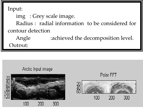
In preprocessing stage, the shadow artifacts are enhanced and the descriptions of radial and tangential characteristics extracted from the IVUS local image region. So, the local IVUS image region is transformed into polar coordinate conversion. In Polar coordinate conversion rows and columns are arranged into a frequency domain format and corresponding angles and distance are calculated, which is denoted as image Iimage (r (radial), θ (tangential)) (Figure 2). The number of Contour detection steps is facilitated for Ultrasound medical images such as contour initialization and refining of the obtained contour in IVUS image regions.
Fig 2: Polar coordinate conversion
4.2 Minimizing the false positive findings
At this stage, a vital and an important feature key point is captured and extracted from IVUS images. Segmentation approach normally considering the number of normal objects are portioning it into smaller parts and then extracting the features in an original image, which provides the results detection system in an efficient manner. Here, the most important issues in an IVUS imaging system are the standard feature set selection and classification method, which is mainly used to extract (ROI) Regions Of Interest while minimizing the false-positive findings on images.
Input:
img : Grey scale image.
Radius : radial information to be considered for contour detection
Angle :achieved the decomposition level.
Output:
pcimg : output images are generated
4.3 Hybrid Feature Extraction Techniques
To improve the performance of classification accuracy, more than a few image features are extracted in an effort and also describes the specific characteristics of each object in an image. Specific feature could be extracted, such as statistical features, shape features, Multi region histogram features, neighborhood gray tone difference matrix, statistical feature matrix gray level difference statistics, and Spatial gray level dependence matrices that described the local features such as object scaling and rotation in Intravascular Ultrasound (IVUS) plaque images (table 1).
TABLE 1: TECHNIQUES FOR FALSE POSITIVE
FINDINGS
Feature extraction techniques Features
Statistical features Mean, median skewness, and kurtosis. Standard deviation,
Spatial dependence matrices in gray level
Entropy difference, inverse difference moment, sum, average, Angular second moment, sum variance.
Shape features
The X-coordinate maximum length of the plaque image, y-coordinate, area (ROI), Perimeter of ROI.
Gray level difference statistics Angular second moment, entropy, mean, contrast. Neighborhood gray tone
difference matrix
Coarseness, contrast, complexity and strength
Statistical features matrix Periodicity and roughness.
The above specific features could be extracted from the images and then clubbed together into an array for classification purpose.
4.4 Reduction of Misclassification Error
A classification system is an essential part of an IVUS Imaging system. During the entire process, SVM kernel modifier is used to map the input vector into higher dimensional space where a hyper plane can separate the data into different classes. To avoid the misclassification error, the training classifier finds out the optimal hyper plane. This work considers the Radial basis function to handle the nonlinear relations with feature vectors and their classes. Extracted feature vectors were normalized into [0, 1] before applying the classifier. The Radial basis kernel with standard deviation was selected. There are two parameters in kernel function, namely K and γ. The γ is the error term and K is the kernel parameter. The reported results were obtained in order to set up the kernel parameter. The Modified kernel function used to map the samples, namely and , represented as feature vectors in some input space are defined as
K = (x,x') = exp√(- (((|(|x-x^' | )| ))⁄2σ))
Therefore γ>0 (1)
The classifiers that are utilized in the area of the detection of plaque disease are those employed in most of the medical image analysis procedures. Different types of plaque cases are categorized based on the following classes such as Coronary Lumen diameter Measurement (Figure 3 and 4) and Nonlinear SVM kernel modifier.
Classify input patterns using the constructed nonlinear SVM classifier as shown in Figure 5.
Pseudo Code 1. % begin classification...
2. [Labels, Classes, DecisionValue, predication value]= SVMkernel(predicted, αY, SVs, Parameters, Bias, NSV, nLabel); 3. % end of the classification
4. Pause % Strike any key to continue
5. % Compare with resultant labels and true labels of the data
6. Plot (1: length (True Labels), True Labels,'b-',1: length (True Labels), Labels,'r.');
7. Ylabel ('Class Index'); 8. Xlabel ('Pattern Index');
9. Legend ('True Labels','Resultant Labels', 0);
Fig 3: Comparison Graph
4.5 Statistical analysis / Validation for Improvement of Classification Accuracy
In this section the true positive, true negative, false positive and false negative and accuracy of classifier validation studies is calculated. Additionally, the kappa statistic and Confidence Interval level are also computed from the above said parameters for quantifying the degree of agreement.
CI (0.86%) = X ±1.96 x X x (1 – X)/ N (2) Kappa = ((observed – expected) Proportion / 1 –
expected proportion). (3)
The Kappa value 0.92 indicates the good agreement test.
5
RESULTS AND DISCUSSION
In this section, the performance of the proposed approach is presented. First, the performance of shadow detection procedure is investigated and corresponding segmented IVUS images are characterized by the proposed approach. True Positive, False Positive, False Negative, True Negative values are obtained by comparing manually and automatically detected regions. After shadow detects the Shape, statistical gray level difference matrices, neighborhood gray tone and statistical feature matrix were detected from the remaining portion of the plaque area. Finally accuracy was also calculated in order to assess the performance of the proposed method.
7 years man imaged after LAD Stanton –
(Branch Stent)
Lumen area
Lumen diameter
Vessel area
Max
723 Min
2.4 mm
mm2
Case: Dense calcium
7.7 mm2 Max 3.2 mm
16.2 mm2 Min
2.8 mm
Case: small vessel
4.7 mm2 Max
2.5 mm 7.2 mm 2 Min
2.4 mm
Fig 4 Sample Plaque measurements
Figure 5: Input pattern classification
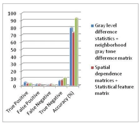
According to the feature extraction, results based classifications are validated and also the algorithm performance are reported and compared with the existing parameter and are shown in table 2.
TABLE 2: ACCURACY IMPROVEMENT BASED ON THE
HYBRID FEATURE EXTRACTION TECHNIQUES
True Positiv e
False Positiv e
False Negativ e
True Negativ e
Accurac y (%)
Gray level difference statistics + neighborhoo d gray tone difference matrix
5 2 1 7 80
Spatial dependence matrices + Statistical feature matrix
3 2 2 8 73.3333
3
Shape +
statistical 3 0 1 11
93.3333 3
Figure 6 shows accuracy is calculated based on the Parameters like true positive, false positive, false negative and true negative.
Figure 6: Graphical representation of Accuracy
6
CONCLUSION
In this paper, an effective approach to the challenging problem of plaque classification was presented. Several combining features were produced in this work. The enhanced SVM kernel modifier is used to describe the reliability of the obtained results. The results supported the positive effects of combining the three feature extraction methods. And then, generated the multi - resolution characteristics of more detailed images which performed better. Furthermore, the results obtained might be the (93%) highest achievable accuracy, using the combination of image based feature extraction method. The future research can use an M - mode image instead of B-mode image in order to perform classification of IVUS image.
REFERENCES
[1]. Serruys PW, Garcia-Garcia HM, Buszman P, Erne P, Verheye S, Aschermann M, et al. Effects of the direct lipoprotein-associated phospholipase A(2) inhibitor darapladib on human coronary atherosclerotic plaque. Circulation. 2008 Sep 9;118 (11):1172-82.
[2]. Okamura T, Onuma Y, Garcia-Garcia HM, Bruining N, Serruys PW. High-speed intracoronary optical frequency domain imaging: implications for three-dimensional reconstruction and quantitative analysis. Eurointervention. 2012 Feb;7(10):1216-26.
[3]. Serruys PW, Morice MC, Kappetein AP, Colombo A, Holmes DR, Mack MJ, et al. Percutaneous coronary intervention versus coronary-artery bypass grafting for severe coronary artery disease. N Engl J Med. 2009 Mar 5;360(10):961-72.
[4]. Farooq V, Gogas BD, Okamura T, Heo JH, Magro M, Gomez-Lara J, et al. Three-dimensional optical frequency domain imaging in conventional percutaneous coronary intervention: the potential for clinical application. Eur Heart J. 2011 Nov 21.
using intravascular radiofrequency data analysis: recommendations for acquisition, analysis, interpretation and reporting. EuroIntervention. 2009 Jun;5(2):177-89.
[6]. Gonzalo N, Garcia-Garcia HM, Serruys PW, Commissaris KH, Bezerra H, Gobbens P, et al. Reproducibility of quantitative optical coherence tomography for stent analysis. EuroIntervention. 2009 Jun;5(2):224-32.
[7]. Garg S, Serruys PW, van der Ent M, Schultz C, Mastik F, van Soest G, et al. First use in patients of a combined near infra-red spectroscopy and intra-vascular ultrasound catheter to identify composition and structure of coronary plaque. Eurointervention. 2010 Jan;5(6):755-6.
[8]. Hassen Lazrag, Kamel Aloui, Med Saber Naceur, ―Automatic Segmentation of Lumen in Intravascular Ultrasound images using Fuzzy Clustering and Active Contours‖, International conference on control, engineering & Information Technology (CEIT ’13) Proceedings Engineering & Technology- Vol.1,2013. [9]. K.V. Archana, Dr.R.Vanithamani, ―Review on
Intravascular Ultrasound Image Detection‖, International Journal of Advanced Research in Science, Engineering and Technology, Vol.4, Issue 7, July 2017. [10]. Vaishali Naik, R.S.Gamad et al, ―The Carotid
Intima – media Thickness Measurement of Ultrasound images‖, Proceedings of the 12th INDIACom, INDIACom-2018; IEEE Conference ID: 42835, ISBN 978-93-80544-28-1, 2018.