THE REPRESENTATION OF SPACE IN THE RAT HIPPOCAMPUS
AS REVEALED USING NEW COMPUTER-BASED METHODS
Michael L. Recce
A thesis submitted for the degree of Doctor of Philosophy
Department of Anatomy and Developmental Biology, University College London
ProQuest Number: 10106593
All rights reserved
INFORMATION TO ALL USERS
The quality of this reproduction is dependent upon the quality of the copy submitted.
In the unlikely event that the author did not send a complete manuscript and there are missing pages, these will be noted. Also, if material had to be removed,
a note will indicate the deletion.
uest.
ProQuest 10106593
Published by ProQuest LLC(2016). Copyright of the Dissertation is held by the Author.
All rights reserved.
This work is protected against unauthorized copying under Title 17, United States Code. Microform Edition © ProQuest LLC.
ProQuest LLC
789 East Eisenhower Parkway P.O. Box 1346
We shall not cease from exploration. And the end o f our exploring will be to arrive where we started And know the place for the first time.
ABSTRACT
While there is agreement that the hippocampus plays an important role in brain function, the details are hotly debated. Extracellular recordings from freely moving rats have provided significant but not conclusive evidence that the rodent hippocampus is specific to map based spatial navigation. The spatial hypothesis is supported by a well developed theory, and can be readily tested. These experiments require methods for accurately and simultaneously measuring the activity patterns of multiple single hippocampal neurons with high resolution in both space (location of the animal) and time of spike firing.
Computer based methods are described which improve the accuracy of
chronic hippocampal recording. Experiments using these methods reveal
differences, from previous studies, in the characteristics of hippocampal single neurons and the relationship between extracellular electrical activity and the animal's spatial location. In particular, the receptive field of each putative hippocampal pyramidal cell (place cell) is smaller and more localised. Smaller firing regions improve the performance of an associative memory for places, perhaps located in the hippocampus (region CA3 ).
During displacement behaviours in the rat, hippocampal EEG has a striking sinusoidal activity pattern, the theta rhythm. In contrast to published data the frequency of the theta rhythm is shown to be correlated with the speed of locomotion of the rat. In addition the frequency of the theta rhythm is shown to predict the speed of the animal's movement. The correlation between speed and EEG frequency may explain how place cell firing maintains spatial specificity with changes in movement parameters.
CONTENTS
Contents 3
Acknowledgements 5
Abstract 6
Chapter 1. Background
1.1 The role of the hippocampus 8
1.2 Outline of this research 13
Chapter 2. Hippocampal Anatomy
2.1 Gross anatomy of the hippocampus 16
2.2 Hippocampus proper 18
Numbers of neurons and synapses Intrinsic projections within the
20
hippocampus proper 21
2.3 Dentate gyrus 25
Mossy fibres 26
2.4 Entorhinal cortex 28
2.5 Subicular complex 29
2.6 Projections to and from the septum 30
2.7 Additional hippocampal efferents/afferents 32
2.8 Summary 32
Chapter 3. Hippocampal Physiology
3.1 Introduction 37
3.2 Neuronal firing patterns 37
Place cells 39
3.3 Methodological issues 39
Tasks and environments 39
Extracellular recording 43
Single electrode methods 44
Multiple electrodes 46
Measuring spatial firing patterns 48
3.4 Properties of place fields in static environments 49
Firing rate of place cells 51
Size and shape of place fields 52
Multiple place fields
Topographic coding: correlates of
53
anatomically nearby cells 53
Homogeneity of place cells 54
3.5 Sensory control and memory in place cells 56
Spatial firing 56
Spatial memory 58
3.6 Environment dependent firing 60
3.7 Other properties of place cells 62
3.8 Other correlates o f hippocampal complex
spike cells 63
3.9 Spatial activity in other hippocampal cells 65
3.10 Hippocampal EEG 67
3.11 Summary 70
Chapter 4. Methods
4.1 General methods 72
Surgery 72
Microdrives 72
Tetrodes 74
Data collection 77
Behavioural testing 79
Data analysis 80
4.2 Design decisions 80
Design of the filters 80
Digitised waveforms 81
Chapter 5. Properties of Hippocampal Place Cells
5.1 Introduction 85
Using place cells to represent spatial
location 86
5.2 Methods 89
Isolating the activity patterns of single
neurons 89
Shape of place fields 90
5.3 Results 91
Tetrodes improve the resolution of
extracellular recording 92
Place cells in a "one"-dimensional space 100 Place cells activity patterns in
two-dimensional space 106
5.4 Discussion 118
Chapter 6. The Frequency of Hippocampal Rhythm is Correlated with Speed of Movement
6.1 Introduction 121
6.2 Methods 123
Results 126
Theta rhythm frequency predicts speed o f
movement 134
6.3 Discussion 137
Predicting the speed and location The nature of the theta rhythm
Implications for the cognitive map model
Chapter 7. Phase Relationship between the Rhythm and Hippocampal Single Cells
Introduction Methods Results
Basic observations
Phase correlate of the place cells Discussion
Representation of spatial location Mechanism underlying the phase shift 7.1 7.2 7.3 7.4 139 140 140 144 144 148 148 150 153 154 155
Chapter 8. Discussion and Summary
8.1 How is space represented in the activity of
hippocampal cells? 161
8.2 Tetrode based recording 161
Have single neurons been isolated yet? 161
Place fields are smaller and more localised 162
Performance o f clustering algorithms 163
More improvements are needed 164
8.3 Correlation between movement speed and the theta
rhythm 164
Movement episodes, saccades and motor
programs 165
8.4 The phase shift of place cell firing 166
Synaptic changes 167
8.5 Hidden frequency models 169
References 171
Acknowledgements
Chapter 1: Background
W hen first the shaft into his vision shone O f light anatomized! Euclid alone Has looked on Beauty bare. Fortunate they Who, though once only and then but far away. Have heard her massive sandal set on stone.
1.1 The role of the hippocampus
The hippocampus is one of the most studied areas of the brain. Interest stems from: (1) its position deep in the brain, many synapses removed from transducers or motor-effectors, (2) its putative role in human memory, (3) the discovery of long term potentiation (LTP) , the most widely studied model of synaptic plasticity), and (4) the discovery that cells are spatially coded.
Structurally the hippocampus is the simplest form of cortex. It contains one major cell type, confined to a single layer (compared with the large number of cell types and six principal layers of the neocortex). This simplicity is in stark contrast with its role in processing information from sensory systems. Swanson (1983) grouped brain regions according to the level of sensory processing they perform. He proposed a sequence of hierarchically organised sensory areas in which later stages abstract and integrate information from earlier stages. Early stages (e.g. V I) are specialised for processing a single modality, whereas later stages handle poly modal and supramodal associations. The hippocampus is one of the most complex supramodal association areas, receiving inputs from other supramodal association areas and accumulating input from all sensory systems. The anatomy of the rat hippocampus and related brain structures is reviewed in Chapter 2.
Each of the brain areas upstream from the hippocampus can, and probably does, change the representation of the sensory information collected from the animal's environment. In a sensory area, the representation of information is strongly linked to the characteristics of the signals that are being transduced. For example, the auditory system must be arranged to manage the physical properties of sound just as a computer keyboard is arranged to fit the characteristics of our hands.
If the electronic signals in a keyboard are examined, they reflect the key layout as much as any information managing process. The hippocampus is sufficiently far from the sensory transduction process that the representation of information can be assumed to be in a format independent of the peculiarities of any input modality. For this reason, among others, the representation o f information in the hippocampus may reveal more clearly the nature of neuronal function.
and less the characteristics o f the animal's environment. With this view it is quite surprising that activity patterns of hippocampal neurons were found to correlate with observable behaviour (O'Keefe and Dostrovsky 1971). The firing properties of these putative pyramidal cells, called place cells, provided the first evidence that the hippocampus plays a role in spatial information processing. Place cells have a spatially specific and an environment specific firing pattern. The previously measured properties of these cells are explored in Chapter 3, and new measurements form a central part o f this research (Chapter 5).
Place cells, along with three other lines o f evidence, led O'Keefe and Nadel (1978) to propose that the hippocampus is a cognitive map. Cognitive maps were first introduced by Tolman (1932,1948) to explain place learning in rats. Use of maps provided an explanation for the rat's ability to find short-cuts, and to learn goal locations in the absence of behavioural drives, suchas hunger (latent learning). An alternative view, suggested by Hull (1932), is that navigation is achieved by following a sequence of stimulus-response-stimulus steps. O'Keefe and Nadel (1978) developed a more concrete and explicit distinction between these two paradigms for navigation, one based on routes and the other based on maps (for a current synopsis see O'Keefe 1991), and proposed that independent neural systems exist in the brain to support these two types of navigation. They called these systems the taxon system for route navigation, and the locale system for map-based navigation, and they proposed that the locale navigation system resides in the hippocampus.
occurs during displacement movements, but not stationary movements. The published correlations between movement and the 0 rhythm are reviewed in Chapter 3, and new results are described in Chapter 6.
The hippocampus has also been intensively studied for its role in neurological disorders including epilepsy, schizophrenia and Alzheimer's disease. Bilateral removal of the hippocampus (and nearby structures) in patient H.M., as treatment for epilepsy, produced a profound retrograde and anterograde amnesia (Scoville and Milner 1957). This finding has led to extensive lesion experiments on various species to uncover the specific memory deficit that results from hippocampal damage. It has also led to numerous functional block level models in which the hippocampus has a memory role. One partitioning of memory function, suggested by Olton et al. (1979), subdivides into reference memory (e.g. the rules for performing a task) and working memory (e.g. scratch pad for the current conditions in a task). In most cases these models do not predict the activity patterns of single neurons, and they are difficult to test with methods other than lesions.
Lesion experiments have played an essential role in the development of the current understanding of brain function, but the removal of one brain region can both affect overall behaviour and change the level and form of activity in another brain region. Other methods, which provide a means to observe normal brain function in natural circumstances, will be required to determine the function of the brain at the neuron level. Currently, extracellular recording in freely moving animals is as close as it is possible to get to the ideal method of observing brain function.
In 1971 Marr proposed a theory which describes how the hippocampus could provide a memory function. The strength of this theory is that it provides an explicit role for each type of neuron found in the hippocampus, and it provides a rationale for the numbers and connectivities of these neurons. However Marr's model does not describe how the proposed memory function of the hippocampus fits into the overall processing of information in the brain, which makes it difficult to test.
Extensions to this model have been proposed which either describe ways to make the memory function more explicit (Gardner-Medwin 1976; W illshaw and Buckingham 1990), or suggest how the putative memory role of the hippocampus fits into the overall function of the brain (McNaughton and Nadel 1990; Treves and Rolls 1992). In general this type of memory model proposes that the hippocampus is a short term memory store, which requires only a single presentation of the event to remember, and then plays an important role in constructing the longer term memory trace.
O'Keefe and Nadel re-interpreted the hippocampal lesion literature, and found further evidence for the hippocampal cognitive map. Following the theory, tasks were divided into those involving and those not involving the locale system, and predictions were made about the expected performance in each task following a hippocampal lesion. Spatial tasks including the Morris water maze and the Olton 8-arm maze show clear deficits in performance after hippocampal lesions (Morris et al. 1982; Olton et al. 1978; for review see Jarrard 1993).
Finally, the fourth piece of evidence was the interpretation of the amnesic syndrome as the loss of episodic memory (memory for specific events set in a spatio-temporal context). This hypothesis is supported by the finding that patients with hippocampal damage are impaired on spatial memory tasks (Milner 1965). More recently Pigott and Milner (1993) found that patients, following right anterior temporal lobectomy, were deficient in recognising changes in objects in a complex scene. However, only the subset of patients with hippocampal damage were unable to detect the case in which two objects were interchanged.
1991 vol. 1). As described above, there is empirical evidence and theoretical support for both the hypothesis that the hippocampus is a memory store and that it is important for processing spatial maps of environments. It could be argued that without the measurable correlate of neuronal activity, hippocampal function is too difficult to understand until the earlier stages o f neuronal processing are better understood.
The cognitive map theory, and the navigation role that it proposes for the hippocampus, is a detailed functional level model that has not yet been tied to the function of single neurons. However, the place cell phenomenon brings the cognitive map model closer to a neuron level model than achieved by other function block level models. Out of the set of possible functions for the hippocampus, the spatial hypothesis is the most directly testable using extracellular recording.
The spatial function hypothesis also makes specific predictions about the neuron level representation of space. The cognitive map was taken to be an explicit Euclidean description of an environment in a coordinate system that was based on the world and not on some part of the animal's body or sensory surfaces. In addition, it was proposed that the map is a complete and homogeneous representation for an environment, rather than a collection of independent fragments or sets of associations between movements and local views. For example, this predicts that the place cell representation of an environment should change holistically rather than in parts.
The predictions of the cognitive map model have not yet been fully tested. From the literature review in Chapter 3, it is clear that there is wide variation in the published qualitative and quantitative properties of place cells. In part, this is due to limitations in the technology for extracellular recording. Higher resolution measurements of the representation of space in the activity of groups of hippocampal neurons should lead either to an extension of the hippocampal cognitive map theory to the neuronal level or to a revision o f the hypothesised spatial role.
In addition, measurement of the detailed properties of individual
function. The spatial coding could be present mostly in the dynamic interaction between active cells. This requires simultaneous recordings from as many single neurons as possible.
1.2 Outline of this research
The goal of the research described in this dissertation is to record accurately and simultaneously the extracellular firing properties of several single hippocampal place cells and the hippocampal EEG, and to correlate these electrical signals with the animal's location in an environment. The emphasis is on increasing the spatial and temporal resolution of these recordings and the number of simultaneously recorded single neurons.
In 1983 McNaughton, O'Keefe and Barnes proposed a method that uses two extracellular electrodes, called a stereotrode, to improve the chances for isolating the activity of the individual neurons and to increase the number of simultaneously recorded neurons. In the same paper they suggested that four electrodes could be used to improve the recording further. This suggestion has led to the development of the new apparatus, described in Chapter 4, based on a four electrode bundle, called the tetrode. The tetrode has the additional advantage that it may enable identification of the relative anatomical location of active cells. Chapter 4 includes a description of the computer based methods that are the heart o f the new method, and presents the important design decisions.
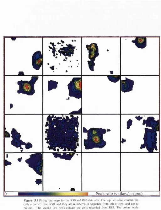
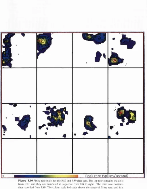
Chapter 5 contains results from experiments that have been designed to measure the basic properties of place cells with the tetrode and the new apparatus. In these experiments, measurements of place cell receptive field size, shape and distribution were made in several different environments including an open field, a walled box, an open circular area, a plus maze and a linear track. Most of the data were collected while the animal searched for randomly scattered food (open field, walled box, and open circular area), but in other experiments the animal ran back and forth for a food reward on a linear track. Also, some o f the data were collected while the animal performed a more complex arm selection task in a plus maze.
hippocampal EEG. The correlation between the 0 rhythm and displacement movements was a key argument given in support o f the cognitive map theory, but the published data are far from conclusive. As described above, the primary question is the relationship between the 0 rhythm frequency and the animal's speed of movement. In these experiments (Chapter 6) the animal is run on a linear track or a plus maze.
Finally, the spatial and temporal relationship between the hippocampal 0 rhythm and the activity of hippocampal place cells is examined. Prior published experimental results examined only the average phase relationship between the 0 rhythm and the activity of single hippocampal neurons. The new methods make it possible to analyse the phase relationship independently for each cycle of the 0 rhythm and for many cells in the same electrode placement. In these experiments (Chapter 7) the animal ran back and forth on a linear track for a food reward at both ends of the track.
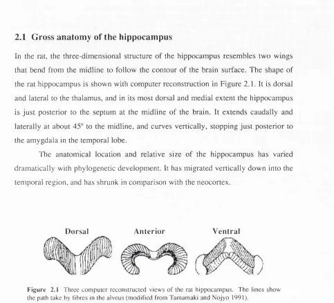
2.1 Gross anatomy of the hippocampus
In the rat, the three-dimensional structure of the hippocampus resembles two wings that bend from the midline to follow the contour of the brain surface. The shape of the rat hippocampus is shown with computer reconstruction in Figure 2.1. It is dorsal and lateral to the thalamus, and in its most dorsal and medial extent the hippocampus is just posterior to the septum at the midline of the brain. It extends caudally and laterally at about 45° to the midline, and curves vertically, stopping just posterior to the amygdala in the temporal lobe.
The anatomical location and relative size of the hippocampus has varied dramatically with phylogenetic development. It has migrated vertically down into the temporal region, and has shrunk in comparison with the neocortex.
Dorsal Anterior Ventral
F igu re 2.1 Three com puter reconstructed v iew s o f the rat hippocam pus. The lin es show the path take by fibres in the alveus (m od ified from Tamamaki and N o jy o 1991).
Ramon y Cajal (1911) observed, based on connectivity patterns, that the hippocampus is highly associated with the adjoining cortical regions. These cortical areas, called the parahippocampal region, contain the transition from the six layers of neurons in neocortex to the single cell layer in the hippocampus. The hippocampal formation includes the hippocampus and the parahippocampal region. Most of the connections between the hippocampus and the rest of the neocortex are made through the parahippocampal region, which includes the entorhinal cortex and the subicular complex. As suggested above, the functional characteristics of the hippocampus can probably not be fully separated from those of the parahippocampal region. Therefore, the anatomy of the entire hippocampal formation is examined in this dissertation.
the curve of the hippocampus to the temporal pole. Along this axis, each perpendicular slice defines a transverse plane, which is shown unrolled into an axis in Figure 2.2B.
f V rv.
F ig u re 2 .2 The hippocam pal form ation and the primary subregions are show n unrolled. Part A is a N issl stained longitudinal segm en t o f the hippocam pus. Part B sh o w s relative anatom ical location o f the subregions [fd (fascia dentata), sub (su b icu lu m ), psub (parasubiculum ), ecm (m edial entorhinal cortex), eel (lateral entorhinal cortex)] (reprinted from M cN aughton 1989).
In the transverse plane, the hippocampus is defined by the two interlocking C-shaped layers of cells and the surrounding white matter. The shorter, thicker C shape with a smaller radius of curvature is the dentate gyrus (also called the fascia dentata), and the larger C shape is the hippocampus proper (also called the cornu ammonis). These layers, are visible as bands in the transverse section, that is shown in Figure 2.2 and 2.3. These layers are extend to form sheets o f cells in Figure 2.2B.
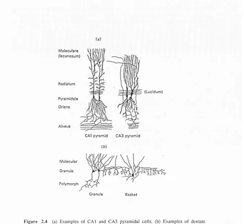
2.2 Hippocampus proper
M o lecu la re V
(la cu n o su m )
Radiatum
Pyram idale
O riens
A lv eu s
(L ucidum )
CAl pyram id C A 3 p yram id
(6)
M olecular
G ranule
P olym orph
Granule B a sk et
F ig u r e 2 ,4 (a) E xam ples o f C A l and C A 3 pyram idal c e lls, (b) E xam p les o f dentate granule c ells and a basket cell. (R eprinted from O 'K eefe and N adel 1978 p .l 10)
All of the subregions of the hippocampus proper contain roughly the same cell types and anatomical layers. Over 99% of the neurons are pyramidal cells, and the cell bodies are in a single cell layer. Pyramidal cells have both apical and basal dendrites, which are oriented nearly parallel to each other. In addition to the cell body layer, the stratum pyramidale, four to five additional layers are distinguished (see Figure 2.4), which contain the axons and dendrites of the pyramidal cells and a small number of interneurons. The basal dendrites divide into many branches just below the cell soma in the stratum oriens, but the apical dendrites extend for several hundred microns through the stratum radiatum before dividing into many sub-branches in the
lucidum, between the stratum pyramidale and the stratum radiatum, which is the termination region for the mossy fibres. Axons arising from hippocampal pyramidal cells divide into branches in the alveus and usually travel perpendicular to the orientation of the dendrites (Ishizuka et al. 1990). These cells make excitatory (Gray's type I) connections, and glutamate is the putative neurotransmitter.
In addition to the pyramidal cells, several interneurons have been identified in the hippocampus proper (Ramon y Cajal 1911; Lorente de No 1934). The most abundant interneurons are called basket cells, and have cell bodies either in the stratum pyramidale or just below in the stratum oriens. Like pyramidal cells, basket cells have both apical and basal dendrites, and are oriented in the same direction as pyramidal cells. However, unlike pyramidal cells, basket cell dendrites have no spines and their axons form a dense plexus extending for several hundred microns in the stratum pyramidale. There is approximately one basket cell for every 200 pyramidal cells in the hippocampus proper, and G ABA is the putative neurotransmitter.
Other interneurons are present in the stratum oriens, stratum radiatum and stratum moleculare, but their role is less well understood. It has been suggested that one group of interneurons in the stratum oriens may provide a second inhibitory control system (Lacaille et al. 1987).
N um bers o f neurons and synapses
There is considerable variation in the number and distribution o f neurons among strains of rats, and unfortunately the available data cross both strains and species. In each of the two hippocampi of a Sprague-Dawley rat there are 420,000 pyramidal neurons in the C A l field, 26,000 in the CA2 field and 304,000 in the CA3 field (Amaral et al. 1990). The size of the cell bodies decreases from CA3 to C A l, and the density of cells roughly doubles (Boss et al. 1985). Even within a region of the hippocampus the pyramidal cells are not uniformly distributed. The density o f CA3 pyramidal cells increases three to four times from the septal to the temporal pole (Gaarskjaer 1978).
dendritic branches. Accurate quantitative measurements have only recently become possible with the development of new methods in intracellular labelling and computer reconstruction. These methods have been used (Ishizuka et al. 1990, Tamamaki and Nojyo 1991, Amaral et al. 1990) to re-examine the shape and extent o f neuronal processes within the hippocampus. The dendritic length in the C A l field is largely uniform throughout the region, with approximately one third o f the dendritic length in the basal dendrites, and two thirds in the apical dendrites (mostly concentrated in the stratum radiatum). It is estimated that these cells receive 11,000 synapses from CA3 pyramidal cells and 2500 from the entorhinal cortex. In contrast, the length and distribution of the dendrites of CA3 neurons varies with their position in the transverse plane. The largest dendritic arbors are located close to the CA2 field. CA3 cells are estimated to receive 12,000 synapses from other CA3 pyramidal cells and as many as 3500 inputs from the entorhinal cortex. The axons o f pyramidal cells in the hippocampus proper branch into several intrinsic and extrinsic projections, which are described in more detail below.
The distribution of intemeurons within the hippocampus proper is not known in detail, but it is often assumed to be uniform. However the structure of the axons and dendrites of basket cells suggests that they correlate activity across large regions of the hippocampus. Basket cell axons make strong inhibitory connections with the soma of approximately 3000 pyramidal cells (estimated from Schwartzkroin and Kunkel 1985 and Boss et al. 1985). One method for analysing the anatomical evidence for the influence of inhibitory interneurons is to examine the relative spread of their axons and dendrites in the two-dimensional surface defined by the cell body layer. Along this surface the basket cell dendrites have much less spread than their axons. This implies that the group of pyramidal cells influenced by basket cells is more widespread than, and somewhat different from, the set that provides their inputs. Of course, this influence also depends on the distribution o f pyramidal cell axons and dendrites.
Intrinsic pro jectio n s within the hippocam pus p ro p e r
feed-forward and feedback inhibitory connections. Some of these projections are included in the description of the cells above. They are summarised in Figure 2.5 which contains a block diagram of principal projections between regions of the hippocampal formation.
The axons of the CA3 and CA2 pyramidal cells provide a feed-forward excitatory pathway to CAl which innervates both ipsilateral and contralateral regions. The ipsilateral projection is called the Schaffer collaterals, and the contralateral fibres form the commissural projection. Each CAl pyramidal cell receives approximately 5500 synapses from the collaterals originating in each hemisphere, which course through the stratum radiatum. Assuming one contact per neuron, each CA3 pyramidal cell randomly contacts 1.8% of the CAl pyramidal neurons (Amaral et al. 1990). However, the axonal plexus of pyramidal cells does not extend over the entire region and therefore the synaptic contact is not randomly distributed. From data on the size of the axonal plexus (Tamamaki and Nojyo 1991) the probability of contact is six to eight times higher, or about 15%, within the projection zone.
subicular region CAl
\ r
sensory cortex
dentate gyrus CA3
entorhinal
cortex
In addition to the Schaffer collaterals, axons from CA3 pyramidal cells branch to produce direct excitatory feedback to ipsilateral and contralateral CA3 pyramidal cells, called the association projection. As with the feed-forward projections, the association fibres are presumed to produce equal numbers of synapses in both CA3 fields. In comparison with other brain regions, the density of excitatory feedback in CA3 is high; each pyramidal cell innervates the dendrites of approximately 6000 others in each hemisphere, which terminate in the stratum radiatum, on dendritic spines. As with the Schaffer collaterals, the probability of contact is higher than expected by random contact, since the axonal plexus o f each neuron reaches only part o f the hippocampus. Random contact would result in each pyramidal cell projecting to 1.9% of the others. However constraints on the area covered by the branching of pyramidal cell axons (Tamamaki and Nojyo 1991) may increase the probability of contact to over 6%.
The organisation of the Schaffer collateral and association fibre projections has been the focus of considerable recent study. However these studies have not produced completely consistent results. Using extracellular labelling and the extended hippocampus, Ishizuka et al. (1990) and Chan et al. (1992) agreed on the organisation in the termination zones of Schaffer collaterals, in which a band of CA3 cells projects to a band of C A l cells. In three dimensions the two bands correspond to a single slice of the hippocampus. This slice is not perpendicular to the longitudinal axis, but is tilted towards the temporal pole as if it were hinged at the fimbria. Unfortunately this view is not consistent with complete cells reconstructed from intracellular injection of HRP (Tamamaki and Nojyo 1991).
the Schaffer collaterals are largely within the same slice, then there is considerably more modularity in the function of the hippocampus.
This level of structural detail is very important in guiding the construction of neuron level models for hippocampal function, but is very difficult to obtain. In the first place, the axes used to described the gross anatomy are not sufficient for analysing the low level structure of the projections. To improve the definition of axes, some researchers are using the mossy fibres as an axis (Andersen et al. 1971), while others propose using the alvear fibres (Tamamaki and Nojyo 1991) or the CA3-CA2 border (Ishizuka et al. 1990). Secondly, unlike projections in the neocortex, the collaterals of hippocampal pyramidal cells are not restricted to the vicinity of the cell body. The functional implications o f this fact are discussed in the next chapter, but from the anatomical point of view the fact makes measurements of the axonal branching patterns more difficult. Currently there are either detailed data for a small number of cells or less precise data from extracellular dye injection or degeneration studies.
Within a transverse slice there is an ordering in the termination pattern o f the association projection and the Schaffer collaterals along the dendrites of hippocampal pyramidal cells (reviewed in Ishizuka et al. 1990). In general it is not clear how much of this structure is associated with variation in function. Some o f the structure results from the sites available during the development of the fibre projections, and may not have a functional role.
Recently feedback connections with much lower density have been demonstrated both anatomically (Chan 1992) and physiologically (Christian and Dudek 1988) between C A l pyramidal cells.
2.3 Dentate gyrus
In transverse section, the dentate gyrus is divided into three principal layers. One of these, the granule layer, stands out as a C shape in a Nissl stain, and is densely packed (>75k per m m \ four times the density of CA3) with the soma o f the principal projection cells, the granule cells. These small cells (<10 |im soma) have only apical dendrites, which are directed outward from the bend o f the granule layer and arborize in the molecular layer. Within the concave bend of the granule layer there is a second cell-rich layer called the polymorph layer or the dentate hilus. The granule layer bends around the proximal part o f the CA3 field. It is often divided into the
suprapyramidal blade between C A l and CA3, and the infrapyramidal blade below the CA3 pyramidal layer. Among strains of rat there is variation in the number of granule cells (Boss et al. 1985), but on average there are approximately one million. This accounts for 97% of the neurons in the dentate gyrus, and over 99% of the granule layer neurons. The axons of the granule cells make excitatory synapses in the dentate hilus and project to the CA3 field. The projection to CA3, called the mossy fibres, is described in detail below.
The remainder of the neurons in the cell body layer are intemeurons (approx. 4500), which receive excitatory projections from the granule cells and provide inhibitory feedback. Most of the intemeurons are basket cells and their cell bodies lie just below the granule layer. Basket cells have both apical and basal dendrites, and their axons extend over one third o f the longitudinal and transverse extent of the granule layer (Struble et al. 1978), forming inhibitory synapses on approximately 1000 granule cells (McNaughton 1989). There are on average 180 granule cells for every basket cell, and each receives about 10 basket cell synapses.
The dentate hilus contains a large number (>21) of morphologically distinct types of neuron (Amaral 1978). The most abundant type of neuron, called the mossy cell, is a large spiny multipolar cell that makes extensive excitatory synapses along the longitudinal axis of the hippocampus. There are approximately 32,500 neurons in the polymorph layer, out of which 10,000 are immunoreactive for somatostatin and GAD, and 20,000 are presumed to be mossy cells (Amaral et al. 1990). The hilus receives extensive inputs from the brain stem, which are discussed below.
As in the hippocampus proper, there are many important neuronal projections within the dentate gyrus. Unlike the hippocampal pyramidal cells, there is no direct excitatory feedback connection between granule cells (Claiborne et al. 1986), but there is an excitatory feedback pathway in the interaction between mossy cells and granule cells. Each granule cell receives approximately 2000 contacts from mossy cells. There is also a suggestion of an excitatory feed-forward pathway, as the mossy cell dendrites extend into the molecular layer and may receive direct entorhinal input. Inhibitory feedback exists between basket cells and granule cells, and between mossy cells and intemeurons in the hilus. There is also evidence for inhibitory feed-forward connections in a direct projection from the entorhinal cortex to the granule cell layer basket cells.
The dentate gyrus is the primary target for projections from the upper layers of the entorhinal cortex. The afferent fibres terminate in the outer two thirds of the molecular layer in a highly ordered manner, and each granule cell receives about 6000 synapses. The inner third receives synapses from the ipsilateral and the contralateral mossy cells. The projection from the entorhinal cortex is discussed in more detail below. The dentate gyrus also receives substantial projections from sub-cortical regions.
M ossy fib re s
transverse band (<400|im; Gaarskjaer 1981) into the CA3 region. When the mossy fibres reach the CA2-CA3 border they turn sharply towards the temporal pole of the hippocampus, where there is a larger density of CA3 pyramidal cells. Each mossy fibre contacts a very small fraction of the population of CA3 pyramidal cells (4.6 x 10’^), with one synapse for every 135 |im of length (Claiborne et al. 1986), resulting in contact with 14 pyramidal cells along its approximately 2mm projection in the CA3 field.
These synapses are unique in the brain, with a large size (up to 10 |im ) that envelops complex spines of CA3 pyramidal cells (Kjaerheim and Blackstad 1961). Each mossy fibre bouton synapses on four or five dendritic spines, but they are all from the same pyramidal cell (Frotscher et al. 1991). The mossy fibres are well known to exert an excitatory influence on CA3 pyramidal cells, and there is evidence that the neurotransmitter used is glutamate (Crawford and Connor 1972; Storm-Mathisen et al. 1983). However there is also evidence from histochemical staining that the terminals contain the inhibitory neurotransmitter GABA (Ottersen and Storm-Mathisen 1985) and that it coexists in the same terminals with glutamate (Sandler and Smith 1991). In addition the terminals contain several neuropeptides and unusual quantities of zinc (Stengaard-Pedersen et al. 1983).
There is considerable structure in the mossy fibre projection. Claiborne et al. (1986) found that portions of the CA3 field nearest the dentate hilus are preferentially reached by fibres in the infrapyramidal blade, while the parts of field CA3 farthest from the dentate hilus are reached by mossy fibres from the granule cells in the tip of the suprapyramidal blade. Again it is not clear how much of this is due to the availability of connection sites and how much has a direct effect on the function of the mossy fibre projection.
The striking organisation in the mossy fibre projection has inspired several interpretations o f its role in hippocampal function. Andersen et al. (1971) proposed that the entire hippocampus is organised in a series of transverse slices that are largely functionally independent. This suggestion, called the lamella hypothesis,
role for this projection, proposed also in 1971 by Marr, is described in detail at the end of this chapter.
2.4 Entorhinal cortex
The entorhinal cortex, which is part of the parahippocampal gyrus, receives and integrates projections from large parts of the cortical mantle (Insausti et al. 1987a) and relays the input into the hippocampus. It is the principal source of neocortical information for the hippocampus, and from the connectivity patterns its role must be functionally linked with the hippocampus. The output from the entorhinal area is distributed to all the major regions of the hippocampus, but the majority of the entorhinal projections to the hippocampus terminate on the dendrites of neurons in the dentate gyrus. This important fibre tract is called the perforant path (Ramon y Cajal 1911).
The entorhinal cortex is divided into the medial entorhinal (MEA) and the
lateral entorhinal areas (LEA) (Lorente de No 1933), based on morphological differences (Steward and Scoville 1976) and on their projection patterns (Haug 1973). The anatomy and connection patterns of the entorhinal cortex have recently been reviewed (Witter et al. 1989) and are only summarised here. It has six cell layers, of which layers I-III are called superficial and IV-VI are called deep. The cortex differs from neocortical regions in that the deep and superficial layers are separated by a cell-sparse layer called the lamina dessicans, which disappears at the distal extent of the entorhinal cortex. There are numerous morphologically distinct cell types in the entorhinal cortex. Their structure and distribution were recently re-examined by Germroth et al. (1991).
more widespread over the longitudinal axis. McNaughton et al. (1981) showed that 2-3% o f the perforant path synapses generated an activation which was greater by a factor o f 10 to 20 than that caused by other synapses. It has been suggested (McNaughton 1989) that these synapses have an important role in constructing memory traces. In 1973, Bliss and Lpmo discovered that tetanic stimulation of this pathway caused a long lasting potentiation of the synapses, called LTP.
The entorhinal cortex also projects to the C A l and CA3 fields. The projection to the CA3 field is similar to the projection to the dentate gyrus. There is a clear proximal to distal gradient along the apical dendrites, and they both arise from layer II neurons in the entorhinal cortex (Steward and Scoville 1976). The more distal synapses in the dendritic arborisations are from the most distal region of the LEA.
The projection to the C A l field is quite different, however, and originates from a separate group of neurons in layer III o f the entorhinal cortex. The region of the C A l field nearest to the subicular border receives an input from the LEA only, and the part of the C A l field nearest to the CA3 field receives input from the MEA only (Witter et al. 1989). A few neurons in layer V o f the entorhinal cortex also send axons to the hippocampus (Kohler 1985a).
The major input to the entorhinal cortex is from the perirhinal area, which in turn makes reciprocal connections with sensory and parasensory areas of the cortex. The only direct sensory input to the entorhinal area is from the olfactory bulb (Heimer 1968; Kosel et al. 1981), which projects to the LEA. The inputs from the perirhinal cortex project to the lateral aspects of the LEA and MEA. The inputs to the more medial aspects o f the LEA and MEA are not known, but it is suspected that they get inputs primarily from the aforementioned lateral aspects. The pattern of termination of the inputs to the entorhinal cortex is highly topographic.
There is also a projection to the entorhinal cortex from the C A l field and the subiculum. In most cases, the same C A l pyramidal cell projects to the subiculum, the medial septum and the entorhinal cortex (Swanson et al. 1981).
2.5 Subicular complex
The subicular complex is subdivided into the subiculum, the presubiculum and the
is disagreement among researchers about further subdivision of the subicular complex. However, based on differences in connectivity, van Groen and Wyss (1990) suggest that the postsubiculum is distinct from other subicular regions. The postsubiculum has attracted significant recent attention due to the discovery o f head direction cells (Taube et al. 1990a,b) in that area. The subiculum is closest to the C A l field and resembles most closely the layered structure o f the hippocampus proper. It is a five-layered cortex, and layer II is similar in structure to C A l (Lorente de No 1934). Presubiculum and parasubiculum more closely resemble the structure of the neocortex.
Comparatively little is known about the subicular complex, perhaps because the cortical fields are small in the rodent brain and they have a high degree of diversity and complexity. There are approximately 128,000 pyramidal cells in the subiculum (Amaral et al. 1990), but no numbers of cells are available for the other areas of the subicular complex.
This area has an important role in hippocampal function, as it receives a massive projection from the CA l and relays the information to the entorhinal cortex. CA l pyramidal neurons project principally to the ipsilateral subiculum (Finch and Babb 1980,1981; Finch et al. 1983). The projection forms a column that is 250-300 |im wide, 550 |im high, and 2 mm long, and parallel to the border between the C A l and the subiculum (Tamamaki et al. 1987; Tamamaki and Nojyo 1990). The subicular complex completes a loop in an information pathway that leads from the entorhinal cortex to the dentate gyrus, on to the CA3, then to the C A l and the subiculum, and back to the entorhinal cortex. However, this is not the only pathway for information to return to the entorhinal cortex (reviewed in W itter et al. 1989). The projections from the subiculum are solely ipsilateral (Kohler 1985b) and reach all layers o f the entorhinal cortex. The subiculum also projects to the retrosplenial cortex, and to sub-cortical regions including the thalamus and nucleus accumbens.
2.6 Projections to and from the septum
The projections are concentrated in the dentate gyrus and the CA3 region, but are comparatively sparse in the C A l region (Swanson and Cowan 1979). In return, there is a projection back to the septum from all parts of the hippocampal formation (in particular from C A l) (Raisman et al. 1966), but these fibres largely terminate in the lateral septum. A surprising recent finding is that there are very few projections between the lateral and the medial septum (Leranth et al. 1992).
The densest termination of the input from the medial septum is in the dentate gyrus polymorph layer. A substantial projection also exists to the subiculum and to the entorhinal cortex (Milner and Amaral 1984). This projection is mostly to the MEA, and terminates in the lamina desiccans and in layer II.
The projection from the medial septum is topographic. The cells near the midline project to MEA and to septal parts of the remainder of the hippocampal formation, but more lateral parts project to lateral parts of the entorhinal cortex and to temporal parts of the hippocampal formation (Meibach and Siegel 1977; M onmaur and Thomson 1983; Saper 1984). Input from the septum is more dense in more temporal hippocampal regions (Milner et al. 1983). Fibres from the medial septum terminate on both pyramidal cells and basket cells in the hippocampus proper. The projection has been thought to be entirely cholinergic (Lewis and Shu te 1967), but more recently it has been shown that at least 30% of the projection cells are GABAergic (Kohler et al. 1984) and form an independent group from the cholinergic cells (Brashear et al. 1986).
2.7 Additional hippocampal efferents/afferents
The primary projections to and from the hippocampus pass through either the parahippocampal region or the septum, and have been discussed above. There are however several projections from brainstem areas, which primarily innervate the dentate hilus. These include a noradrenergic input from the locus coeruleus (Fuxe 1965) and a serotonergic input from the median and dorsal raphe (Kohler and Steinbusch 1982). From the hypothalamus there is a cholinergic projection to the hippocampal formation (Wyss et al. 1979), and a histaminergic input from the supramammillary region (Segal 1979) which terminates in the hilus.
In addition to the hippocampal efferents discussed above there is a projection from the C A 1 and the subiculum to the nucleus accumbens (Phillipson and Griffiths 1985) and a projection from the CA3 to the hypothalamus (Swanson and Cowan 1977).
2.8 Summary
This presentation of the anatomy of the hippocampal formation is necessarily incomplete for several reasons. Firstly, it is impossible in a short space to include all the detail that is available. Secondly, some of this detail has a less significant impact on the function of the region. Most importantly, only a small part of the anatomical detail is known.
For example, it would be very useful to know the distribution o f basket cells, which may help to explain how groups of neurons interact. One possible distribution would predict independent control of cell assemblies; another would predict a mechanism for integrating activity levels over a large region.
The second difficulty with anatomical detail is how to manage so large a body o f information. As the quantitative information becomes available, a better way must be found to communicate the detail unambiguously. The important information spans several scales, from the high-level convergence and divergence characteristics to low-level synaptic contact for individual neurons. An efficient communication mechanism for anatomical details should incorporate all the scales, and it should be unambiguous where a new piece of information fits in. Other fields of scientific research, such as molecular biology, have developed this kind of common communication scheme.
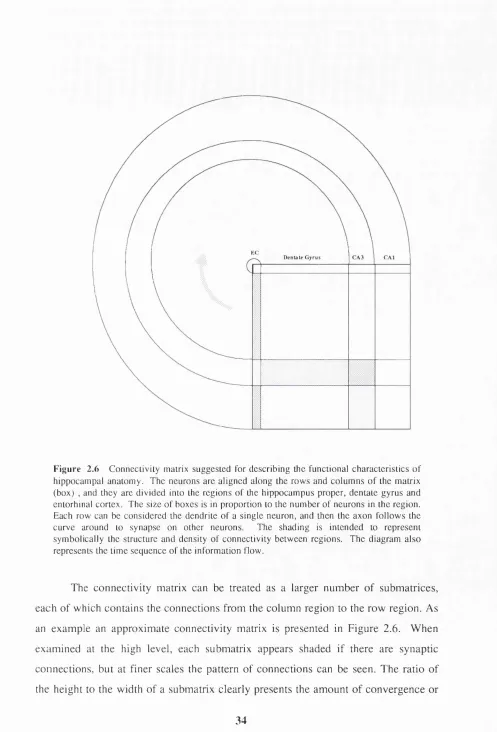
E C
D e n ta te G y r u s C A 3 C A l
F ig u re 2 .6 C on n eciiviiy malrix suggested for describing the functional characteristics o f hippocam pal anatom y. The neurons are aligned along the row s and colu m n s o f the matrix (box) , and they are divided into the regions o f the hippocam pus proper, dentate gyrus and entorhinal cortex. The siz e o f b oxes is in proportion to the number o f neurons in the region. Each row can be considered the dendrite o f a sin gle neuron, and then the axon fo llo w s the curve around to synapse on other neurons. The shading is intended to represent sym b olically the structure and density o f con n ectivity betw een regions. T he diagram a lso represents the tim e seq u en ce o f the inform ation How.
divergence. If this ratio is large there is divergence, and if it is small there is convergence. Information flow is indicated by the stippled arrow in the figure. The input arrives at the top of the connection matrix and is projected through the connections to a second region. It then follows the arrow to become an input to the next region. In this way the diagram can incorporate both the connectivity and the time of occurrence for events. The propagation delays in the neuronal circuits correspond to the time to flow from one region to another.
For example, it is very easy with this diagram to show both the divergence between the entorhinal cortex and the dentate gyrus, and the specific nature o f the mossy fibre projection to the CA3. Using this kind of diagram it is possible to test the impact of different connection patterns. For example, using the CA3 association projection in one configuration (Tamamaki and Nojyo 1991), the activation of synapses from one mossy fibre can reach all CA3 pyramidal cells by the second cycle around the diagram. At this same point in time, the activation will have reached only a fraction of the C A l pyramidal cells.
This kind of diagram is very useful for presenting the detailed anatomy of a region. Firstly, it clearly shows the convergence and divergence from one region to another. Secondly, it illustrates the extent to which regions are interconnected (number o f shaded submatrices). Finally, it contains a mechanism for representing the temporal properties. All these properties can be examined at many different scales within the same diagram.
3.1 Introduction
The discovery of place cells (O’Keefe and Dostrovsky 1971) and the cognitive map model (O'Keefe and Nadel 1978) have provided a major new inroad into the study of hippocampal function. However, after two decades of active research there is not yet general agreement that hippocampal pyramidal cell activity is primarily correlated with spatial position (e.g. Eichenbaum et al. 1992). This chapter reviews the properties of place cells, which are divided into: (1) a set of "static" properties in unchanging environments, (2) the effect of sensory input and memory on cell activity patterns, and (3) the changes in activity between environments.
A large part of the variation in the published properties of place cells is a direct result of variation in methods and the design of experiments. For this reason, the place cell characteristics must be reviewed in the context of methodological issues.
Most of the research on place cells does not distinguish between cells in the C A l-3 fields, and in this review they are discussed together. Spatially biased firing patterns have also been seen in some of the neurons that project to, or receive inputs from, place cells. These results are summarised in Section 3.8.
In addition, this chapter briefly reviews the characteristics of the hippocampal BEG. In most of the literature the hippocampal EEG is classified into two forms: the theta rhythm, and large irregular activity (LIA). In this review particular attention is paid to the theta rhythm, and the evidence for its part in the hippocampal navigation system.
3.2 Neuronal firing patterns
decreasing. Firing of complex spikes was found to have no simple relation to presence or absence of a slow wave 0 rhythm.
F ig u re 3.1 Com plex-spike recorded in the C A l region, (reprinted from Fox and Ranck 1981)
The second type of neurons is more tonic, has a more uniform and higher frequency firing rate, and never produces complex spike bursts. Ranck called these neurons theta cells, due to the correlation between their activity and the presence of the hippocampal 0 rhythm. Theta cells and complex spike cells are also identified by the shape of their extracellular action potentials. Theta cells produce shorter duration and smaller amplitude spikes compared with complex spike cells (Ranck
1973; Fox and Ranck 1975).
Fox and Ranck (1975) also demonstrated that theta cells and complex spike cells have different anatomical distributions. These data led them to suggest that theta cells are intemeurons, and that complex spike cells correspond to pyramidal neurons (Fox and Ranck 1975).
Place cells
In 1971, O'Keefe and Dostrovsky demonstrated that some of the cells recorded from C A l in a freely moving rat code for the animal's spatial location. Place cells have a low overall firing rate, which is significantly higher in the active region (place field) than in the remainder of the environment. In another study (O'Keefe 1976), place cells in the dorsal C A l field were found to be a subset of the complex spike cells, and therefore putative pyramidal cells. In agreement with Ranck, O'Keefe (1976) identified a physiologically separate group of cells, correlated with the 0 rhythm and movements, which he called displace cells. In the recent literature, and this dissertation, displace cells are called theta cells.
The data from several research groups are consistent in the report of spatial firing in hippocampal complex spike cells (see Table 3.1, p.50). However, there is much less agreement on the qualitative and quantitative details of the firing pattern of these cells. The variations in properties include: (1) the size and shape of the place field of a cell, (2) the extent to which spatial location alone controls the firing of the cells, and (3) the persistence of the firing pattern over time. Differences in observed properties result from the structure of the environment and the design of the experiments, and these differences are compounded by the difficulty in isolating the activity of single neurons with extracellular electrodes.
3.3 Methodological issues
Tasks and environments
One o f the key characteristics of place cells, demonstrated in all of the initial studies, is that their firing pattern is largely independent of specific behaviour patterns or the task that the animal is performing. In spite o f this evidence, the design of hippocampal recording experiments can affect the characteristics of the recorded place cells. In this section these design issues are discussed, in order to provide the context for a review of the published properties of place cells.
reasonably balanced coverage of the environment, and the second is for the animal to display a set of behaviours (e.g. selecting a turn in a maze) to indicate that it knows its spatial location. The defining characteristic of a place cell is comparatively higher firing in one part of an environment, which can be reliably measured only if the animal's behaviour is almost uniformly distributed in space. In many o f the initial studies there was no measurement of the time that the rat spent in parts o f the environment. In the extreme, if the rat has occupied only one place, then any active cell could be interpreted as a place cell. Some o f the place cell data have been recorded during stereotyped movements that do not cover the environment, and to this extent the degree of spatial preference cannot really be determined. In a recent quantitative study (Muller and Kubie 1987) the task was designed to optimise coverage o f the environment. In this task the animal searches for randomly placed food in a walled cylinder.
There are two problems with this type of task. The first problem is that there is no guarantee that the hippocampus is being used at any particular point in time. This issue is difficult to separate from a discussion of the investigator's model of
hippocampal function. In models favouring a simple sensory correlate of
hippocampal activity the issue is less critical. However if the cognitive map model is correct then there will be a useful hippocampal firing pattern only if the animal has an active mental map of the current environment and knows its location. The solution to this problem (O'Keefe and Conway 1978; Olton et al. 1978) is to record from place cells, while challenging the animal with a task that is known, from lesion studies, to require the hippocampus. During these tasks the animal demonstrates through its behaviour that it knows its spatial location.
The requirement for a behavioural measure of the rat’s awareness of location is even greater in changing environments. In the reviewed literature changing environments are often used. This type of experiment is, of course, the only means for determining the inputs that are driving place cell activity. If there is no method for testing the animal's use of the hippocampus then the activity patterns recorded from the place cells could be inaccurate. O'Keefe and Conway (1978, 198' ) pioneered a cue-controlled task in which the environment could be manipulate , while maintaining a behavioural test of hippocampal function. However, mo t experiments that require choice on the part of the animal have difficulty in achievir i
a uniform behaviour pattern over the environment. This problem is addressed in tl 2
cue-controlled task, since there is largely uniform coverage in the start art . However, a second problem is that even in a cue-controlled task the hippocampi s may be used for spatial processing during only part of the time (i.e. when the rat s making the choice).
r;-;-.-, Goal \ W /
I ' . V" , V . X
• i,
f. >
Some of the published properties of place cells have been collected in tasks that have neither uniform coverage of the environment nor any indication of the concurrent use of the hippocampus. For example, Breese et al. (1989) recorded from thirsty rats while they ran in stereotyped movements between identical water dishes, which were placed in four corners and the centre of a platform. Initially the water dishes contained a drop of water in a random pattern, and during this period the firing of place cells was recorded. Following this period only one of the dishes was baited and the change in place cell firing was measured. Unfortunately, this task does not provide coverage of the entire environment, and since there is no measure of the rat's awareness of location the place cell firing could be very difficult to interpret.
In some of the published data the rat is coaxed or pushed around the environment. This is a less controlled form of the randomly distributed food task (Muller and Kubie 1987) discussed above, but since the behaviour is being disturbed, then the spatial representation could be disturbed as well.
Data on hippocampal single units have been collected using a range of different rewards and penalties to shape the rat's behaviour. Given the general plasticity of the nervous system, there is a risk that artificial and repetitive behavioural paradigms will lead to artificial representations in the neuronal activity patterns. For this reason, less credence is given to data collected during extreme food deprivation, water deprivation or shock penalties. Secondly, although useful data can result from lesion studies, recordings from cells in these circumstances must be treated differently.
Extracellular recording
All o f the known properties of place cells have been found by recording the firing patterns of putative single neurons using extracellular electrodes, as the animal moves within an environment. There is great variation in the recording methods, which have produced corresponding variation in results. The important issues leading to this variation are briefly described here. Again, this analysis is used as context for the literature review that follows.
All membrane potential changes of neurons are in principle observable with an electrode outside the cell membrane. When the internal potential changes, one part of the cell is depolarised more than the adjacent areas and a current flows; an extracellular field is generated as a result o f this current. The amplitude of the voltage outside the cell is significantly attenuated by the membrane resistance, and it decreases at least as fast as the inverse square of the distance from the cell membrane. The wave shape of action potentials is determined by several factors, including the type and distribution of the electrical sources and sinks present in the cellular membrane and the morphology of the cell's dendritic arborisation. If the extracellular medium behaves as a perfect ohmic resistor (i.e. no change in this resistance with current flow), with an isotropic impedance (i.e. no variation of the current flow with direction), then the extracellular wave shape at a point near the cell body is roughly the derivative with respect to time of the internal potential (Hubbard et al. 1969).
In the extracellular space, each action potential is superimposed on the background voltage, which results from the cumulation of many dendritic spikes, synaptic potentials and the firing of nearby neurons. The wave shape o f a single unit, recorded from the hippocampus of a freely moving rat, is a small (10-20%) fluctuation superimposed on the accumulated electrical activity (EEG). If two or more neurons near the recording electrode are active at the same time, then the extracellular spikes can be distorted beyond recognition. In fact, it would be essentially impossible to record the activity of individual hippocampal neurons if there was a higher incidence of simultaneous activity.