
doi: 10.11648/j.iji.20140203.11
ISSN: 2329-177X (Print); ISSN: 2329-1753 (Online)
General approach for isolation of immunoglobulins
fragments with the core hinge
Maria Timchenko, Vladimir Tischenko
*Laboratory of NMR of Biosystems, Institute of Theoretical and Experimental Biophysics RAS, Pushchino, Russia
Email address:
[email protected] (V. Tischenko)
To cite this article:
Maria Timchenko, Vladimir Tischenko. General Approach for Isolation of Immunoglobulins Fragments with the Core Hinge. International Journal of Immunology. Vol. 2, No. 3, 2014, pp. 16-23. doi: 10.11648/j.iji.20140203.11
Abstract:
The original limited proteolysis approach was proposed for large (multidomain) proteins with post-translational modifications for obtaining of their novel fragments. It was realized for human immunoglobulins representing two subclasses IgG2 and IgG3. This approach was based on two techniques: masking of protein regions which are normally susceptible to proteolytic enzymes and increasing the possibility of proteolysis for sites which are not ordinarily accessible to these enzymes. The masking of immunoglobulin part which is sensitive to proteolysis was performed by Fab fragments (Fv subfragments) of antibodies and the increase of lability of stable regions was realized by pH change and mild reduction of disulfide bonds.Keywords:
Limited Proteolysis, Immunoglobulin, Hinge, Fc Fragment, Facb Fragment1. Introduction
The common practice to study the structure and functions of large proteins is isolation of their fragments. This approach enables to investigate structural organization of such proteins and their active centers by more informative methods such as X-ray analysis and NMR which are not applicable for multidomain or multisubunit macromolecules with high internal mobility. The same concept was widely used for the study of immunoglobulins.
The main discovery clarifying the role of immunoglobulins as bivalent molecules was made by Porter [1] during the limited proteolysis of rabbit IgG. The fragments of two types were obtained: two Fab fragments responsible for interactions with antigens and Fc fragment in which, as it was shown later [2,3], multiple active centers involved in effector functions are located. Thereafter several other fragments of Ig were isolated by limited proteolysis [2-4]. The efficiency of the used approach was defined by the presence of flexible and labile regions in Ig polypeptide chain between domains or subunits which are accessible to proteases.
Porter et al. [5,6] also showed that upon changes of certain conditions the conformational transition could be induced in IgG resulting in weakening of interactions between concrete domains and appearance of new site for protease cleavage. Namely Facb fragment of IgG was obtained by such procedure. It was formed as a product of plasmin proteolysis
of peptide bond between CH2 and CH3 domains after
preliminary incubation of IgG at acidic pH [5,6].
Further studies have shown that after such incubation with the following increase of pH the protein goes to metastable long-lived state [7,8]. The conformational transition with weakening of inter-domain interactions can be induced not only by pH but both using denaturants and varying a temperature [2-4,9].
At the same time the increase of protein stability, e.g. upon binding with different ligands, enhances the resistance to protease cleavage. For instance, some ligands can directly mask the regions of polypeptide chain containing recognition and restriction sites for proteolytic enzymes [2,10,11].
It should be noted that the study both Ig proteolytic stability and its proteolytic fragments is important due to the fact that a number of infectious agents produce various proteases cleaving Ig to neutralize the action of immune system [12-15]. As a result it promotes not only the decrease of humoral immunity but the formation of fragments of cleaved Ig which are dangerous for infected organism.
modifications in host cell which are necessary for fully functional protein [20,21]. The usage of eukaryotic cells also can’t guarantee the obtaining of native protein structure because the expression occurs in “foreign” cell [21,22] where the level of enzyme activity can differ from native one [22-26]. This could result in serious impacts even for myeloma cell [27]. Besides, several proteins cannot be isolated in native state both in bacterial and eukaryotic cells due to the failure of correct folding of mature protein (not proprotein) [28-31]. Therefore the method of limited proteolysis is still actual and relevant for fundamental and medical studies.
In this work we propose the original approach for preparation of new IgG fragments by limited proteolysis. The essence of this method is the destabilization of some regions of polypeptide chain for increasing their accessibility to protease cleavage along with the stabilization of regions with a labile structure to avoid their cleavage by proteolytic enzymes.
2. Materials and Methods
2.1. Isolation, Purification, Typing and Analysis of Samples Homogeneity
Myeloma IgG2 (LOM and SIN) and IgG3 (PET and SUR) were isolated from blood serum of multiple myeloma patients by standard procedure using subsequent fractionation by ammonium sulfate, ion-exchanging chromatography and gel-filtration [32,33]. Ig subclasses were identified by monospecific antisera in a double agar gel immunodiffusion test [34]. The homogeneity of studied samples was checked by SDS-PAGE [35].
2.2. Preparation of Fc Fragments, CH2 and CH3 Domains of
IgG, Hinge Region and Upper Hinge
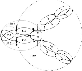
Ig structure is represented on Fig.1. Fc fragments from IgG2 LOM and SIN were obtained by standard procedures of limited proteolysis with papain [32,36] and trypsin [37], hFc fragments were also isolated by trypsin proteolysis according to our approach. CH2 domains were obtained from
papain-digested Fc fragments of IgG2 by trypsinolysis as described earlier [38,39]. However, firstly, the incubation before the proteolysis was at pH 2.8 instead of pH 2.5 [10,11]. Secondly, the reaction was carried out in the presence of Fab fragments from rabbit antibodies against CH2 domains at CH2
to Fab ratio of 1:1 for the stabilization of labile structure of these domains [10,40,41]. After the completion of reaction this complex was dissociated at pH 4.0 (10 mM glycine buffer), and two proteins were separated by gel filtration at the mentioned conditions. Dimers of CH3 domains
(pFc’-fragments) were obtained by pepsinolysis at enzyme to substrate ratio of 1:100 in 100 mM acetate buffer, рН 4.5 according to the standard procedure [42]. Hinge region of IgG3 (pFh fragment) was obtained as described previously [43,44]. The sh (short hinge) fragment representing only a part of the hinge was obtained by successive action of proteolytic enzymes. Firstly, the mild hydrolysis by papain resulting in
the cleavage of peptide bond Cys241G-Asp241H [45,46] and the formation of sFch fragment containing about half of all hinge [47] was carried out.
Figure 1. General scheme of IgG structure. It should be noted that hinge of human IgG3 is significantly longer than that of IgG2. The overlapping area of domains corresponds to the intensity of their interaction. The interaction between CH2 domains is realized only by a few contacts between carbohydrate moiety, localized in Asn297. Arrows indicate the places of cleavage by: (1) - trypsin, resulting in hFc fragment of IgG2 formation; (2) - trypsin, resulting in Facb fragment of IgG3 formation; (3) – papain, resulting in Fch fragment of IgG3 formation. VL and CL – variable and constant domains of light chain; VH,CH1,CH2,CH3 - variable and constant domains of heavy chain; lh, ch, uh – lower, core and upper hinge segments, correspondingly. Fab subunit is a globular structure formed by a pair of variable (VL and VH) and a pair of constant (CL and CH1) domains and responsible for the interaction with antigens. Fс subunit is a globular structure formed by a pair of CH2 and CH3 domains and responsible for effector functions. The corresponding fragments are formed by the action of proteases on different hinge segments.
Secondly, the Leu234-Leu235 peptide bond in the lower hinge segment was mildly hydrolyzed by pepsin in acetate buffer, pH 4.5, 1:100) in a contrast to [63]. Then the desired sh fragment was purified by gel filtration in the same buffer.
Immunodiffusion was performed in 10 mM Tris-HCl, 150 mM NaCl according to Ellerson et. all [39]. To precipitate the studied specimen F(ab’)2 fragments, obtained from
corresponding antibodies with pepsin [2], were used.
2.3. Preparation of Antibodies, their Fab Fragments and Fv Subfragments Against Fab and Fc Fragments, CH2,
CH3 Domains, Hinge (pFh) and upper Hinge
Fragments of IgG3
The antibodies against different regions of IgG3 PET were obtained by immunization of rabbit or goat by corresponding fragments and domains as described previously [10,11]. Fab fragments of these antibodies were obtained by standard papain digestion [32,36]. Different Fv subfragments were obtained from rabbit or goat antibodies by low-temperature pepsinolysis as described in our paper [20].
Its VH and VL domains were cross-linked by
heterobifunctional and homobifunctional reagents by procedures described previously [48,49]. For identification of obtained fragments the immunodiffusion was performed in 10 mM Tris-HCl, 150 mM NaCl according to Ellerson et. all [39].
segment of IgG3 from the total pool of antibodies produced against hinge region affinity chromatography were carried out. The desired antibodies were purified by affinity chromatography with the column with immobilized sh, where antibody against core hinge and low hinge region, but not against the upper part, were sorbed.
2.4. Determination of Molecular Mass and Sedimentation Constants
The molecular masses and sedimentation constants of obtained fragments and proteins were determined by high-speed (weight average molecular mass Mw) and
low-speed (Z-average molar mass Mz) equilibrium
centrifugation by Yphantis method [50] and Van Holde - Baldwin method [51] using the analytical ultracentrifuge Beckman Spinco model E (Beckman Coulter, USA) and МОМ (МОМ, Hungary) with interference or schlieren optics at 200C.
2.5. Calorimetric Measurements and Calculation of Thermodynamic Parameters
All experiments were carried out on microcalorimeters DASM-4A with 1 ml gold capillary cell and on DASM-4 with 0.43 ml platinum coiled capillary cell. The concentration of proteins in solution was changed in the range of 1-6 mg/ml. The measurements were carried out at a rate of 0.25-2 0C/min. Calorimetric curves were processed as described in [52].
3. Results
3.1. Preparation of HFC Fragment Containing Core Hinge
To date it was managed to obtain Fc fragment from IgG2 only without core hinge due to some reasons. Firstly, IgG2 has the shortest hinge region formed by twelve-membered peptides. The main part of it appears to be in poly-L-proline double helix conformation. According to preliminary theoretical analysis it was predicted that this helix should have a rather rigid structure [53] which is typical for fibrillar structures [54-56]. This assumption was confirmed experimentally by X-ray analysis [57-59], electron microscopy [60] and thermodynamic studies [43,61-63]. Additionally the helix is stabilized by disulfide bonds [47] and IgG2 has four disulfide bridges [64].
Secondly, the flexible upper hinge region providing Fab mobility in molecule and in which the limited proteolysis is occurred resulting in the formation of mentioned fragment has the smallest size and consists of only three amino acids residues [82]. The specified circumstances determine more rigid structure of molecule as a whole and proximity of Fab and Fc subunits which probably result in difficulties in obtaining of ordinary Fc fragments and significantly influence on biological activity of IgG2 (ability to activate the complement system), which differs from those of IgG1 and IgG3 [2,3,65]. Finally the stability of IgG2 CH2 domains
especially with pH decrease is the lowest among those of
human IgG [10,11,32,42,66]. All these details impede the obtaining of full-length Fc fragment of IgG2 by standard procedures. In most cases the limited proteolysis of human immunoglobulins IgG2 using different proteases results in hydrolysis of peptide bond in lower hinge region with formation of non-typical Fc fragment [37,47] in contrast with Fc fragments of other subclasses [47,67]. As a result, two newly formed N-terminal parts of heavy chains are no longer linked by covalent bonds and dissociate under denaturating conditions in the absence of reducing agents. Based on represented facts and our data obtained earlier in the studies of IgG of different animals [2,9-11,32,60,68,69] we propose the new strategy of IgG2 limited proteolysis which could result in hydrolysis of peptide bond in upper hinge region with the formation of full-length Fc fragment containing the core hinge (so-called hFc fragment).
To obtain required fragments the conditions for trypsinolysis of IgG2 were chosen empirically at first. In practice, the reaction was carried out as follows. The sample was incubated in 1 mM phosphate buffer pH 5.8 containing 20 mM cysteine at enzyme to substrate ratio of 1:20 at 35-37ºС during two hours. To prevent undesirable degradation of CH2
domains the different concentrations of Fv subfragments from anti-СH2 antibodies were present in solution. Both the part of
formed fragments and intact IgG2 were separated from other products of proteolysis by affinity chromatography on the column with immobilized anti-CH2,3 antibodies. Proteins
were eluted from the column with 10 mM glycine buffer, pH 4.0, whereupon the pH was increased to 7.0 with 250 mM phosphate buffer. On next step the obtained fragments and remaining intact IgG2 were separated by gel-filtration on АСА-34 column equilibrated with 10 mM phosphate buffer. The presence of Fc fragments in low molecular weight fraction was identified using antisera to them. The electrophoresis both of these fragments and fragments after final affinity chromatography on a column with immobilized anti-CH2 antibodies is shown in Fig.2.
The results of electrophoresis for novel Fc fragment in the presence of reducing agent are represented on the first lane. The reaction of hydrolysis by trypsin itself was carried out in the presence of two-fold excess of Fv subfragments from rabbit antibodies against CH2 domains. Molecular weight of
the fragment under these conditions is about 25 kDa. On the second lane the results for Fc fragment of IgG2 in the absence of reducing agent are shown. This fragment was obtained by standard proteolysis of peptide bond in lower hinge region by papain according to standard procedure [32,36]. Its molecular weight is also about 25 kDa but little less than for the first sample.
At the same time in the absence of reducing agent the mobility of the first sample strongly decreases and its molecular weight is about 50 kDa. These facts indicate that polypeptide chains of obtained Fc fragment are bound by disulfide bonds. It means that at least the part of core hinge is present in its structure in addition to CH2 and CH3 domains.
changes of proteolysis conditions [70]. According to obtained results it can be assumed that a trypsin cleaves the peptide bond in upper hinge region where there is a sequence Arg218-Lys219-Cys220 which is a potential proteolysis site of enzyme. This was to be expected due to the fact that after reduction of disulfide bonds between the two heavy chains, the accessibility of this site to proteases increases. Thus, the obtained fragment is full-length Fc fragment or hFc fragment. It should be noted that its yield in trypsinolysis reaction strongly depends on the presence of Fv subfragments, which stabilize the labile structure of CH2 domains and protect them
from proteolysis (see Fig.2, lanes 3-5).
Figure 2. SDS-PAGE of proteolytic products of IgG2 LOM: 1 – hFc fragment with reducing agent, the fragment was obtained by trypsinolysis at the IgG2 to rabbit Fv IgG against CH2 domains ratio of 1:2; 2 – Fc fragment without reducing agent, the fragment was obtained by standard papain digestion; 3,4,5 – Fc fragments obtained by trypsinolysis at the IgG2 LOM to Fv subfragments ratio of 1:1, 1:0,5, 1:0,2, correspondingly; 6 – hFc fragment without reducing agent, the fragment was obtained by trypsinolysis at the IgG2 to rabbit Fv IgG against CH2 domains ratio of 1:2; 7 – molecular weight markers. The results for IgG2 SIN are similar.
3.2. Preparation of Facb Fragment of IgG3
The hinge size of the third subclass of human immunoglobulins (IgG3) significantly exceeds the similar regions of any other human IgG [19,47,71] and therefore this region has the additional proteolysis sites.
In view of the above data the optimal procedure of limited proteolysis was as follows. IgG3 PET (20 mg/ml) diluted in 1 mM phosphate buffer, pH 7.0, was titrated to pH 2.8 by 2 M HCl and trypsin (10 mg/ml in 1 mM HCl) was added to an enzyme to substrate ratio of 1:100. Then the pH was rapidly readjusted to pH 7.0 with 2 M Tris buffer (pH 7.0) containing Fv subfragments both to core hinge (equimolar amount) and to CH2 domains (two-fold excess). The reaction was carried out
for 75 s then it was stopped by equimolar amount of soybeen inhibitor.
The obtained Facb fragments were purified using following approach. The gel-filtration using AcA-34 equilibrated with 10 mM glycine buffer, pH 4.0 was applied at the first stage. As a result of this stage the most high-molecular weight components of reaction mixture (formed Facb fragments and remaining intact IgG3) were separated from all other components. The second stage was the separation of Facb fragments from intact IgG3 using the column with immobilized antibodies against CH3 domains. As a result the
fragment uniquely identified as Facb fragment was obtained as it is seen from the data represented in Fig.3. The obtained
samples were homogenous according to the electrophoretic data and the analysis of C-terminal amino acid residues [94]. It is important to note that the samples were homogenous also from the physical point of view according to analytical ultracentrifugation data [67,68] which enabled to study the intramolecular conformational transitions.
Figure 3. Immunodiffusion analysis of Facb IgG3 PET. The center well contains antibodies against upper hinge. The wells 1,2 contain Facb fragment, obtained from IgG3 PET, in well 3 there is Facb fragment, obtained from IgG3 SUR, which also has a compact hinge [43,44,65]. The wells 4,5,6 contain Fab, Fch and Fc fragments of IgG3 PET.
3.3. Study of Structural Peculiarities and Conformational Flexibility of Obtained Fragments and Initial IgG
The hFc fragment obtained for the first time from IgG2 LOM and SIN, novel Facb fragments from IgG3 PET enable to receive information both about the structure of fragments themselves and their “parent” molecules.
The calorimetric melting curves for IgG3 LOM and its hFc fragment are represented in Fig.4a. The same pattern was observed for IgG2 SIN.
Upon melting of hFc fragments of indicated proteins the first heat absorption peak occurs at 67.3ºС and not at 44.8ºС. The first peak on calorimetric melting curves for IgG and Fc fragments always corresponds to the melting of CH2 domains
as it follows from the numerous data obtained by different methods for IgG and Fc fragments [10,11,32,60,69,70].
Sedimentation data for freshly prepared Facb fragment and its monomeric form after storage during 72 hours at рН 7.0 and 20ºС are represented in Fig.4b.
4. Discussion
To date there are no data about the obtaining from human IgG both full-length Fc fragment [37,47] and Facb fragment [5,6]. In the first case the potential site for proteolysis is unavailable while in the other case there are several alternative sites.
Here we propose a modified method of proteolysis for the preparation of the Fc fragment of IgG2 with core hinge (hFc fragment) and Facb fragment of IgG3.The limited proteolysis was carried out under the following two rules: the stabilization of native labile parts of protein and the artificial increase of lability of the region where the potential cleavage site is located. In Ig the labile regions include, first of all, the main part of upper and low hinge and to less degree interdomain segments of polypeptide chains and unpaired CH2 domains. The core hinge, small regions of upper and low hinge, which are adjacent to it, and paired domains are the stable regions (Fig. 1). Therefore it is necessary to take into account the specific features of structure for each protein, which undergoes the proteolysis.
Thus, for successful carrying out of limited proteolysis reaction using trypsin for obtaining of hFc fragment IgG2 there are following prerequisites. Firstly, there is Arg218-Lys219 sequence in the upper hinge region [64] which contains potential cleavage sites for trypsin action. Secondly, the core hinge is destabilized at moderate concentrations of cysteine [11,44]. Thirdly, we have reduced the interaction between Fab and Fс subunits by рН decrease and by increase of the temperature [7,11,60,68,69]. Finally, Fab fragments or Fv subfragments, obtained from anti-СH2 antibodies, are able to stabilize a labile structure of СH2 domains [11,70].
As in the case of IgG2 the successful proteolysis of IgG3 is based on the consideration of general principles of IgG structural organization, peculiarities of human IgG3 structure and specific features of IgG3 PET.
Due to the huge size of the hinge in IgG3 there are additional sites which could potentially be attacked by different proteolytic enzymes. Notably that the proteolysis by papain first of all results in a cleavage of Cys241V-Asp241W peptide bond (Kabat numbering [45]) in core hinge of IgG3 [46], differing from proteolysis of peptide bond in upper hinge region for IgG1. Further proteolysis resulting to Fab fragments formation occurs as in a case of human IgG1 due to
the availability of identical blocks
Thr223-His224-Thr225-Cys226 in both proteins. Besides,
IgG3 contains a number of other sites that are suitable for exposure to various proteases [72,73]. However, the main problem is that in IgG3 there is a site for trypsinolysis, while the absence of it in a rabbit IgG enabled to obtain Facb fragment [5,6].
Thus, in a neutral pH proteolytic enzymes are able not only to cleave the intact protein in the upper hinge region, but also to attack the core hinge. With pH decrease, first of all, CH2 domains are hydrolyzed [42] due to their destabilization [11], as well. On the basis of these data it can be assumed that it is difficult to obtain Facb fragment of IgG3 without certain preliminary procedures to stabilize labile regions containing potential cleavage sites.
But there are a number of prerequisites for carrying out of this reaction. First, there is the peptide bond Lys338-Ala339 between CH3 and CH2 domains of IgG3 as for rabbit IgG, the selective hydrolysis of which by trypsin results in Facb formation in a case of intact protein [5,6]. It should be noted that the similar hydrolysis is possible in a case of the treatment of Fc fragment of human IgG1 containing this peptide bond [38,39]. The selective cleavage occurs with the retention of structure of formed fragments [38,39] including the most susceptible to proteolysis CH2 domains. It is also worth to underline that the most labile and susceptible to hydrolysis CH2 domains of IgG3 are not less stable than those of rabbit IgG and human IgG1 [10,11,32,48,60,68]. Furthermore it was shown in [10,11,40,41], that there is a possibility to additionally stabilize these domains.
Second, the proteolysis of intact rabbit IgG was occurred after their preliminary preincubation at acidic pH [5,6,38,39]. As we have shown earlier [7,8] after such treatment IgG1 goes into a metastable state, which is characterized by the weakening of the interactions between the CH2 and CH3 domains. The contact zone between CH3 and CH2 domains was studied in [74] on the basis of X-ray analysis for Fc fragment of IgG1. Due to the high homology of primary structures of Fc subunits of rabbit IgG, IgG3 and human IgG1 [45] the location of amino acid residues in contact zone one can analyze. Such analysis indicates that six of the seven amino acids residues in CH2 domain interacting with CH3 domain are conserved for both proteins, and only Arg340 in rabbit IgG is substituted for Lys in IgG3. This circumstance gives a good reason to assume that this protein could be in a metastable state during carrying out of appropriate procedures. The available experimental data [75] confirm the ability of both intact IgG3 and its Fc fragment to be in metastable state.
Thus, the problem of successful obtaining of Facb fragment apparently will be solved if we’ll manage: а) to stabilize the structure of CH2 domains of IgG3 in such manner that trypsin predominantly cleaved peptide bond Lys338-Ala339; b) to hide peptide bond Lys218-Thr219 in the upper hinge of the protein which is potentially accessible to tryptic hydrolysis; c) to hide the bonds in core hinge Lys241E-Ser241F, Lys241T-Ser241U, Lys241II-Ser241JJ [47] (Kabat numbering [45]) which are potentially accessible to tryptic hydrolysis.
Facb IgG3 PET fragment increasing it only by 20-25% at equimolar ratio of the two proteins. In the case of polyclonal IgG3 the presence of Fv subfragments becomes critical [76]. Probably, this is due to the fact that the hinge of IgG3 PET is able to be not only in a rod-shape [57,71,77,78], but also in a compact form. The presence of the latter we showed by several methods including electron microscopy [44,60-63].
5. Conclusions
Thus in this study it was shown that it is possible to obtain the fragments containing the regions which are the most labile and sensitive to the action of proteolytic enzymes. The proposed approach enables to obtain novel fragments from IgG with unanticipated properties (stabilization of CH2
domains in Fc of IgG2, high resistance of IgG3 PET hinge to the protease action, metastability of the Facb structure of IgG3 PET). It should be noted that the properties of Fc IgG2, expressed in eukaryotic but not human cell, differ from properties of the fragment obtained by us [66]. Thus this approach could be considered as general especially for the multidomain proteins with a large number of post-translation modifications playing the key role in structure stabilization. For example by this approach the fragments of complement factor С1q with predetermined number of globular heads (intact molecule has six heads) were obtained [79].
Acknowledgements
The work was supported by RFBR (grant 11-04-00064-а and 14-04-01682-а).
Authors are grateful to Alexander Timchenko for comments and helpful suggestions.
References
[1] R.R .Porter, “The hydrolysis of rabbit y-globulin and antibodies with crystalline papain,” Biochem. J., vol.73, pp. 119–126, 1959.
[2] D.R. Burton, and J.M. Woof, “Human antibody effector function,” Adv. Immunol., vol.51, pp.1-84, 1992.
[3] H.W. Jr. Schroeder, and L. Cavacini, “Structure and function of immunoglobulins,” J. Allergy Clin. Immunol., vol. 125, pp. 41-52, 2-10.
[4] A.N. Weir, A. Nesbitt, A.P. Chapman, A.G. Popplewell, P. Antoniw, and A.D. Lawson, “Formatting antibody fragments to mediate specific therapeutic functions,” Biochem. Soc. Trans., vol. 30, pp. 512-516, 2002.E.C. Franklin, “Structural units of human 7S gamma globulin,” J. Clin. Invest., vol.39, pp. 1933-1941, 1960.
[5] G.E. Connell, and R.R. Porter, “A new enzymic fragment (Facb) of rabbit immunoglobulin G,” Biochem. J., vol.124, p. 53P, 1971.
[6] M. Colomb, and R.R. Porter, “Characterization of a plasmin-digest fragment of rabbit immunoglobulin gamma that binds antigen and complement,” Biochem. J., vol.145, pp.
177-183, 1975.
[7] V.M. Tischenko, “Metastable state of the Fc fragment,” J. Therm. Anal. Calorimetry, vol.62, pp. 6368, 2000.
[8] V.M. Tischenko, and V.P. Zav’yalov, “Long-term metastable conformation of human Fc gamma subunit,” Immunol. Lett., vol.84, pp. 241-245, 2002.
[9] V.P. Zav’yalov, and V.M. Tishchenko, “Mechanisms of generation of antibody diversity as a cause for natural selection homeothermic animals in the process of evolution,” Scand. J. Immunol., vol.33, pp. 755-762, 1991.
[10] V.M. Tischenko, V.M. Abramov, and V.P. Zav’yalov, “Investigation of the cooperative structure of Fc fragments from myeloma immunoglobulin G,” Biochemistry, vol.37, pp. 5576-5581, 1998.
[11] M.A. Timchenko, and V.M. Tischenko, “The destabilization of CH2 domains in intact IgG2 is accompanied by reduced ability to inhibit complement system factor C1,” Biochemistry (Mosc.), vol.78, pp. 759-766, 2013.
[12] C.R. Caffrey, S. Scory, and D. Steverding, “Cysteine proteinases of trypanosome parasites: novel targets for chemotherapy,” Curr. Drug. Targets., vol.1, pp. 155-162, 2000. [13] J.J. Cazzulo, V. Stoka, and V. Turk, “The major cysteine proteinase of Trypanosoma cruzi: a valid target for chemotherapy of Chagas disease,” Curr. Pharm. Des., vol.7, pp. 1143-1156, 2001.
[14] 14. J.J. Cazzulo, “Proteinases of Trypanosoma cruzi: patential targets for the chemotherapy of Changas desease,” Curr. Top. Med. Chem., vol.2, pp. 1261-71, 2002.
[15] 15. P. Berasain, C. Carmona, B. Frangione, J.J. Cazzulo, and F. Goñi, “Specific cleavage sites on human IgG subclasses by cruzipain, the major cysteine proteinase from Trypanosoma cruzi,” Mol. Biochem. Parasitol., vol.130, pp. 23-29, 2003.
[16] . R. Chen, “Bacterial expression systems for recombinant protein production: E. coli and beyond,” Biotechnol. Adv., vol.30, pp. 1102-1107, 2012.
[17] D. Mattanovich, P. Branduardi, L. Dato, B. Gasser, M. Sauer, and D. Porro, “Recombinant protein production in yeasts,”
Methods Mol. Biol., vol.824, pp. 329-358, 2012.
[18] T. Burnouf, “Recombinant plasma proteins,” Vox. Sang., vol.100, pp. 68-83, 2011.
[19] S. Huck, P. Fort, D.H. Crawford, M.P. Lefranc, and G. Lefranc, “Sequence of a human immunoglobulin gamma 3 heavy chain constant region gene: comparison with the other human C gamma genes,” Nucleic Acids Res., vol.14, pp. 1779-1789, 1986).
[20] I.R. Correia, “Stability of IgG isotypes in serum,” MAbs., vol.2, pp. 221-232, 2010.
[21] I. Ritamo, M. Cloutier, L. Valmu, S. Neron, and J. Räbinä, “Comparison of the glycosylation of in vitro generated polyclonal human IgG and therapeutic immunoglobulins,” Mol. Immunol., vol.57, pp. 255-262, 2014.
[23] I. Chantret, T. Duprè, C. Delenda, S. Bucher, J. Dancourt, A. Barnier, A. Charollais, D. Heron, B. Bader-Meunier, O. Danos, N. Seta, G. Durand, R. Oriol, P. Codogno, and S.E. Moore, “Congenital disorders of glycosylation type Ig is defined by a deficiency in dolichyl-P-mannose:Man7GlcNAc2-PP-dolichyl mannosyltransferase,” J. Biol. Chem., vol.277, pp. 25815-25822, 2002.
[24] C. Thiel, M. Schwarz, M. Hasilik, U. Grieben, F. Hanefeld, L. Lehle, K. von Figura, and C. Körner, “Deficiency of dolichyl-P-Man:Man7GlcNAc2-PP-dolichyl
mannosyltransferase causes congenital disorder of glycosylation type Ig,” Biochem. J., vol.367, pp. 195-201, 2002.
[25] C. Fagioli, and R. Sitia, “Glycoprotein quality control in the endoplasmic reticulum. Mannose trimming by endoplasmic reticulum mannosidase I times the proteasomal degradation of unassembled immunoglobulin subunits,” J. Biol. Chem., vol.276, pp. 12885-12892, 2001.
[26] K. Ko, Y. Tekoah, P.M. Rudd, D.J. Harvey, R.A. Dwek, S. Spitsin, C.A. Hanlon, C. Rupprecht, B. Dietzschold, M. Golovkin, and H. Koprowski, “Function and glycosylation of plant-derived antiviral monoclonal antibody,” Proc. Natl. Acad. Sci. U S A, vol.100, pp. 8013-8018, 2003.
[27] F.A. Gala, and S.L. Morrison, “V region carbohydrate and antibody expression,” J. Immunol., vol.172, pp. 5489-5494, 2004.
[28] J. Eder, M. Rheinnecker, and A.R. Fersht, “Folding of subtilisin BPN': role of the pro-sequence,” J. Mol. Biol., vol.233, pp. 293-304, 1993.
[29] J.L. Sohl, S.S. Jaswal, and D.A. Agard, “Unfolded conformations of alpha-lytic protease are more stable than its native state,” Nature, vol.395, pp. 817-819, 1998.
[30] J.C. Whisstock, and S.P. Bottomley, “Molecular gymnastics: serpin structure, folding and misfolding,” Curr. Opin. Struct. Biol., vol.16, pp. 761-768, 2006.
[31] T.O. Baldwin, J.A. Christopher, F.M. Raushel, J.F. Sinclair, M.M. Ziegler, A.J. Fisher, and I. Rayment, “Structure of bacterial luciferase,” Curr. Opin. Struct. Biol., vol.5, pp. 798-809, 1995.
[32] A.I. Denesiuk, V.M. Tishchenko, V.M. Abramov, and V.P. Zav’yalov, “Theoretical and experimental studies on the conformation of the hinge region in human immunoglobulin subclasses,” Mol. Biol. (Mosk.), vol.17, pp. 1257-1266, 1983. [33] K.W. Hedlund, R.Jr. Wistar, and D. Nichelson, “The
identification of the subclasses of human IgG by analytical isotachophoresis,” J. Immunol. Methods, vol.25, pp. 43-48, 1979.
[34] R.G. Navalkar, M. Norlin, and O. Ouchterlony, “Characterization of leprosy sera with various mycobacterial antigens using double diffusion-in-gel analysis-II,” Int. Arch. Allergy Appl. Immunol, vol.28, pp. 250-260, 1965.
[35] K. Weber, and M. Osborn, “The reliability of molecular weight determinations by dodecyl sulfate-polyacrylamide gel electrophoresis,” J. Biol. Chem., vol.244, pp. 4406-4412, 1969. [36] B. Frangione, E.C. Franklin, H.H. Fudenberg, and M.E. Koshland, “Structural studies of human gamma-G-myeloma proteins of different antigenic subgroups and genetic specificities,” J. Exp. Med., vol.124, pp. 715-732, 1966.
[37] R.A. Vogt, and T.E. Michaelsen, “Enzymatic fragmentation of an unusual human IgG2 (Kva) myeloma protein,” Scand. J. Immunol., vol.26, pp. 59-69, 1987.
[38] J.R. Ellerson, D. Yasmeen, R.H. Painter, and K.J. Dorrington, “A fragment corresponding to the C(H)2 region of immunoglobulin G (IgG) with complement fixing activity,”
FEBS Lett., vol.24, pp. 318-322, 1972.
[39] J.R. Ellerson, D. Yasmeen, R.H. Painter, and K.J. Dorrington, “Structure and function of immunoglobulin domains. III. Isolation and characterization of a fragment corresponding to the Cgamma2 homology region of human immunoglobin G1,”
J. Immunol., vol.116, pp. 510-517, 1976.
[40] V.M. Tishchenko, V.S. Khristoforov, and O.P. Blizniukov, “Thermodynamic and hydrodynamic study of Bence-Jones proteins,” Mol. Biol. (Mosk.), vol.43, pp. 148-156, 2009. [41] V.M. Tishchenko, “Effect of interdomain interaction on
amyloidogenic properties of Bence-Jones proteins,” Mol. Biol. (Mosk.), vol.45, pp. 1055-1064, 2011.
[42] M.W. Turner, H.H. Bennich, and J.B. Natvig, “Pepsin digestion of human G-myeloma proteins of different subclasses. I. The characteristic features of pepsin cleavage as a function of time,” Clin. Exp. Immunol., vol.7, pp. 603-625, 1970.
[43] V.M. Tishchenko, “The hinge region of human IgG3 is rod-like under acidic pH condition,” Mol. Biol. (Mosk.), vol.34, pp. 95-100, 2000.
[44] V.M. Tishchenko, “The unusual thermodynamic properties of compact forms pFh fragments (hinge region) IgG3 Kuc and Sur,” Mol. Biol. (Mosk.), vol.45, pp. 1065-1072, 2011. [45] E.A. Kabat, T.T. Wu, M. Reid-Miller, H.M. Perry, and K.
Gottesman, “Sequences of Proteins of Immunological Interest,” 4th ed., National Institutes of Health, Bethesda, Md., 1987.
[46] B. Sjoberg, E. Rosenqvist, T. Michaelsen, S. Pap, and R. Osterberg, “The solution shapes of IgG3 immunoglobulin and its Fch and Fc fragments. A small-angle X-ray scattering study,” Biochim. Biophys. Acta, vol.625, pp. 10-17, 1980. [47] T.E. Michaelsen, B. Frangione, and E.C. Franklin, “Primary
structure of the “hinge” region of human IgG3. Probable quadruplication of a 15-amino acid residue basic unit,” J. Biol. Chem., vol.252, pp. 883-889, 1977.
[48] V.M. Tischenko, “Effect of hinge region state on interaction of human IgG3 with complement system,” Biochemistry (Mosc.), vol.66, pp. 1352-1355, 2001.
[49] O.P. Bliznyukov, L.D. Kozmin, L.L. Vysotskaya, A.K. Golenkov, V.M. Tishchenko, M.P. Samoylovich, and V.B. Klimovich, “Human immunoglobulin light chains lambda form amyloid fibrils and granular aggregates in solution,”
Biochemistry (Mosc.), vol.70, pp. 458-466, 2005.
[50] D.A. Yphantis, “Equilibrium ultracentrifugation of dilute solutions,” Biochemistry, vol.3, pp. 297-317, 1964.
[51] K.E. Van Holde, and R.L. Baldwin, “New approach for molecular weight determination,” J. Phys. Chem., vol.62, pp. 734-743, 1958.
[53] C. Renneboog-, “Conformational study of the human immunoglobulin G1 hinge peptide,” J. Mol. Biol., vol.64, pp. 221-236, 1972.
[54] P.L. Privalov, E.I. Tiktopulo, and V.M. Tischenko, “Stability and mobility of the collagen structure,” J. Mol. Biol., vol. 127, pp. 203-216, 1979.
[55] V.M. Tischenko, A.M. Ichtchenko, C.V. Andreyev, and A.V. Kajava, “Thermodynamic studies of the collagen-like region of human subcomponent C1q. A water-containing structural model,” J. Mol. Biol., vol.234, pp. 654-660, 1993.
[56] S.A. Potekhin, and P.L. Privalov, “Co-operative blocks in tropomyosin,” J. Mol. Biol., vol.159, pp. 519-535, 1982. [57] М. Marquart, J. Deisenhofer, R. Huber, and W. Palm,
“Crystallographic refinement and atomic models of intact molecule Kol and its antigen-binding fragment at 3.0 Å and 1.9 Å resolution,” J. Mol. Biol., vol.141, pp. 369-391, 1980. [58] L.J. Harris, S.B. Larson, K.W. Hasel, J. Day, A. Greenwood,
and A. McPherson, “The three-dimensional structure of an intact monoclonal antibody for canine lymphoma,” Nature, vol.360, pp. 369-372, 1992.
[59] L.J. Harris, E. Skaletsky, and A. McPherson, “Crystallographic structure of an intact IgG1 monoclonal antibody,” J. Mol. Biol., vol.275, pp. 861-872, 1998
[60] S. Ryazantsev, V. Tishchenko, V. Vasiliev, V. Zav'yalov, and V. Abramov, “Structure of human myeloma IgG3 Kuc,” Eur. J. Biochem., vol.190, pp. 393-399, 1990.
[61] V.M. Tishchenko, “Hydration of the compact and extended forms of pFh fragments from IgG3 Kuc and Sur,” Biofizika, vol.56, pp. 5-6, 2011.
[62] V.M. Tischenko, and V.P. Zav’yalov, “Core hinge of human immunoglobulin G3 as a system of four independent co-operative blocks,” Immunol. Lett., vol.86, pp. 281-285, 2003.
[63] V.M. Tischenko, G.A. Zav'yalova, and V.P. Zav’yalov, “Folding of the human immunoglobulin G3 Kus core hinge into the thirteenth globular domain,” Immunol. Lett., vol.90, pp. 43-47, 2003.
[64] C. Milstein, and B. Frangione, “Disulphide bridges of the heavy chain of human immunoglobulin G2,” Biochem. J., vol.121, pp. 217-225, 1971.
[65] J.L. Dangl, T.G. Wensel, S.L. Morrison, L. Stryer, L.A. Herzenberg, and V.T. Oi, “Segmental flexibility and complement fixation of genetically engineered chimeric human, rabbit and mouse antibodies,” EMBO J., vol.7, pp. 1989-1994, 1988.
[66] R.F. Latypov, S. Hogan, H. Lau, H. Gadgil, and D. Liu, “Elucidation of acid-induced unfolding and aggregation of human immunoglobulin IgG1 and IgG2 Fc,” J. Biol. Chem., vol.287, pp. 1381-1396, 2012.
[67] A.C. Wang, and H.H. Fudenberg, “Fc and Fab fragments from IgG2 human immunoglobulins characterized,” Nature New Biol., vol.240, pp. 24-26, 1972.
[68] V.M. Tischenko, V.P. Zav’yalov, G.A. Medgyesi, S.A. Potekhin, and P.L. Privalov, “A thermodynamic study of cooperative structure in rabbit immunoglobulin G,” Eur J. Biochem., vol.126, pp. 517521, 1982.
[69] V.M. Tishchenko, L. Lund, M. Goodall, and R. Jefferis, “Cooperative structures within glycosylated and aglycosylated mouse IgG2b,” in Labbury J.E, Chowhry BZ, Eds., Biocalorimetry, Applications of calorimetry in the biological sciences. Wiley, London, 1998, pp 267-275.
[70] V.M. Tishchenko, “Correlation between macro- and micro-stability СН2 domains of human IgG2 and their biological activity. I. Analysis the calorimetric and optical melting curves,” Mol. Biol. (Mosk.), vol.48, pp. 480-490, 2014. [71] R. Pumphrey, “Structure human IgG subclasses,” Immunol.
Today, vol.7, pp. 174-178, 1986.
[72] A. Molla, T. Kagimoto, and H. Maeda, “Cleavage of immunoglobulin G (IgG) and IgA around the hinge region by proteases from Serratia marcescens,” Infect. Immun., vol.56, pp. 916-920, 1988.
[73] M.A. Kerr, L.M. Loomes, and B.W. Senior, “Cleavage of IgG and IgA in vitro and in vivo by the urinary tract pathogen Proteus mirabilis,” Adv. Exp. Med. Biol. Vol.371A, pp. 609-611, 1995.
[74] E.A. Padlan, “Fc receptors and the action of antibodies: X-ray diffraction studies of antibody constant regions,” in Metzger H, Eds., American Society for Microbiology, Washington D.C., 1990, pp 12-30.
[75] V.M. Tischenko, “Metastable state of the Fc fragment from myeloma IgG3,” Mol. Biol. (Mosk.), vol.48, 2014, in press. [76] V.M. Tishchenko, “The properties of Fc fragment IgG3 PET
with compact hinge region,” Biochemistry (Mosc.), vol.79, 2014, in press.
[77] B. Sjoberg, E. Rosenqvist, T. Michaelsen, S. Pap, and R. Osterberg, “The solution shapes of IgG3 immunoglobulin and its Fch and Fc fragments. A small-angle X-ray scattering study,” Biochim. Biophys. Acta, vol.625, pp. 10-17, 1980. [78] L. Gregory, K.G. Davis, B. Sheth, J. Boyd, R. Jefferis, C. Nave,
and D.B. Bartom, “The solution conformations of the subclasses of human IgG deduced from sedimentation and small angle X-ray scattering studies,” Mol. Immunol., vol.24, pp. 821-829, 1987.
![Figure 3. Immunodiffusion analysis of Facb IgG3 PET. The center well IgG3 SUR, which also has a compact hinge [43,44,65]](https://thumb-us.123doks.com/thumbv2/123dok_us/8480227.1713745/4.595.336.519.451.710/figure-immunodiffusion-analysis-facb-igg-center-compact-hinge.webp)
