Open Access
Research
Light-dependent magnetoreception in birds: increasing intensity of
monochromatic light changes the nature of the response
Roswitha Wiltschko*
1, Katrin Stapput
1, Hans-Joachim Bischof
2and
Wolfgang Wiltschko
1Address: 1Fachbereich Biologie der J.W. Goethe-Universität, Siesmayerstraße 70, D-60054 Frankfurt a.M., Germany and 2Fakultät Biologie,
Universität Bielefeld, Lehrstuhl Verhaltensforschung, Postfach 100131, D-35501 Bielefeld, Germany
Email: Roswitha Wiltschko* - [email protected]; Katrin Stapput - [email protected]; Hans-Joachim Bischof - [email protected]; Wolfgang Wiltschko - [email protected]
* Corresponding author
Abstract
Background: The Radical Pair model proposes that magnetoreception is a light-dependent process. Under low monochromatic light from the short-wavelength part of the visual spectrum, migratory birds show orientation in their migratory direction. Under monochromatic light of higher intensity, however, they showed unusual preferences for other directions or axial preferences. To determine whether or not these responses are still controlled by the respective light regimes, European robins, Erithacus rubecula, were tested under UV, Blue, Turquoise and Green light at increasing intensities, with orientation in migratory direction serving as a criterion whether or not magnetoreception works in the normal way.
Results: The birds were well oriented in their seasonally appropriate migratory direction under 424 nm Blue, 502 nm Turquoise and 565 nm Green light of low intensity with a quantal flux of 8·1015 quanta s-1 m-2, indicating unimpaired magnetoreception. Under 373 nm UV of the same
quantal flux, they were not oriented in migratory direction, showing a preference for the east-west axis instead, but they were well oriented in migratory direction under UV of lower intensity. Intensities of above 36·1015 quanta s-1 m-2 of Blue, Turquoise and Green light elicited a variety of
responses: disorientation, headings along the east-west axis, headings along the north-south axis or 'fixed' direction tendencies. These responses changed as the intensity was increased from 36·1015
quanta s-1 m-2 to 54 and 72·1015 quanta s-1 m-2.
Conclusion: The specific manifestation of responses in directions other than the migratory direction clearly depends on the ambient light regime. This implies that even when the mechanisms normally providing magnetic compass information seem disrupted, processes that are activated by light still control the behavior. It suggests complex interactions between different types of receptors, magnetic and visual. The nature of the receptors involved and details of their connections are not yet known; however, a role of the color cones in the processes mediating magnetic input is suggested.
Published: 15 February 2007
Frontiers in Zoology 2007, 4:5 doi:10.1186/1742-9994-4-5
Received: 17 October 2006 Accepted: 15 February 2007
This article is available from: http://www.frontiersinzoology.com/content/4/1/5 © 2007 Wiltschko et al; licensee BioMed Central Ltd.
Background
The Radical Pair model of magnetoreception by Ritz and colleagues [1] proposes that directional information from the geomagnetic field is obtained with the help of radical pair processes taking place in the eye. By photon absorp-tion, molecules are elevated to an excited state, where they form singlet and triplet radical pairs, with the ratio between singlets and triplets depending on the alignment of the respective molecules in the magnetic field. A com-parison of, e.g., the triplet yield in the various spatial directions would indicate magnetic North [for details, see [1]]. For birds, this model is now supported by experi-mental evidence demonstrating the involvement of radi-cal pair mechanisms [2-4] and identifying the right eye as site of magnetoreception [5]. The initial step of the proc-esses leading to magnetoreception, the absorption of a photon by a suitable photopigment, makes the detection of magnetic directions light-dependent and raises the question of how magnetoreception is affected by different light regimes.
The light-dependency of magnetoreception was analyzed in a series of experiments using migratory orientation of passerines as a criterion whether or not birds could derive directional information from the magnetic field in a given situation. The test lights were monochromatic lights pro-duced by LEDs (light-emitting diodes) with a bandwidth in the range of 30 to 50 nm at half the maximum inten-sity. Tests with Australian silvereyes, Zosterops l. lateralis, [6,7], European robins, Erithacus rubecula [8-10] and Euro-pean garden warblers, Sylvia borin [11] revealed that mag-netoreception requires light from the blue-to-green part of the visual spectrum: the birds were well oriented in their seasonally appropriate migratory direction under wave-lengths up to 565 nm green light, whereas they were diso-riented under 590 nm yellow and beyond. In addition to these passerines, a similar wavelength-dependency is indi-cated for homing pigeons [12], so that it can be assumed to be rather widespread among birds.
The initial tests with migrants were performed under the rather low light intensity of 6 to 9·1015 quanta s-1m-2, a
light level found in nature more than half an hour before sunrise or after sunset. When the light intensity was increased sevenfold, which still corresponds to light levels before sunrise or after sunset, a surprising phenomenon became evident: while the birds continued to be disori-ented under yellow and red light [10,13], they ceased to prefer their migratory direction as they had done before under the blue-to-green range of the spectrum. Instead, they showed axial headings along the east-west axis or occasionally unimodal tendencies in 'fixed' directions that did not change between spring and autumn [4,7,10,14]. These were unexpected findings. During migration season, birds are highly motivated to head into
their migratory direction; hence their altered behavior implies that the magnetic compass system was disrupted and could no longer provide the directional information required to locate the migratory direction – brighter mon-ochromatic lights seem to interfere with magnetorecep-tion.
The nature of the observed responses is unclear and raises the question about the factors controlling this behavior. Exposing European robins to a broad-band oscillating magnetic field (0.1 to 10 MHz) indicated that their orien-tation under high intensity turquoise light was no longer based on the radical pair mechanism underlying the nor-mal magnetic compass [4]. A case that might involve a similar phenomenon has been described in amphibians: after pre-treatment with certain light regimes, salaman-ders showed an axial preference that in some animals was associated with the orientation of magnetite crystals in their heads [15]. So it seemed possible that the behavior of the robins under monochromatic light of higher inten-sities was no longer controlled by light, but instead by magnetite or magnetite-based receptors.
As a first step to test this hypothesis, we performed a sys-tematic study on the orientation of European robins under monochromatic light of different intensities under blue, turquoise and green light, with additional tests under UV. If the birds' responses were still controlled by a light-dependent mechanism, one would predict that a fur-ther increase in light intensity should continue to affect their behavior. – Our findings show that this is indeed the case.
Results
The robins were tested in spring under 424 nm Blue, 502 nm Turquoise and 565 nm Green at four different inten-sities. The tests under a quantal flux of 8·1015 quanta s-1
m-2, a light level where birds had always shown good
ori-entation in migratory direction, served as controls. The other test intensities were 36, 54 and 72·1015 quanta s-1
m-2. Under these higher light levels, we observed a
phe-nomenon that rarely occurred under dim light: the behav-ior became axially bimodal, with the birds preferring a direction and it's opposite. This was true for the distribu-tion of activity of individual recordings as well as for the headings of individual birds. To take this axiality into account, we used in case of axiality the preferred end of the axes for further analysis (for details, see method sec-tion).
Orientation of European robins under 424 nm Blue, 502 nm Turquoise and 565 nm Green light of different quantal flux (given on the left side in quanta s-1m-2)
Figure 1
Orientation of European robins under 424 nm Blue, 502 nm Turquoise and 565 nm Green light of different quantal flux (given on the left side in quanta s-1m-2). The triangles at the periphery of the circles mark the mean headings of
Under the low Blue, Turquoise and Green light of 8·1015 quanta s-1 m-2, the birds headed in their seasonally appropriate spring migratory direction slightly east of North. These three distributions are not different from each other (P > 0.05), with long vectors, indicating excel-lent agreement among the 12 birds tested.
Under brighter light, the behavior depended on the wave-length as well as on the intensity of light:
(1) Under 424 nm Blue, the birds preferred the north-south axis in all three remaining intensities. The axial pref-erences are significant for 36 and 72·1015 quanta s-1 m-2;
the axis observed under 54·1015 quanta s-1 m-2, although
not significant, has a considerable length and suggests a similar tendency. The three distributions are not signifi-cantly different from each other (P > 0.05). – In another series performed in autumn under 424 nm Blue at 30·1015 quanta s-1 m-2, slightly lower than the 36·1015
quanta s-1 m-2 used in spring, robins showed a significant
preference for the east-west axis (n = 16, 98°–278°, rN = 0.82, P < 0.001)
(2) Under 502 nm Turquoise, the birds preferred the east-west axis under 36·1015 quanta s-1 m-2 and showed
uni-modal northerly tendencies at the two higher intensities. The axial preference is significantly different (at least P < 0.01) from the unimodal headings which do not differ from each other (P > 0.05).
(3) Under 565 nm Green, the most diverse behavior was observed, with all three samples significantly differing from each other (at least P < 0.05): disorientation under 36·1015 quanta s-1 m-2, a preference for the east-west axis
under 54·1015 quanta s-1 m-2 and a preference for the
north-south axis under 72·1015 quanta s-1 m-2.
Table 1 also includes the percentage of axial recordings, which was low under the low intensity of the three colors and also under Turquoise where the birds showed unimo-dal headings, but markedly higher in the other condi-tions. A similar relationship is found with the percentage
of axial vectors of individual birds, which reached more than 50% in some cases when the birds showed axial pref-erences. The median vector lengths of the individuals, reflecting the intra-individual variance, were fairly high in all conditions.
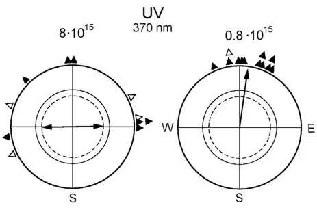
In autumn, we also performed tests under 373 nm UV at 8·1015 quanta s-1 m-2, which is the light level that
repre-sented the lowest intensity in the above-mentioned exper-iments. We observed an axial preference for the east-west axis (n = 16; 74°–254°, rN = 0.58, P < 0.01). The results of the following spring experiments under this light regime again produced an axial preference for the east-west axis under 8·1015 quanta s-1 m-2 (Fig. 2, left, and Table 3),
which looked similar to the respective distributions under Blue, Turquoise and Green at higher intensities. However, in tests under 373 nm UV at 0.8·1015 quanta s-1 m-2, i.e. at
only one tenth of the quantal flux, the robins preferred their seasonally appropriate northerly migratory direction (Fig. 2, right). The directional choices of the individual birds are given in Table 4.
Discussion
The orientation responses observed under UV, Blue, Tur-quoise and Green light at the various intensities clearly fall into two distinct categories: under the lowest light
level, which was 0.8·1015 quanta s-1 m-2 for UV and
8·1015 quanta s-1 m-2 for Blue, Turquoise and Green, the
robins showed a strong preference for their migratory direction. Under 8·1015 quanta s-1 m-2 UV light and under
increased light intensities of the other colors, a variety of responses, mostly axial preferences, was observed. The northerly headings observed under bright Turquoise, although superficially similar to the migratory direction, also represent responses of a different nature [4] (see below). Together, the data clearly show that even when the birds were no longer heading in their migratory direc-tion, their behavior continued to change as the light inten-sity increased. This implies that it is still controlled by light-dependent processes.
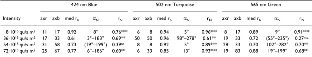
Table 1: Orientation data of three groups of 12 birds, recorded under various intensities of blue, turquoise or green light
424 nm Blue 502 nm Turquoise 565 nm Green
Intensity axr axb med rb αN rN axr axb med rb αN rN axr axb med rb αN rN
8·1015 qu/s m2 11 17 0.92 8° 0.76*** 6 8 0.94 5° 0.96*** 8 17 0.89 9° 0.91***
36·1015 qu/s m2 17 33 0.61 3°–183° 0.69** 50 50 0.96 98°–278° 0.61** 19 33 0.72 (55°–235°) 0.27n.s. 54·1015 qu/s m2 31 58 0.73 (19°–199°) 0.39ns 8 8 0.92 5° 0.89*** 28 33 0.70 102°–282° 0.70** 72·1015 qu/s m2 25 67 0.77 6°–186° 0.60** 6 33 0.85 13° 0.93*** 19 83 0.88 19°–199° 0.68**
The following considerations will focus on (i) the inten-sity-dependent pattern of responses indicated, (ii) possi-ble reasons for the change in the nature of the responses, (iii) the possible involvement of the color cones and (iv) the origin of directional information for the axial and 'fixed direction' responses.
Compass orientation and other responses
The behavior under the low light intensity Blue, Turquoise and Green represent true migratory orientation: under this and similar light levels, birds prefer their migratory direction in spring as well as in autumn. They showed the
expected seasonal reversal, and the compass mechanism involved is the normal avian inclination compass [4,7,16]. Tests with oscillating magnetic fields indicate that magnetoreception under Green and Turquoise light is based on radical pair processes [2-4] as proposed by the model of Ritz and colleagues [1]. The same can be assumed for dim Blue light, where the orientation corre-sponds to that under Green and Turquoise in all other aspects [17], and presumably also for UV light of very low intensity. That is, under low monochromatic lights from 373 nm UV to 565 nm Green, magnetoreception works in the normal way as under 'white' light, providing birds
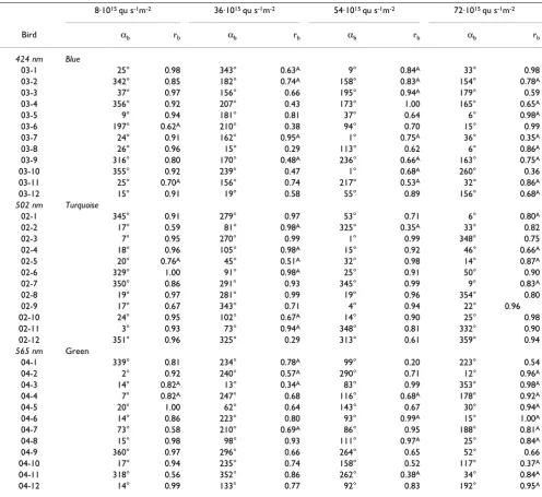
Table 2: Vectors of individual birds based on 3 recordings each
8·1015 qu s-1m-2 36·1015 qu s-1m-2 54·1015 qu s-1m-2 72·1015 qu s-1m-2
Bird αb rb αb rb αb rb αb rb
424 nm Blue
03-1 25° 0.98 343° 0.63A 9° 0.84A 33° 0.98
03-2 342° 0.85 182° 0.74A 158° 0.83A 154° 0.78A
03-3 37° 0.97 156° 0.66 195° 0.94A 179° 0.59
03-4 356° 0.92 207° 0.43 173° 1.00 165° 0.65A
03-5 9° 0.94 181° 0.81 37° 0.64 6° 0.98A
03-6 197° 0.62A 210° 0.38 94° 0.70 15° 0.99
03-7 24° 0.91 162° 0.95A 1° 0.75A 36° 0.35A
03-8 26° 0.96 15° 0.29 113° 0.62 6° 0.86A
03-9 316° 0.80 170° 0.48A 236° 0.66A 163° 0.75A
03-10 355° 0.92 239° 0.47 1° 0.68A 260° 0.36
03-11 25° 0.70A 156° 0.74 217° 0.53A 32° 0.86A
03-12 15° 0.91 19° 0.58 55° 0.89 156° 0.68A
502 nm Turquoise
02-1 345° 0.91 279° 0.97 53° 0.71 6° 0.80A
02-2 17° 0.59 81° 0.98A 325° 0.35A 33° 0.82
02-3 7° 0.95 270° 0.99 1° 0.99 348° 0.75
02-4 18° 0.96 105° 0.98A 15° 0.92 46° 0.66A
02-5 20° 0.76A 45° 0.51A 32° 0.98 14° 0.87A
02-6 329° 1.00 91° 0.98A 25° 0.91 50° 0.90
02-7 350° 0.86 291° 0.93 345° 0.99 9° 0.83A
02-8 19° 0.97 281° 0.99 19° 0.96 354° 0.80
02-9 17° 0.67 343° 0.71 4° 0.94 22° 0.96
02-10 24° 0.95 102° 0.67A 14° 0.90 25° 0.98
02-11 3° 0.93 73° 0.94A 348° 0.81 332° 0.90
02-12 351° 0.96 325° 0.29 313° 0.61 359° 0.94
565 nm Green
04-1 339° 0.81 234° 0.78A 99° 0.20 223° 0.54
04-2 2° 0.92 240° 0.57A 290° 0.71 12° 0.96A
04-3 14° 0.82A 13° 0.34A 83° 0.99 353° 0.98A
04-4 7° 0.82A 247° 0.68 116° 0.68A 178° 0.92A
04-5 20° 1.00 62° 0.64 143° 0.67 30° 0.94A
04-6 14° 0.86 223° 0.80 93° 0.99A 15° 1.00A
04-7 73° 0.58 210° 0.69A 86° 0.95 188° 0.81A
04-8 15° 0.98 98° 0.93 111° 0.97A 25° 0.84A
04-9 360° 0.97 296° 0.66 264° 0.65 52° 0.66
04-10 17° 0.94 235° 0.74 158° 0.52 117° 0.37A
04-11 318° 0.56 352° 0.86 262° 0.38A 34° 0.84A
04-12 14° 0.99 133° 0.77 92° 0.83 192° 0.95A
with directional information from the magnetic field that they can use to locate their migratory direction and prob-ably also any other direction they may wish to pursue.
The behavior under monochromatic light of higher inten-sity is different. Increased monochromatic lights do not simply cause a switch from migratory orientation to another specific response; instead, they elicit a variety of different responses. This is most conspicuous under green
light: Here, the birds show disorientation at 36·1015
quanta s-1 m-2, then, as intensity increases, a preference for
the east-west axis and finally a preference for the
north-south axis. Muheim and colleagues [18], also testing rob-ins under green light, observed axial behavior in the migratory direction and in the opposite direction under intensities of 14 and 29·1015 quanta s-1 m-2, which may
be a first step away from normal migratory orientation.
Together, the responses observed under 8·1015 quanta s-1
m-2 UV and at higher intensities of Blue, Turquoise and
Green suggest that the pattern observed under Green might be a general one. Under all wavelengths, we found a preference of the east-west axis that, under increased intensity, was followed by a preference of the north-south
Table 3: Orientation of 12 birds recorded under UV light of two intensities
373 nm UV
Intensity axr axb med rb αN rN
8·1015 qu/s m2 47 50 0.76 89°–269° 0.50*
0.8·1015 qu/s m2 11 8 0.95 8° 0.96***
axr, axb, percentage of axial recordings and axial mean vectors of birds, respectively (both as defined in text); med rb, median vector length of the individual birds, representing the intraindividual variance; αN, rN, grand mean vector or axis (non-significant data in parentheses), with asterisks at rN indicating significance by the Rayleigh test, see Table 1
Orientation of European robins under 373 nm UV light of different quantal flux (in quanta s-1m-2 above the circle)
Figure 2
Orientation of European robins under 373 nm UV light of different quantal flux (in quanta s-1m-2 above the circle).
axis, with the modification that under Turquoise, a uni-modal 'fixed' northerly direction [4] replaces the axial north-south tendency. However, where normal migratory orientation ends and random and axial behavior begins appear to depend on the wavelength as well as on the intensity of light.
The different types of responses at brighter light imply a disrupted function of the magnetoreception system under monochromatic light of higher intensity. It raises a number of questions: What causes the magnetoreception system to cease functioning in the normal way? Is there a functional significance of the axial and 'fixed' direction responses? And: what is the nature of the directional infor-mation for the 'fixed' directions and axial responses?
What causes the change in magnetoreception?
An effect of the brighter monochromatic lights on circa-dian patterns and motivation (e.g. [19,20]) is rather unlikely in view of the fact that even the brightest lights used in the present study were of intensities found well after sunset (see Method section), i.e., at a time of day when nocturnal migration is in progress. Also, the disrup-tion of the normal magnetic percepdisrup-tion process cannot be attributed to the higher intensity of light itself or to satu-ration of the crucial receptors. The avian magnetic com-pass works under bright sun light (see e.g. [21-24] for homing pigeons and day migrants), and cage tests showed that also nocturnal migrants use their magnetic compass for migratory orientation under natural day light when migratory behavior was induced by food deprivation [25]. Caged robins, too, were well oriented under 'white' test lights of higher intensity [13]. However, the 'white' test lights, like day light, were composed of wavelengths from all parts of the spectrum. Therefore, it seems to be the nar-row bandwidth of the monochromatic test lights used
rather than their brightness that gives rise to the observed effects.
The same wavelengths of light allow very good orienta-tion at low intensities, but disrupt the magnetic compass orientation as the intensity of the monochromatic light increases. Our experiments were performed under light levels that, in humans, are mesopic conditions, i.e., where both the rod and the cone system is active. In humans, this transition zone covers at least 3 log units [26]; its extension in birds is unknown. Note that the light levels where we observed a change from compass orientation to an axial response along the east-west axis increased with increasing wavelengths, from UV over Blue and Turquoise to Green, suggesting a similar relationship for the end of normal perception of magnetic directions. This is a strik-ing parallel to the sensitivity of the color cones, which decreases with increasing wavelength, with the UV-sensi-tive-cone type being more sensitive than the short-wave-length-sensitive cone, this type being more sensitive than the medial-wavelength-sensitive cone and the long-wave-length-sensitive cone type being least sensitive [see e.g. [27]]. This implies an involvement of the color cones under the higher light intensities, suggesting that the per-ception of magnetic directions works properly under monochromatic light as long as the test lights do not acti-vate the cones above a certain level.
A possible role of the color cones?
The radical pair model [1] proposes that photon absorp-tion causes a photopigment to form radical pairs and gen-erate the signals that mediate magnetic compass information. However, at present, neither the nature of the relevant photopigment nor the type of cells where the reception processes take place are precisely known. A role of the rods and color cones in avian magnetoreception is
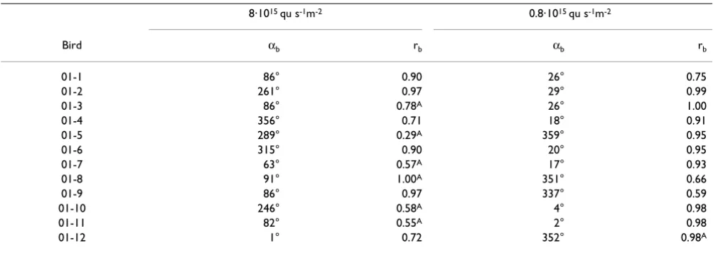
Table 4: Vectors of individual birds under 373 nm UV light based on three recordings each
8·1015 qu s-1m-2 0.8·1015 qu s-1m-2
Bird αb rb αb rb
01-1 86° 0.90 26° 0.75
01-2 261° 0.97 29° 0.99
01-3 86° 0.78A 26° 1.00
01-4 356° 0.71 18° 0.91
01-5 289° 0.29A 359° 0.95
01-6 315° 0.90 20° 0.95
01-7 63° 0.57A 17° 0.93
01-8 91° 1.00A 351° 0.66
01-9 86° 0.97 337° 0.59
01-10 246° 0.58A 4° 0.98
01-11 82° 0.55A 2° 0.98
01-12 1° 0.72 352° 0.98A
usually not considered, because rhodopsin and the other opsins do not form the required radical pairs. Ritz et al. [1] therefore proposed that these could be formed by crypto-chrome, a novel photopigment first known from plants (see [28]), but also found in the retina of chickens [29] and of passerine birds [30,31]. Cryptochromes have recently been shown to mediate magnetic effects in plants [32]. This implies the possibility of specialized photore-ceptors for magnetoreception. The disruptive effect of intense monochromatic light, on the other hand, suggests that these receptors may interact with the normal visual perception system, in some complex way.
One possibility is that cryptochrome does not directly absorb the light, but receives the energy from a light-har-vesting system of other pigments, as it is proposed, e.g., for photosynthesis [33]. Yet under this assumption, it is hard to explain why higher light intensities should lead to a change in the nature of response away from normal compass orientation to axial responses and 'fixed direc-tion'.
However, the behavior suggesting an involvement of the cones is only observed under higher intensity monochro-matic light. This has to be included in the considerations on the role of cones in magnetic perception. The answer may lie in the fact that the output of a given cone is affected by both, the wavelength of the incident light and its intensity. Both parameters together give only one out-put value. Color perception is then based on the balance of the outputs of the three (mammals) or four (birds) cone types, as it is measured, for example, by the retinal ganglion cells where the input from the photoreceptors converges. Natural light will always excite several types of cones, since even objects that appear unicolored to us usu-ally reflect a multitude of different wavelengths. Hence all cone types normally receive at least a certain amount of excitation by photons. In view of this, it is quite conceiva-ble that the color system is tuned to perceive a mixture of almost all visual wavelengths under normal conditions.
Monochromatic light would cause an imbalance between the different receptors, and there is a lot of evidence that a strong imbalance in the color of the visual scene lead to strong habituation of selected cones, which in turn causes the appearance of aftereffects like the sensation of the countercolor when the imbalance is eliminated [34]. By using monochromatic light with only a narrow spectral band, but of a relatively high intensity, the difference between the excitation of the cones projecting to one opponent color ganglion cell might become too large to be accepted by the system as normal, and the ganglion cell will no longer produce the appropriate activity. This may cause the visual system to also reject the magnetic infor-mation because it could be erroneous. In other words, the
visual system may be able to gate, i.e. control the transfer, of the magnetic input somewhere on its way to the brain area where it is processed. Although there is ample evi-dence for the existence of such gating systems – almost all sensory information, for example, is thought to be gated in the thalamic nuclei on its way to the forebrain [35] – this is a mere assumption in the case of magnetic informa-tion. The activation of a visual brain area only at night recently described [36] might be an example of a gating process that allows the transfer of information towards this area only under certain conditions.
Whether an imbalance between the different color recep-tors is the correct explanation for the responses observed under high intensity monochromatic light must remain open at present. It means, however, that these responses need not necessarily be of functional significance. Instead, they might be by-products of a perception system driven beyond its functional limits, reflecting a complex relation-ship between various receptors and units that awaits fur-ther analysis.
Where does the polar magnetic information originate?
The unimodal response at 54·1015 quanta s-1 m-2
Tur-quoise was found to be polar, not involving the normal avian inclination compass, and tests applying high fre-quency fields showed that it is not based on radical pair processes [4]. It seems likely that the axial responses under intense monochromatic light share these characteristics. This raises the question where this type of directional information comes from, if it does not originate in radical pair processes.
A magnetite-based receptor seems to be a logical assump-tion, as magnetite-based receptors could convey polar directions (see e.g. [37]). Magnetite has been found in the ethmoid region and in the upper beak of birds [38,39], but electrophysiological recordings from the correspond-ing branch of the trigeminal nerve [40] as well as behavio-ral studies [41-43] seemed to suggest that magnetite-based receptors in birds provide information on magnetic inten-sity rather than directional information. However, it can-not be excluded that they additionally mediate directional information. The relationship between the axial prefer-ence and the orientation of magnetite particles described in salamanders [15] appears to suggest a role of magnetite in these responses, and a recent study [44] indicates that another 'fixed' direction response in birds was indeed mediated by the iron-based receptors in the upper beak [39].
wavelength of light. This is not only true for the present study, but also for various 'fixed' directions observed in other studies [7,14,17,45]. The control of the axial and 'fixed' direction responses by the ambient light regime is difficult to explain by attributing it to a magnetite-based mechanism without auxiliary assumptions, like e.g. inter-action between the magnetite-based and light-dependent mechanisms.
Conclusion
Our study with monochromatic Blue, Turquoise, Green and UV light revealed two distinct types of orientation behavior depending on the intensity of light: normal compass orientation under very low light, and a variety of different responses under light of higher intensity, proba-bly changing according to a specific pattern as intensity increases. The occurrence of these responses indicates a complex interaction of various receptors and for the first time suggests an involvement of the color cone system in magnetoreception.
The interpretation of our findings suffers greatly from the fact that the cells representing the magnetoreceptors have not yet been reliably identified (but see [1,31]) and that, as a consequence, we do not know how they are intercon-nected with other units. In general, we know too little about the wiring of the avian retina, especially the color processing system, to come up with an explanation that is not entirely speculative. As to a possible involvement of magnetite-based receptors in the axial and 'fixed' direction responses, future tests will have to clarify their role.
Materials and methods
The experiments reported here were performed in the gar-den of the Zoological Institute in Frankfurt a.M., Germany (50°08'N 8°40'E) in pre-spring during a six week period each from the first week of January to about mid-February.
Test birds
The test birds were European robins, a night-migrating passerine species. Groups of 12 robins each were mistnet-ted as transmigrants in the Botanical garden in Frankfurt during the first two weeks of September of the year before the tests. The birds were juveniles identified as being of Scandinavian origin by their relatively long wings. They were kept indoors in individual cages under white light
from a fluorescent lamp in a photoperiod that simulated the natural one outside until the beginning of December, when their photoperiod was decreased to L:D 8:16. Around New Year, it was changed in two steps to L:D 13:11. This induced premature spring migratory restless-ness so that the tests could begin in the first week of Janu-ary. During testing and thereafter, the photoperiod was maintained at L:D 13:11, and in the last week of March, when this photoperiod was reached outside, the birds were released at the site of capture.
Test lights
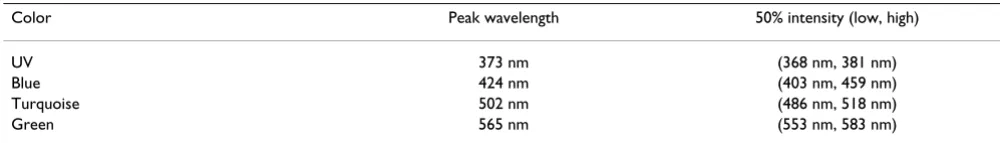
The test lights were produced using the same (Blue, Green) or similar (Turquoise) LEDs (light-emitting diodes) as in earlier studies (e.g. [7,10]). Their spectra and that for UV used here for the first time are given in Table 5. Sets of 24 or 48 LEDs, or, for UV, of 3 LEDs, mounted on a plastic disc were suspended above the test cages (see Fig. 3), with the light intensities controlled by adjusting current and the numbers of LEDs activated to produce test lights of equal quantal flux. The four light levels were about 8, 36, 54 and 72·1015 quanta s-1 m-2, for Blue,
Tur-quoise and Green and 8 and 0.8·1015 quanta s-1 m-2 for
UV. The blue to green lights were measured in the test cages as irradiance using Optometer P9710-1 (Gigahertz-Optik, Puchheim, Germany) with the radiometric probe "Visible" RW-3703-2, a silicon photoelement for the wavelength range 400 – 800 nm, and the UV light with the corresponding UV probe. The light levels used here are generally slightly higher than the respective ones used in the previous studies (e.g. [7,10]).
For an idea what the four light levels mean compared with the natural light outside, let us state that e.g. the green light corresponded to light found outside under largely clear sky more than 45 min, about 38 min, 34 min and 32 min before sunrise and after sunset, or, if only the green part of the spectrum between 553 and 583 nm is consid-ered, to light about 28 min, 20 min, 17 min and 15 min before sunrise and after sunset (estimates based on our own measurements).
Test apparatus and performance
The tests were performed in wooden huts in the garden of the Zoological Institute, where the local geomagnetic field was undisturbed with an intensity of 46 000 nT, and + 66°
Table 5: Emission spectra of the LEDs used in the present paper
Color Peak wavelength 50% intensity (low, high)
UV 373 nm (368 nm, 381 nm)
Blue 424 nm (403 nm, 459 nm)
Turquoise 502 nm (486 nm, 518 nm)
inclination. The directional tendencies of the birds were recorded in funnel cages [46] lined with coated paper (typewriter correction paper BIC, Germany; formerly Tipp-Ex), where the birds were tested one at a time (see [6]). Each funnel cage was placed inside an aluminum cyl-inder, which isolated the cages against each other, with the top of the cylinder consisting of the plastic disk
carry-ing the LEDs (Fig. 3). The light passed through two sets of diffusers before it reached the test bird.
Recording the robins' orientation began in the evening at about the time when the light went off in the housing cages and lasted for 75 min. When active, the birds left scratch marks on the coating of the inclined walls of the
Test apparatus for testing individual birds under monochromatic lights
Figure 3
cages, which documented the distribution of their activity. Each bird was tested under the various test conditions until they had produced three recordings with sufficient activity (= 35 scratches) in each.
Data analysis
After removal from the cage, the coated paper was divided into 24 sectors, and the scratch marks in each sector were counted. From the distribution of the scratches, the bird's heading and the concentration of activity (expressed by the vector length) of the respective test were calculated. From the three headings of each bird, we calculated the mean vector of this bird under each condition, with direc-tion αb and length rb. Based on the mean headings αb of the 12 test birds, the grand mean vector for each condi-tion, with the direction αN and the length rN, was calcu-lated.
Under higher light levels, the birds often showed axial behavior, with the scratches within the cage bimodally distributed along an axis, as indicated by a higher concen-tration (longer vector) obtained when the angles were doubled (modulo 360°) so that opposite sectors fall
together [47]. At the same time, many birds' vectors rb
increased considerably when they were calculated by dou-bling the angles (modulo 360°), indicating axial choices with two of the three headings on one side and one at the other. To take this axiality adequately into account, we fol-lowed the procedure used in [[48], [49]]: all recordings with the axial vector at least 0.03 longer than the respec-tive unimodal vector were treated as axial. Likewise, when the three recordings of each bird were comprised, the mean vector as well as the mean axis was calculated, and the bird's behavior was considered axial when the axial vector length rb was at least 0.10 longer than the unimodal vector length rb. In these cases, we used the preferred end of the axis (i.e. in case of the single recordings, the end with more activity and, in case of a bird's axial vector, the end with more headings, expressed by its being closer to the direction of the unimodal vector) for further calcula-tions. The data from all test conditions are treated this way, and the percentage of axial recordings as well as the percentage of axial bird vectors rb is included in Table 1. When calculating the grand mean vectors from the mean headings of the individual birds, αb, we also calculated the grand axis by doubling the angles to test for an axial dis-tribution of means, and used the longer vector for decid-ing between unimodal and axial preferences.
Statistical analysis
The grand mean vectors or grand axes were tested by the Rayleigh test for directional preferences [47]. The birds' mean headings under the low light levels were compared with the parametric Watson Williams test for differences in direction. Within each color, the distribution of the
mean headings was compared with the non-parametric Mardia Watson Wheeler test [47]. Since here most vectors were axial, we applied this test to the transformed distri-butions resulting from doubling the angles (modulo
360°). From the vector lengths rb per bird (considering
the axial vector length when the vectors was defined as axial), we calculated the grand medians.
Competing interests
The author(s) declare that they have no competing inter-ests.
Authors' contributions
RW designed the study, did the statistical analysis and wrote the manuscript. KS took a large part in providing the test birds and performing the experiments, in particu-lar in controlling the various light regimes. HJB provided expertise on the avian visual system and wrote part of the discussion. WW contributed significantly to designing the study, had designed test apparatus and testing procedure, had a major part in the data analysis and modified the manuscript.
Acknowledgements
Our work was supported by the Human Frontier Science Program (grant to RW) and the Deutsche Forschungsgemeinschaft (grant to WW). We sincerely thank P. Galland, Universität Marburg, and T. Ritz, University of California at Irvine, for helpful suggestions and comments. Thanks are also due to S. Denzau, F. Galera, D. Geiß, S. Hilmer, C. Koschella, N. Moldan, S. Münzer, C. Nießer, K. Patzolt, T. Pavkovic and M. Stavermann for their val-uable help with keeping the birds and conducting the experiments. The experiments were performed in accordance with the rule and regulations for animal welfare in Germany.
References
1. Ritz T, Adem S, Schulten K: A model for vision-based magne-toreception in birds. Biophys J 2000, 78:707-718.
2. Ritz T, Thalau P, Phillips JB, Wiltschko R, Wiltschko W: Resonance effects indicate a radical-pair mechanism for avian magnetic compass. Nature 2004, 429:177-180.
3. Thalau P, Ritz T, Stapput K, Wiltschko R, Wiltschko W: Magnetic compass orientation of migratory birds in the presence of a 1.315 MHz oscillating field. Naturwissenschaften 2005, 92:86-90. 4. Wiltschko R, Ritz T, Stapput K, Thalau P, Wiltschko W: Two
differ-ent types of light-dependdiffer-ent responses to magnetic fields in birds. Curr Biol 2005, 15:1518-1523.
5. Wiltschko W, Traudt J, Güntürkün O, Prior H, Wiltschko R: Later-alization of magnetic compass orientation in a migratory bird. Nature 2002, 419:467-470.
6. Wiltschko W, Munro U, Ford H, Wiltschko R: Red light disrupts magnetic orientation of migratory birds. Nature 1993,
364:525-527.
7. Wiltschko W, Munro U, Ford H, Wiltschko R: Magnetic orienta-tion in birds: non-compass responses under monochromatic light of increased intensity. Proc R Soc Lond B 2003,
270:2133-2140.
8. Wiltschko W, Wiltschko R: Migratory orientation of European robins is affected by the wavelength of light as well as by a magnetic pulse. J Comp Physiol A 1995, 177:363-369.
9. Wiltschko W, Wiltschko R: The effect of yellow and blue light on magnetic compass orientation in European Robins, Erith-acus rubecula. J Comp Physiol A 1999, 184:295-299.
Publish with BioMed Central and every scientist can read your work free of charge "BioMed Central will be the most significant development for disseminating the results of biomedical researc h in our lifetime."
Sir Paul Nurse, Cancer Research UK
Your research papers will be:
available free of charge to the entire biomedical community
peer reviewed and published immediately upon acceptance
cited in PubMed and archived on PubMed Central
yours — you keep the copyright
Submit your manuscript here:
http://www.biomedcentral.com/info/publishing_adv.asp
BioMedcentral under monochromatic light of various wavelengths. J Exp Biol
2001, 204:3295-3302.
11. Rappl R, Wiltschko R, Weindler P, Berthold P, Wiltschko W: Orien-tation of Garden Warblers, Sylvia borin, under monochro-matic light of various wavelengths. Auk 2000, 117:256-260. 12. Wiltschko R, Wiltschko W: Pigeon homing: Effect of various
wavelengths of light during displacement. Naturwissenschaften 1998, 85:164-167.
13. Wiltschko W, Möller A, Gesson M, Noll C, Wiltschko R: Light-dependent magnetoreception in birds: analysis of the behav-iour under red light after pre-exposure to red light. J Exp Biol 2004, 207:1193-1202.
14. Wiltschko W, Wiltschko R, Munro U: Light-dependent magne-toreception in birds: the effect of intensity of 565-nm green light. Naturwissenschaften 2000, 87:366-369.
15. Phillips JB, Borland SC, Freake MJ, Brassart J, Kirschvink JL: "Fixed-axis" magnetic orientation by an amphibian: non-shoreward-directed compass orientation, misnon-shoreward-directed homing or posi-tioning a magnetite-based map detector in a consistent alignment relative to the magnetic field? J Exp Biol 2002,
205:3903-3914.
16. Wiltschko W, Gesson M, Wiltschko R: Magnetic compass orien-tation of European robins under 565 nm green light. Natur-wissenschaften 2001, 88:387-390.
17. Stapput K, Gesson M, Wiltschko R, Wiltschko W: Light-dependent magnetoreception: behavior of migratory birds under mon-ochromatic and bichromatic lights. In Orientation & Navigation. RIN 05 Proceedings Reading, England: Royal Institute of Navigation; 2005.
18. Muheim R, Bäckman J, Åkesson S: Magnetic compass orientation in European Robins is dependent on both wavelength and intensity of light. J Exp Biol 2002, 205:3845-3856.
19. Deutschlander ME, Phillips JB, Borland SC: The case of light-dependent magnetic orientation in animals. J Exp Biol 1999,
202:891-908.
20. Johnsen S, Lohmann KJ: The physics and neurobiology of mag-netoreception. Nature Review Neuroscience 2005, 6:703-712. 21. Keeton WT: Magnets interfere with pigeon homing. Proc Natl
Acad Sci 1971, 68:102-106.
22. Wiltschko R, Nohr D, Wiltschko W: Pigeons with a deficient sun compass use the magnetic compass. Science 1981, 214:343-345. 23. Wiltschko W, Wiltschko R, Keeton WT, Madden R: Growing up in an altered magnetic field affects the initial orientation of young homing pigeons. Behav Ecol Sociobiol 1983, 12:135-142. 24. Munro U, Wiltschko R: Magnetic compass orientation in the
yellow-faced honeyeater, Lichenostomus chrysops (Meliphagi-dae), a day migrating bird from Australia. J Exp Biol 1993,
181:233-244.
25. Thalau H-P, Wiltschko W: Einflüsse des Futterangebots auf die Tagesaktivität von Trauerschnäppern (Ficedula hypoleuca) auf dem Herbstzug. Courier Forschungsinstitut Senckenberg 1987,
97:67-74.
26. Chien SH, Teller DY, Palmer J: The transition from scotopic to photopic vision in 3-months-old infants and adults: an evalu-ation of the rod dominance hypothesis. Vision Research 2000,
40:3853-3871.
27. Maier EJ: Spectral sensitivities including the ultraviolet of the passeriform bird Leiothrix lutea. J Comp Physiol A 1992,
170:709-714.
28. Sancar A: Structure and function of DNA photolyase and cryptochrome blue-light photoreceptors. Chem Rev 2003,
103:2203-2237.
29. Bailey MJ, Chong NW, Xiong J, Cassone VM: Chicken' Cry2: molecular analysis of an avian cryptochrome in retinal and pineal photoreceptors. FEBS Letter 2002, 513:169-174. 30. Möller A, Sagasser S, Wiltschko W, Schierwater B: Retinal
crypto-chrome in a migratory passerine bird: a possible transducer for the avian magnetic compass. Naturwissenschaften 2004,
91:585-588.
31. Mouritsen H, Janssen-Bienhold U, Liedvogel M, Feenders G, Stal-leicken J, Dirks P, Weiler R: Cryptochromes and neuronal-activ-ity markers colocalize in the retina of migratory birds during magnetic orientation. Proc Nat Acad Sci USA 2004,
10:14294-14299.
32. Ahmad M, Galland P, Ritz T, Wiltschko R, Wiltschko W: Magnetic intensity affects cryptochrome-dependent responses in Ara-bidopsis thaliana. Planta in press. DOI 10.1007/s00425-006-0383-0 33. Berg JM, Tymoczko JL, Styer L: Biochemistry New York, WH Freeman
& Co; 2006.
34. Stromeyer CF: Form-Color Aftereffects in Human Vision. In Handbook of Sensory Physiology. PerceptionVolume 8. Edited by: Held R, Leibowitz HW, Teuber HL. Berlin: Springer; 1978:97-142.
35. Singer W: Central core control of visual cortex functions. In The Neuroscience Fourth Study Program Edited by: Schmitt FO, Worden G. Cambridge, Mass: MIT Press; 1979:1093-1110.
36. Mouritsen H, Feenders G, Liedvogel M, Wada K, Jarvis ED: Night-vision brain area in migratory songbirds. Proc Nat Acad Sci USA 2005, 102:8339-8344.
37. Kirschvink JL, Gould JL: Biogenetic magnetite as a basis for magnetic field detection in animals. BioSystems 1981,
13:181-201.
38. Beason RC, Brennon WJ: Natural and induced magnetization in the bobolink, (Dolichonyx orycivorus). J Exp Biol 1986,
125:75-80.
39. Fleissner G, Holtkamp-Rötzler E, Hanzlik M, Winklhofer M, Fleissner G, Petersen N, Wiltschko W: Ultrastructural analysis of a puta-tive magnetoreceptor in the beak of homing pigeons. J Comp Neurol 2003, 458:350-360.
40. Semm P, Beason RC: Responses to small magnetic variations by the trigeminal system of the Bobolink. Brain Res Bull 1990,
25:735-740.
41. Beason RC, Semm P: Does the avian ophthalmic nerve carry magnetic navigational information? J Exp Biol 1996,
199:1241-1244.
42. Munro U, Munro JA, Phillips JB, Wiltschko R, Wiltschko W: Evi-dence for a magnetite-based navigational 'map' in birds.
Naturwissenschaften 1997, 84:26-28.
43. Mora CV, Davison M, Wild JM, Walker MM: Magnetoreception and its trigeminal mediation in the homing pigeon. Nature 2004, 432:508-511.
44. Wiltschko R, Stapput K, Ritz T, Thalau P, Wiltschko W: Magnetite-based 'fixed direction responses' in migratory birds. HFSP Journal in press.
45. Wiltschko W, Gesson M, Stapput K, Wiltschko R: Light-dependent magnetoreception in birds: interaction of at least two differ-ent receptors. Naturwissenschaften 2004, 91:130-134.
46. Emlen ST, Emlen JT: A technique for recording migratory ori-entation in captive birds. Auk 1966, 83:361-367.
47. Batschelet E: Circular Statistics in Biology London: Academic Press; 1981.
48. Helbig AJ, Wiltschko W: The skylight polarization at dusk affects the orientation behavior of blackcaps, Sylvia atrica-pilla. Naturwissenschaften 1989, 76:227-229.