1535-9778/03/$08.00⫹0 DOI: 10.1128/EC.2.6.1187–1199.2003
Copyright © 2003, American Society for Microbiology. All Rights Reserved.
Mating and Pathogenic Development of the Smut Fungus
Ustilago
maydis
Are Regulated by One Mitogen-Activated Protein
Kinase Cascade
Philip Mu¨ller, Gerhard Weinzierl,† Andreas Brachmann,‡ Michael Feldbru¨gge,
and Regine Kahmann*
Max Planck Institute for Terrestrial Microbiology, D-35043 Marburg, and Institute of Genetics and Microbiology, Ludwig-Maxilians-Universita¨t-Mu¨nchen, D-80638 Munich, Germany
Received 11 April 2003/Accepted 16 September 2003
In the phytopathogenic fungusUstilago maydis, pheromone-mediated cell fusion is a prerequisite for the
generation of the infectious dikaryon. The pheromone signal elevates transcription of the pheromone genes and elicits formation of conjugation hyphae. Cyclic AMP and mitogen-activated protein kinase (MAPK) signaling are involved in this process. The MAPK cascade is presumed to be composed of Ubc4 (MAPK kinase kinase),
Fuz7 (MAPK kinase), and Ubc3/Kpp2 (MAPK). We isolated thekpp4gene and found it to be allelic toubc4.
Epistasis analyses with constitutively active alleles of kpp4 and fuz7 substantiate that Kpp4, Fuz7, and
Kpp2/Ubc3 are components of the same module. Moreover, we demonstrate that Fuz7 activates Kpp2 and shows interactions in vitro. Signaling via this cascade regulates expression of pheromone-responsive genes, presumably through acting on the transcription factor Prf1. Interestingly, the same cascade is needed for
conjugation tube formation, and this process does not involve Prf1. In addition,fuz7as well askpp4deletion
strains are nonpathogenic, whilekpp2deletion mutants are only attenuated in pathogenesis. Here we show that
strains expressing the unphosphorylatable allelekpp2T182A/Y184Fare severely affected in tumor induction and
display defects in early infection-related differentiation.
The phytopathogenic fungus Ustilago maydis, the causal
agent of corn smut disease, displays a complex life cycle, which is linked to distinct morphological changes (27). In its haploid formU.maydisdivides by budding and is nonpathogenic. After fusion of two compatible haploid cells, the pathogenic dikaryon is formed, which grows filamentously. Compatibility is genetically regulated by two mating type loci. The biallelica
locus controls recognition and fusion, while the multiallelicb
locus regulates filamentous growth and pathogenic develop-ment (5). To exert their regulatory function, the bE and bW
homeodomain proteins encoded by theblocus have to
dimer-ize, and a prerequisite for this is that they are derived from
different alleles (20, 28). Thealocus encodes pheromone
pre-cursor and receptor genes that allow recognition and fusion with nonself partners (9). Therefore, the generation of an infectious dikaryon is possible only if cells are compatible, i.e., if they differ at theiraandbloci.
In response to the pheromone signal, conjugation tubes are formed and pheromone-responsive gene expression is ele-vated. Among the induced genes are the pheromone gene
(mfa), the pheromone receptor gene (pra), and the b genes
(54). Transcriptional activation as well as basal expression of these genes requires the high-mobility-group protein Prf1 (22). Prf1 activity is assumed to be controlled by cyclic AMP (cAMP) as well as by mitogen-activated protein kinase
(MAPK) signaling. Adenylyl cyclase (Uac1) is activated
through the G␣subunit of a heterotrimeric G protein (Gpa3)
(29). This in turn leads to the activation of the protein kinase A (Adr1) by triggering dissociation from its regulatory subunit Ubc1 (18). When this signaling route is disturbed,
pheromone-induced transcription of the agenes is blocked (29, 41), and
such strains display filamentous growth that is independent of the b heterodimer (18, 21). Conversely, when this signaling route is activated, e.g., in strains either carrying constitutive alleles ofgpa3or lackingubc1, strongly elevated expression of pheromone genes is observed (29, 41). Interestingly, these mu-tations do not lead to the induction of conjugation tubes. It has been hypothesized that the cAMP cascade acts on Prf1 (23, 29). Prf1 has also been postulated to act downstream of a MAPK module containing the MAPK Kpp2 (37). This
infer-ence stems from the observation that deletion of kpp2/ubc3
abolishes pheromone-dependent expression of themfagenes
as well as conjugation tube formation. Furthermore, deletion
offuz7, encoding a MAPK kinase (MAPKK), also results in
defects in conjugation tube formation while still allowing pher-omone-dependent gene expression (4, 41). On these grounds it has been difficult to place Fuz7 in the pheromone-signaling
cascade. On the other hand, mutations in fuz7/ubc5orubc3/
kpp2 were shown to suppress the filamentous phenotype of
uac1deletion mutants (35). The same screen also led to the
isolation of ubc4, presumed to encode a MAPKK kinase
(MAPKKK), andubc2, encoding a protein with similarities to
Ste50p ofSaccharomyces cerevisiaeand Ste4 of
Schizosaccha-romyces pombe(1, 36). All of these genes were placed in one cascade suppressing filamentous growth caused by low-cAMP conditions (1).
Here we provide genetic as well as biochemical evidence that
* Corresponding author. Mailing address: Max Planck Institute for Terrestrial Microbiology, Karl-von-Frisch-Strasse, D-35043 Marburg, Germany. Phone: 496421178501. Fax: 496421178509. E-mail: [email protected].
† Present address: Vossius & Partner, D-81675 Munich, Germany. ‡ Present address: NIH, NIDDK, LBG, Bethesda, MD 20892.
1187
on September 8, 2020 by guest
http://ec.asm.org/
Kpp2/Ubc3, Fuz7, and Kpp4/Ubc4 act in one cascade that is activated after pheromone perception. Our experiments show that the pathways leading to pheromone-dependent gene ex-pression and conjugation tube formation separate downstream of Kpp2. In addition, the integrity of this MAPK module is also crucial for pathogenic development.
MATERIALS AND METHODS
Strains and growth conditions.TheEscherichia coliK-12 derivatives DH5␣ (Bethesda Research Laboratories) and Top10 (Invitrogen) were used for cloning purposes, andE.coliBL21(DE3)(pLysS) (Novagen) was used for protein ex-pression. TheU.maydisstrains used in this study are listed in Table 1. Prior to transformation into U.maydis, plasmids were digested with DraI (pKpp4-1, pKpp4WT, pKpp4RA, pGFP-Kpp4WT, pGFP-Kpp4RA, pGE1, pKpp2WT, pKpp2AEF, pKpp2K50R, pKpp2WT-GFP, pKpp2AEF-GFP, and pKpp2K50R-GFP),SspI (p123Pcrg1:kpp4PS, p123Pcrg1:kpp4-2, p123Pcrg1:fuz7, and p123), or BsrGI (pOTEF:pra2). In all cases single homologous integration events into the respective loci were verified by Southern analysis. Single homologous integration in theiplocus was verified by PCR and Southern analysis as described previously (32).
U. maydisstrains were grown at 28°C in liquid CM (25), YEPSL (0.4% yeast extract, 0.4% peptone, 2% sucrose), or potato dextrose (PD) (2.4% PD broth [Difco]) medium on a rotary shaker at 220 rpm or on solid PD agar. For induction ofcrg1promoter activity, strains were grown in CM medium contain-ing 1% glucose (CM-Glc) to an optical density at 600 nm (OD600) of 0.5, washed twice with water, and suspended in CM medium with 1% arabinose as a carbon source (CM-Ara).
Hygromycin B was purchased from Roche, nourseothricin (NAT) was chased from the Hans-Kno¨ll-Institute (Jena, Germany), and carboxin was pur-chased from Riedel de Haen (Seelze, Germany). All other chemicals were of analytical grade and were obtained from Sigma or Merck.
Isolation of thekpp4gene.Degenerate primers MEKK4 (GTITAYYTIGGN ATGAAYGC) and MEKK6 (YTTYTTISWDATICCRAARTC) were used for amplification ofU.maydisDNA. Reaction mixtures contained 10 mM Tris-HCl (pH 8.3), 3 mM MgCl2, 50 mM KCl, 50 pmol of primers, and 2 U ofTaq polymerase. Amplification was achieved by 35 cycles of 1 min at 95°C, 1 min at 48°C, and 1 min at 72°C. For sequencing, PCR products of 420 bp were cloned into pCR2.1TOPO. The amplifiedkpp4fragment was used to screen a genomic EMBL3 library (45). From a hybridizing clone,kpp4was subcloned as 5.2-kb
HindIII and 7.4-kbBamHI fragments in pTZ19R, and the resulting plasmids were designated pKpp4H and pKpp4B, respectively. In addition, we cloned a 2.5-kbHindIII-BamHI fragment comprising thekpp4gene into pSP72 to obtain pSP-kpp4H/B.
To isolate cDNA fragments ofkpp4, we produced cDNA by using an oli-go(dT)18primer, Superscript II reverse transcriptase (Life Technologies), and RNA obtained from AB33 (12) as the template. For the subsequent PCR, the following primer combinations were used: kpp4-550 (CGACGCTTCAAGTCG TCC)-OPM45 (GCGTAGCCGGCCGACTG), OPM46 (CGAAGAGGCCAG ATGCGAC)-OPM41 (AACCTTGCGTGCATCCTCAC), OMP40 (CAAAGC TCTCTCCGACAACG)-OPM21 (AGGGCCGTGTCGAGGCAG), OPM46 (C GAAGAGGCCAGATGCGAC)-kpp4rev (GCTTGACCGCCATCAGTAG), and kpp4⫹1490 (CGCGGATCCGCCTCTTCGCTGAACGC)-kpp4⫹974 (CC GGAATTCCGTCGATCGGTCCATGACC).
Plasmids and plasmid constructions. Plasmids pTZ18R (Pharmacia), pTZ19R (Pharmacia), pSP72 (Promega), pSL1180 (Pharmacia), and pBS-SKII(⫹) (Stratagene) were used for cloning, subcloning, and sequencing of genomic fragments, and pCR2.1TOPO (Invitrogen) was used for cloning and sequencing of fragments generated by PCR. pGEX-2T (Pharmacia) was used for protein expression inE.coli. Primers were obtained from Sigma ARK. Sequence analysis of genomic sequences and fragments generated by PCR was performed with an automated sequencer (ABI 377) and standard bioinformatic tools.
pRU11 contains thecrg1 promoter as a 3.5-kbNotI-NdeI fragment, and pSLHyg(⫺) contains a hygromycin resistance cassette as aNotI fragment (12). pCU3 is a pSP72 derivative harboring thetef1promoter as aNotI-NdeI fragment (A. Brachmann and R. Kahmann, unpublished data). pNEBNat(⫹) and pSL-Nat(⫹) are pNEB193 (New England Biolabs) and pSL1180 (Pharmacia) deriv-atives, respectively, both containing a NAT resistance cassette as aNotI fragment (37; A. Brachmann and R. Kahmann, unpublished data). p123 is a pSP72 deriv-ative containing theegfpgene (Clontech) fused to theotefpromoter andnos
terminator and a carboxin resistance cassette (55). pOTEF:pra2 is a p123 deriv-ative. For construction of pOTEF:pra2, we isolated a 1.9-kbHindIII-NotI
frag-ment encompassing theotefpromoter and cDNA frompra2as an ATG fusion from pJG10 (M. Feldbrugge, unpublished data) and ligated it into p123 digested withHindIII andNotI. The resulting plasmid provides for carboxin resistance and harbors thepra2cDNA under the control of theotefpromoter andnos
terminator.
kpp4plasmids.In pkpp4-1 thekpp4open reading frame (ORF) is deleted from bp⫹24 (XmaI site) to bp 74 after the stop codon (AvrII site). For construction, we ligated a 2.1-kbBamHI-XmaI fragment encompassing the 5⬘region ofkpp4
from pKpp2B, a 3-kbAgeI-SpeI fragment from pSLHyg(⫺) containing the hy-gromycin resistance cassette, and a 0.5-kbAvrII-EcoRI fragment from pKpp4B containing the 3⬘region into pTZ19R opened withBamHI andEcoRI.
Plasmid pKpp4WT is a pTZ19R derivative containing the 0.8-kb 5⬘region of
kpp4as aEcoRI-NotI fragment generated by PCR, the hygromycin resistance cassette as a 2.9-kbNotI-NotI fragment from pSLHyg(⫺), and thetef1promoter derived from pCU3 as aNotI-NdeI fragment fused to a 2.7-kb fragment encom-passingkpp4. At position 1 of thekpp4ORF, anNdeI site was introduced by using the annealed oligonucleotides kpp4Linker-I (TATGAGTGCTGCAACA CCTACCAGC) and kpp4Linker-II (CCGGGCTGGTAGGTGTTGCAGCACT CA).
pKpp4RA is identical to pKpp4WT except for the K481E mutation, which was generated by PCR with primers kpp4RAIII (GCCTCTTCGCAGCGCTATGC), kpp4RAV (GCCGCTAGGCGGCTTGCCGAATTCTTTGAGCACGCGCGC CATGAC), and kpp4RAIV (CCACAGGCATGCGCTCACC).
pGFP-Kpp4WT is a pKpp4WT derivative in which thetefpromoter was replaced by a 1.6-kbNotI-NdeI fragment encompassingsgfpunder the control of theotefpromoter (47). This results in a translational fusion of the green fluo-rescent protein (GFP) gene to thekpp4ORF.
pGFP-Kpp4RA is identical to pGFP-Kpp4WT except for the K481E mutation. Plasmid p123Pcrg1:kpp4PS is a p123 derivative in which theotefpromoter and GFP gene were replaced by thecrg1promoter (3.5-kbNotI-NdeI fragment from pRU11) fused tokpp4P681Sincluding the 0.55-kb 3⬘region. To introduce the
P681S mutation, we performed a PCR with primers kpp4PS (AAGATCCGCA ACTTCTTCGGCCAGCGATCGCCCTCAGAACTCATC) and kpp4⫹2239 (G GTGACCATCCATGGAACC).
p123Pcrg1:kpp4-2 is a p123 derivative in which theotefpromoter and GFP gene were replaced by thecrg1promoter (3.5-kbNotI-NdeI fragment from pRU11) fused tokpp4-2including the 0.55-kb 3⬘region. To generate thekpp4-2allele, we ligated a 0.5-kbNheI-PvuII fragment from pKpp4B and a 4.75-kbPvuI
(blunted)-HindIII fragment from pKpp4H into pBS-SKII(⫹) cut withXbaI-HindIII. fuz7plasmids. pHA42 is a pSP72 derivative that contains a 3.3-kb SphI genomic fragment encompassing thefuz7gene obtained from pFuz7 (41).
pGE1 is a pHA42 derivative in which a 0.9-kbNaeI-NsiI fragment encompass-ing bp⫹146 to⫹1057 of the fuz7ORF was replaced by a NAT resistance cassette derived as a 1.5-kbStuI-PstI fragment from pSLNat(⫹).
p123Pcrg1:fuz7DD is a p123 derivative in which theotefpromoter and GFP gene were replaced by thecrg1promoter (3.5-kbNotI-NdeI fragment from pRU11) fused tofuz7DDincluding the 0.2-kb 3⬘region. To introduce the S259D and T263D mutations, we performed a PCR with primers fuz7DD (ACATGT AGGTACTTGTACCAACAAAGTCGTCTGCGATATCGTTGATGAGC) and fuz7⫹1NdeI (CATATGCTTTCGTCCGGTGCG).
pGEX-Fuz7 is derivative of pGEX-2T containing anNcoI-MfeI fragment encoding a His6-tagged version offuz7derived from pET-Fuz7 (P. Muller and R. Kahmann, unpublished data).
kpp2plasmids.p123kpp2 is a p123 derivative in which the GFP gene was replaced by a 1-kbNcoI-NotI fragment that codes for Kpp2 and was generated by PCR with primers kpp2A (CATGCCATGGCACATGCCCACGGACAGC) and kpp2B (ATTTGCGGCCGCAAGATCAACGCATGATCTC).
pKpp2WT is a pTZ19R derivative that contains a 1.3-kb 5⬘region ofkpp2
derived as aHindIII-BglII fragment from pKpp2H, a 0.6-kbBglII-NotI fragment encoding the 3⬘part ofkpp2from p123kpp2, themfa2terminator as a 0.4-kb
NotI-BamHI fragment, a NAT resistance cassette obtained as a 1.5-kbMfe
I-BamHI fragment from pNEBNat(⫹), and a 1.1-kbEcoRI-XhoI fragment from pKpp2H representing the 3⬘region ofkpp2.
pKpp2AEF is identical to pKpp2WT except for the mutations T182A and Y184F. These were introduced by PCRs with primers kpp2C (CCATCGTGTG GCAACGAATTCGGCCATGAAACCC), kpp2D (GGAGCTCTCCGATGAC CAC), and kpp2B (see above).
pKpp2K50R is identical to pKpp2WT except for the K50R mutation intro-duced by PCRs with primers K50RI (CTCGTGTCGCCATCCGGAAGATCA CCCCATTCGATCAC), K50RII (TGACGCGATGCATGTCGG), and K50RIII (CAAAAGACGCGTCGCTGC).
pKpp2WT-GFP is a pKpp2WT derivative containing akpp2-gfpfusion. The GFP gene was isolated as 0.7-kbNcoI-NotI fragment from p123. This fragment
on September 8, 2020 by guest
http://ec.asm.org/
was ligated to a 2.1-kbHindIII-NcoI fragment ofkpp2in which anNcoI site was introduced at codon 351 by PCR with primers kpp2⫹379 (TATCAAACACTG CGTGGCTTG) and kpp2C⬘NcoI (CCATGGTCTCGTTATAAATCAACCTC TTG).
pKpp2AEF-GFP and pKpp2K50R-GFP were constructed by replacing the 2.7-kbBstXI fragments of pKpp2AEF and pKpp2K50R, respectively, with a 3.4-kb BstXI fragment encompassing the kpp2-gfp fusion derived from pKpp2WT-GFP.
pGEX-Kpp2 is a pGEX-2T derivative containing a 1.3-kbNcoI-NotI fragment derived from p123kpp2.
pGEX-Kpp2K50R is a pGEX-Kpp2 derivative in which a 0.5-kbNcoI-BglII fragment was replaced by a 0.5-kbNcoI-BglII fragment harboring the K50R mutation.
DNA and RNA procedures.Standard molecular techniques were used (43). Transformation ofU.maydiswas performed as published previously (45).U.
maydisDNA was isolated as described previously (24). RNA from strains grown
TABLE 1. U. maydisstrains used in this study
Strain Reference Plasmid transformed Integrationlocus Progenitor strain
FB1 (a1 b1) 3
FB2 (a2 b2) 3
SG200 (a1::mfa2 bW2bE1) 8
HA103 (a1 bW2bE1con) 22
FB1⌬kpp2-1 37
FB2⌬kpp2-1 37
FB1⌬prf1 37
FB2⌬prf1 37
FB1⌬kpp4 This study pKpp4-1 kpp4 FB1
FB2⌬kpp4 This study pKpp4-1 kpp4 FB2
SG200⌬kpp4 This study pKpp4-1 kpp4 SG200
HA103⌬kpp4 This study pKpp4-1 kpp4 HA103
FB1kpp4WT This study pKpp4WT kpp4 FB1
FB2kpp4WT This study pKpp4WT kpp4 FB2
FB1kpp4RA This study pKpp4RA kpp4 FB1
FB2kpp4RA This study pKpp4RA kpp4 FB2
SG200GFP-kpp4WT This study pGFP-Kpp4WT kpp4 SG200
SG200GFP-kpp4RA This study pGFP-Kpp4RA kpp4 SG200
FB1⌬fuz7 This study pGE1 fuz7 FB1
FB2⌬fuz7 This study pGE1 fuz7 FB2
SG200⌬fuz7 This study pGE1 fuz7 SG200
HA103⌬fuz7 This study pGE1 fuz7 HA103
FB1kpp2WT-GFP This study pKpp2WT-GFP kpp2 FB1
FB2kpp2WT-GFP This study pKpp2WT-GFP kpp2 FB2
FB1kpp2AEF-GFP This study pKpp2AEF-GFP kpp2 FB1
FB2kpp2AEF-GFP This study pKpp2AEF-GFP kpp2 FB2
FB1kpp2K50R-GFP This study pKpp2K50R-GFP kpp2 FB1
FB2kpp2K50R-GFP This study pKpp2K50R-GFP kpp2 FB2
FB1kpp2WT This study pKpp2WT kpp2 FB1
FB2kpp2WT This study pKpp2WT kpp2 FB2
SG200kpp2WT This study pKpp2WT kpp2 SG200
FB1kpp2AEF This study pKpp2AEF kpp2 FB1
FB2kpp2AEF This study pKpp2AEF kpp2 FB2
SG200kpp2AEF This study pKpp2AEF kpp2 SG200
FB1kpp2K50R This study pKpp2K50R kpp2 FB1
FB2kpp2K50R This study pKpp2K50R kpp2 FB2
FB1Pcrgl:kpp4PS This study p123Pcrgl:kpp4PS ip FB1
FB1Pcrgl:kpp4-2 This study p123Pcrgl:kpp4-2 ip FB1
FB1⌬kpp2-1Pcrgl:kpp4-2 This study p123Pcrgl:kpp4-2 ip FB1⌬kpp2-1
FB1⌬fuz7Pcrgl:kpp4-2 This study p123Pcrgl:kpp4-2 ip FB1⌬fuz7
FB1⌬prf1Pcrgl:kpp4-2 This study p123Pcrgl:kpp4-2 ip FB1⌬prf1
FB1⌬kpp6Pcrgl:kpp4-2 This study p123Pcrgl:kpp4-2 ip FB1⌬kpp6
FB1Pcrgl:fuz7DD This study p123Pcrgl:fuz7DD ip FB1
FB1⌬kpp2-1Pcrgl:fuz7DD This study p123Pcrgl:fuz7DD ip FB1⌬kpp2-1
FB1⌬kpp4Pcrgl:fuz7DD This study p123Pcrgl:fuz7DD ip FB1⌬kpp4
FB1⌬prf1Pcrgl:fuz7DD This study p123Pcrgl:fuz7DD ip FB1⌬prf1
FB2pra2con This study pOTEF:pra2 ip FB2
FB2⌬kpp2-1pra2con This study pOTEF:pra2 ip FB2⌬kpp2-1
FB2⌬fuz7pra2con This study pOTEF:pra2 ip FB2⌬fuz7
FB2⌬kpp4pra2con This study pOTEF:pra2 ip FB2⌬kpp4
FB2⌬prf1pra2con This study pOTEF:pra2 ip FB2⌬prf1
FB1kpp2AEF/Pcrgl:fuz7DD This study p123Pcrgl:fuz7DD ip FB1kpp2AEF
FB1kpp2K50R/Pcrgl:fuz7DD This study p123Pcrgl:fuz7DD ip FB1kpp2K50R
FB1Pcrgl:fuz7DD/kpp2-GFP This study pKpp2WT-GFP kpp2 FB1Pcrgl:fuz7DD
FB1Pcrgl:fuz7DD/kpp2AEF-GFP This study pKpp2AEF-GFP kpp2 FB1Pcrgl:fuz7DD
FB1Pcrgl:fuz7DD/kpp2K50R-GFP This study pKpp2K50R-GFP kpp2 FB1Pcrgl:fuz7DD
SG200Potef:GFP This study p123 ip SG200
SG200⌬kpp4/Potef:GFP This study p123 ip SG200⌬kpp4
SG200kpp2AEF/Potef:GFP This study p123 ip SG200kpp2AEF
on September 8, 2020 by guest
http://ec.asm.org/
in liquid culture was prepared as described previously (29). The following probes were used for Northern analyses: a 0.67-kbEcoRV fragment and a 1.3-kb
EcoRI-EcoRV fragment from pSP4.2EcoRV (9) formfa1andpra1, respectively; a 0.4-kbSpeI-PstI fragment from pTZa2XhoI3.5 (9) formfa2; a 2.6-kbPvuII fragment from pbW2-Nde-bE1 (12) forbEandbW; and a 1.2-kbNdeI-MluI fragment from p123Pcrg1:kpp4-2 and a 1.4-kb NdeI-SphI fragment from p123Pcrg1:fuz7DD forkpp4-2andfuz7, respectively. Radioactive labeling was performed with the NEBlot kit (New England Biolabs). A 5⬘-end-labeled oligo-nucleotide complementary to theU.maydis18S rRNA (10) was hybridized as a loading control in Northern analyses. A PhosphorImager (Storm 840; Molecular Dynamics) and the program ImageQuant (Molecular Dynamics) were used for visualization and quantification of radioactive signals.
Mating, pheromone stimulation, and pathogenicity assays.To test for mating, compatible strains were cospotted on charcoal-containing PD plates (25), and the plates were sealed with Parafilm and incubated at 24°C for 48 h. For pher-omone stimulation, strains were grown in CM-Glc to an OD600of 0.6. Synthetic pheromone (49) dissolved in dimethyl sulfoxide (DMSO) was added to a final concentration of 2.5g/ml, and cells were harvested for microscopic observa-tions and RNA preparaobserva-tions after 5 h of incubation in a 15-ml plastic tube on a tissue culture roller at 28°C. Quantification was done with photomicrographs by manual counting.
Plant infections of the corn variety Early Golden Bantam (Olds Seeds, Mad-ison, Wis.) were performed as described previously (37). For coinoculations of SG200 and derivatives, cells were mixed in equal amounts prior to infection. Fungal structures on the plant surface were visualized by Chlorazole Black E and by Calcofluor staining as described previously (11).
U. maydiscell lysates, glutathioneS-transferase (GST) pulldown, and kinase assay.U.maydis protein extracts were prepared with a French press (gauge pressure of 1,000 lb/in2) and cleared by centrifugation (4°C, 30 min, 33,300 rpm [Sorvall TH-660 rotor]).
GST-Kpp2-, GST-Kpp2K50R-, GST-Fuz7-, or GST-expressing BL21(DE3) (pLysS) cells were grown in dYT containing 1% glucose, ampicillin (100g/ml), and chloramphenicol (34g/ml) at 37°C. At an OD600of⬃0.5, cell suspensions were cooled, 1 mM IPTG (isopropyl--D-thiogalactopyranoside) was added, and
incubation was continued for 16 h at 16°C. Cells were harvested, washed once in buffer A (50 mM Tris-HCl [pH 7.5], 250 mM NaCl, 2.5 mM EDTA, 2.5 mM EGTA, 1% Triton X-100, 1 mM dithiothreitol [DTT]), and resuspended in 1 ml of buffer A containing complete protease inhibitor cocktail (catalog no. 1873580; Roche). Cells were freeze-thawed, and after DNase treatment, insoluble com-ponents were removed by centrifugation (4°C, 30 min, 28,000⫻g). The super-natant was incubated with 50l of glutathione-Sepharose beads (catalog no. 17075601; Amersham Bioscience) for 1 h at 4°C. The beads were washed once with buffer A and five times with 1 ml of buffer B (50 mM Tris-HCl [pH 7.5], 125 mM NaCl, 2.5 mM EDTA, 2.5 mM EGTA, 0.1% Triton X-100, 1 mM DTT).
To assay the kinase activity of GST-Kpp2 and GST-Kpp2K50R, beads were washed once with 1 ml of kinase buffer (KB) (20 mM HEPES [pH 7.4], 15 mM MgCl2, 5 mM EGTA, 1 mM DTT) and split into two portions. One was subjected to kinase assay (see below); the other was resuspended in 50l of sodium dodecyl sulfate-polyacrylamide gel electrophoresis (SDS-PAGE) sample buffer, and after 5 min of boiling, GST-Kpp2 levels were assayed by SDS–10% PAGE and Coomassie blue staining.
For GST pulldown, beads with GST-Fuz7 or GST alone were washed once with GST pulldown buffer (GPB) (50 mM Tris-HCl [pH 7.5], 150 mM NaCl, 0.5% NP-40, 5 mM EDTA, 1 mM DTT) and incubated for 1 h at 4°C with cleared portions ofU.maydiscell extracts (1 mg of protein) prepared in GPB containing complete protease inhibitor cocktail (catalog no. 1873580; Roche). After five washes with 1 ml of GPB, the beads were resuspended in 50l of 2⫻SDS-PAGE sample buffer and boiled for 5 min at 95°C. After centrifugation, 10l was loaded on two SDS–10% polyacrylamide gels to assay GST or GST-Fuz7 levels by Coomassie blue staining and to assay GFP-Kpp2 levels by Western analysis. To obtain extracts from pheromone-treated cells,U.maydisstrains were grown in CM-Glc to an OD600of⬃0.8 with rotary shaking at 28°C. Cultures were transferred to 50-ml plastic tubes, pheromone was added to a final concentration of 2.5g/ml, and cultures were incubated on a tissue culture roller at 28°C. For induction offuz7DD, strains were grown in CM-Glc to an OD600of⬃0.8, washed with water twice, and suspended in CM-Ara. After the cultures were harvested, cells were washed once in immunoprecipitation (IP) buffer (25 mM Tris-HCl [pH 7.5], 10 mM MgCl2, 15 mM EGTA, 75 mM NaCl, 0.1% Tween 20, 1 mM DTT) and resuspended in ice-cold IP buffer containing cocktails of protease and phos-phatases inhibitors (catalog no. 1873580 [Roche] and catalog no. P-2850 and P-5726 [Sigma]).U.maydisprotein extracts were prepared with a French press and cleared by centrifugation.
GFP-tagged Kpp2 was immunoprecipitated by adding 0.5g of rabbit
anti-GFP polyclonal antibody (catalog no. 3999100; Biocat, Heidelberg, Germany), immobilized to Dynabeads protein G (catalog no. 100.03; Dynal) by cross-linking, to portions (1 mg) of cleared cell extracts and mixing for 1 h at 4°C. Precipitated beads were washed once with 1 ml of IP buffer, five times with 1 ml of IP wash buffer (50 mM Tris-HCl [pH 7.5], 5 mM EDTA, 5 mM EGTA, 250 mM NaCl, 0.1% Tween 20, 1 mM DTT), and once with 1 ml of KB; all buffers contained protease and phosphatase inhibitors. The washed beads were split in two portions to assay kinase activity and precipitated Kpp2-GFP levels.
To determine Kpp2-GFP levels, beads were suspended in 20l of SDS-PAGE sample buffer and boiled for 5 min. Ten microliters was separated on an SDS-10% polyacrylamide gel, followed by semidry transfer to a Hybond-P membrane (catalog no. RPN303F; Amersham Bioscience).
GFP-tagged Kpp2 or Kpp4 derivatives were detected by using mouse anti-GFP monoclonal antibody (clones 7.1 and 13.1) (catalog no. 1814460; Roche) at a 1:5,000 dilution, and␣-tubulin (␣-Tub) was detected with anti-␣-Tub monoclo-nal antibody (Oncogene) at a 1:5,000 dilution. To detect phosphorylated Kpp2, the polyclonal phosphoepitope pTEpY-specific antibody 9101 from New En-gland Biolabs was used at a 1:1,000 dilution. As the secondary antibody we used horseradish peroxidase-conjugated goat anti-mouse antibody (diluted 1:10,000) (catalog no. W4021; Promega) or goat anti-rabbit antibody (diluted 1:10,000) (catalog no. 1706515; Bio-Rad) followed by detection with ECL⫹(catalog no. RPN2132; Amersham Bioscience).
To assay kinase activity, residual supernatant was removed and beads were resuspended in 20l of room temperature kinase assay buffer (KB containing 1 mg of myelin basic protein (MBP) (catalog no. 13228-010; Gibco) per ml, 50 mM Na--glycerol phosphate, 5 mM NaVO3, 50M ATP, 5 mM MgCl2, and 0.2M [␥-32P]ATP [6,000Ci/mmol]) and incubated for 20 min at 28°C. Reactions were stopped by adding 20l of 2⫻SDS-PAGE sample buffer and boiling for 5 min. Phosphorylation was analyzed by SDS-15% PAGE, and dried gels were exposed to a PhosphorImager (Molecular Dynamics).
Microscopic observation.For microscopic observation, we used a Zeiss Axio-phot microscope with differential interference contrast optics. Calcofluor fluo-rescence was observed with a standard DAPI (4⬘,6⬘-diamidino-2-phenylindole) filter set. GFP fluorescence was detected with a specific filter set (BP 470/20, FT 493, BP 505-530; Zeiss, Jena, Germany). Pictures were taken with a charge-coupled device camera (catalog no. C4742-95; Hamamatsu, Herrsching, Ger-many). Image processing was done with Image Pro (Media Cybernetics), Adobe Photoshop 6.0, and Canvas 6.0 (Deneba Systems).
Nucleotide sequence accession number.TheU. maydis kpp4gene has been assigned GenBank accession number AF542505.
RESULTS
Isolation of kpp4, encoding a MAPKKK homologue. In a
PCR approach using degenerate primers designed according
to conserved sequences of two MAPKKK genes,STE11ofS.
cerevisiaeandbyr2ofS.pombe, we isolated the genekpp4ofU.
maydis. After sequencing a corresponding genomic clone,kpp4
turned out to be 95% identical toubc4on the nucleotide level.
kpp4is predicted to encode a polypeptide of 1,567 amino acids,
while an ORF coding for 1,166 amino acids had been assigned
toubc4(Fig. 1A) (1). The polypeptides are identical after the
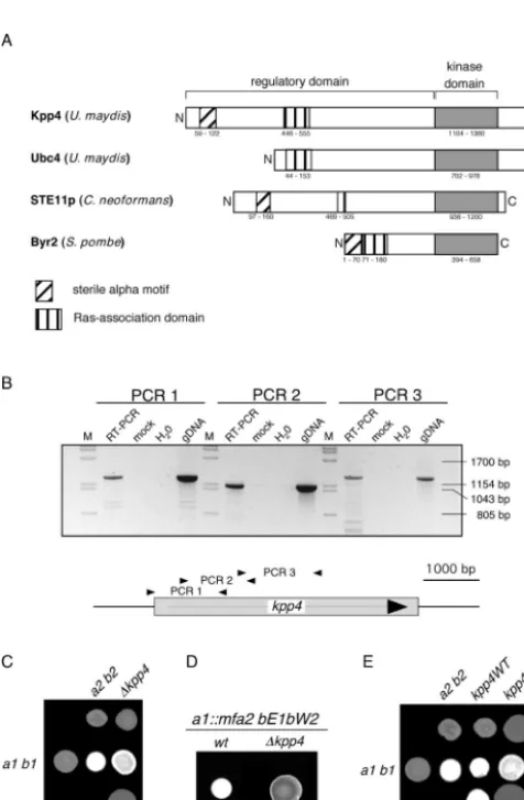
N-terminal 441 and 39 amino acids, respectively (except for a P794S substitution in Kpp4). Reverse transcription-PCR
anal-ysis ofkpp4revealed the absence of introns and placed the 5⬘
end of the mRNA upstream of position⫺100 (Fig. 1B and data
not shown). This reinforces the assertion thatkpp4codes for a
protein of 1,567 amino acids. Therefore, it is likely that the
start codon of the ubc4 ORF was wrongly assigned due to
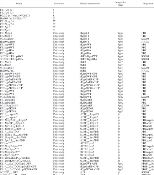
sequencing errors. Inspection of the N-terminal region of Kpp4 with ISREC (http://hits.isb-sib.ch/cgi-bin/hits_motifscan) identified a sterile-alpha motif (SAM) domain which is not present in Ubc4 but can be found in other fungal MAPKKKs,
such as Ste11p of Cryptococcus neoformans and Byr2 of S.
pombe (Fig. 1A). This domain is thought to mediate both homo- and hetero-oligomerization and plays a role in signaling (39, 44). In addition, a Ras association (RA) domain, which is
on September 8, 2020 by guest
http://ec.asm.org/
known to interact with small G proteins (40), could be identi-fied between positions 446 and 555 (Fig. 1A). Despite the substantial differences in the N-terminal domain, we
neverthe-less considerkpp4andubc4to represent the same gene.
Deletion of kpp4 attenuates mating, impairs conjugation
tube formation, and abolishes pathogenic development. The
similarity of Ubc4 to Byr2 ofS.pombeand Ste11p ofS.
cer-evisiaeled Andrews et al. to propose a function ofubc4during the mating process besides suppressing filamentous growth of
auac1deletion mutant (1). To address this question, we have
generated deletion mutations of kpp4 in compatible haploid
strains FB1 (a1 b1) and FB2 (a2 b2) as well as in the haploid
solopathogenic strain SG200 (a1::mfa2 bE1bW2).
In plate mating assays, successful fusion of compatible strains results in the formation of the filamentous dikaryon, which appears as white fuzziness on charcoal plates (Fig. 1C). In this assay we observed no significant reduction in dikaryon
formation of kpp4 deletion strains crossed with compatible
wild-type strains (Fig. 1C). This illustrates that deletion ofkpp4
does not lead to sterility. However, a mixture of two compat-ible⌬kpp4strains failed to develop fuzzy filaments (Fig. 1C). To investigate the mating process in more detail, FB1 and
FB1⌬kpp4 were stimulated with synthetic a2 pheromone.
While the wild-type strain formed conjugation tubes, thekpp4
deletion strain showed no response (Table 2). This demon-strates that the MAPKKK Kpp4 is essential for this
phero-mone-specific change in morphology, as was shown for fuz7
and kpp2/ubc3, coding for a MAPKK and a MAPK,
respec-tively (Table 2) (4, 35, 37). Moreover, deletion of kpp4 in
SG200 (a1::mfa2 bE1bW2), which grows filamentously because
of activeaandbloci, resulted in strongly attenuated filament
formation (Fig. 1D), as described for SG200⌬kpp2-1 and for
diploid⌬fuz7/⌬fuz7 strains (4, 37). This illustrates thatkpp4
affects postfusion processes such as filament formation.
To assay the function of kpp4during pathogenic
develop-ment, corn plants were infected with mixtures of compatible
⌬kpp4mutants or with wild-type strains as a control. We
ob-served tumors in 90% of plants infected with wild-type strains,
whilekpp4deletion strains failed to induce tumors (155 plants
were tested) (Table 3). To exclude the possibility that this
outcome results from cell fusion defects of⌬kpp4strains, we
also performed plant infections with the haploid
solopatho-genic strain SG200 (a1::mfa2 bE1bW2), which induces tumors
without prior fusion (Table 3). Its derivative SG200⌬kpp4 was
FIG. 1. Deletion of kpp4 affects mating and filamentous growth. (A) Schematic representation of Kpp4 domain structure. Domains were identified with ISREC, and domain annotations are PS50105 (SAM do-main), PS50200 (RA dodo-main), and PS500011 (kinase domain). Accession numbers are AAN63948 for Kpp4 (1,567 amino acids), AAF86841 for Ubc4 (1,166 amino acids), AAG30205 for Ste11p ofC. neoformans(1,230 amino acids), and P28829 for Bry2 ofS. pombe(689 amino acids). Inter-estingly, at bp⫹4438 of thekpp4ORF we found hexanucleotide repeats (GCTGCG) which encode an alanine stretch and are present in six copies in FB1 (a1 b1) and FB2 (a2 b2) but only in five copies in RK32 (a1 b3). (B) Reverse transcription-PCR analysis of kpp4. RNA isolated from AB33 was reverse transcribed and then subjected to three different PCRs, i.e., PCR 1 with primers kpp4-550 and OPM45, PCR 2 with primers OPM46 and OPM41, and PCR 3 with primers OPM40 and OPM21, amplifying the regions between positions ⫺99 and ⫹1172, ⫹547 and ⫹1695, and⫹1566 and⫹2872 of thekpp4ORF, respectively. As controls, we performed reactions without reverse transcriptase (mock) or with water or genomic DNA (gDNA) as the template. Lanes M, molecular size markers. (C) The strains indicated on the top were spotted alone and in combinations with the strains indicated on the left on charcoal-containing PD plates. Dikaryotic filaments appear as white fuzziness. (D) The strains indicated the on top were spotted on charcoal-containing PD plates. SG200 developed filaments characterized by white fuzziness, while SG200⌬kpp4 deletion strains are severely affected in filamentation. wt, wild type. (E) The strains indicated on the top were spotted alone (top row) and in combinations with the wild type (middle row) on charcoal-containing PD plates. In the bottom row compatible combinations of eitherkpp4WT(left) orkpp4RA(right) strains were cospotted.
TABLE 2. Conjugation tube formationa
Strain No. of cells % Conjugationtubes
FB1 506 80
FB1⌬kpp4 503 0
FB1kpp4WT 340 96
FB1kpp4RA 318 19
FB1⌬fuz7 508 0
FB1⌬kpp2-1 546 0
FB1⌬prf1 550 0
FB1kpp2AEF 532 0
FB1kpp2K50R 541 0
aCells were exposed to synthetic a2 pheromone, and conjugation tube
forma-tion was determined by microscopic observaforma-tion.
on September 8, 2020 by guest
http://ec.asm.org/
unable to induce disease symptoms (Table 3), demonstrating
an essential function ofkpp4during pathogenic development.
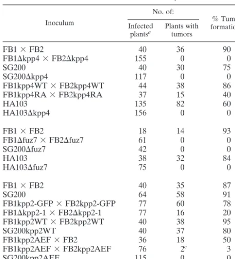
The RA domain of Kpp4 is important for function.To
elu-cidate the role of the RA domain (Fig. 1A) in Kpp4 function,
we introduced a K481E mutation intokpp4and replaced the
endogenous allele in FB1 and FB2 withPtef1:kpp4K481Eas well as withPtef1:kpp4WTas a control. Both alleles were expressed
from the constitutive tef1 promoter (47). The corresponding
mutation in the RA domain ofS.pombeByr2p was shown to
abolish Ras1 binding (52). In a plate mating assay, strains
carryingPtef1:kpp4WT(FB1kpp4WT and FB2kpp4WT) were
indistinguishable from wild-type strains (Fig. 1E), indicating
that expression ofkpp4from thetef1promoter does not
inter-fere with function. In addition, Western analysis with protein extracts from strains expressing either Kpp4 or
GFP-Kpp4RA fusion proteins under the control of the otef
pro-moter (47) revealed that the K481E substitution does not in-fluence Kpp4 protein stability (see Fig. 5A, left panel).
In contrast to the kpp4WTstrains, mixtures of compatible
Ptef1:kpp4K481Estrains (FB1kpp4RA and FB2kpp4RA) did not develop dikaryotic filaments, while the combination of FB2kpp4RA with the compatible wild-type strain FB1 dis-played only a slight reduction in the formation of dikaryotic
hyphae (Fig. 1E). The kpp4K481E strains thus resemblekpp4
deletion strains. To investigate whether conjugation tube
for-mation is also affected by kpp4K481E, we stimulated
FB1kpp4WT and FB1kpp4RA with synthetic a2 pheromone.
Only 59 out of 318 FB1kpp4RA cells (19%) responded to stimulation, whereas in the control, 96% of the cells developed tubes (Table 2). This result indicates that a functional RA domain in Kpp4 is necessary for an efficient response to pher-omone.
To assay the role of the RA domain during pathogenic growth, we infected corn plants with mixtures of FB1kpp4RA
and FB2kpp4RA strains, both carrying thekpp4K481Emutant
allele. While in control experiments with mixtures of FB1kpp4WT and FB2kpp4WT, 86% of infected plants showed
tumor formation, compatiblekpp4K481Estrains induced tumors
in only 40% of the infected plants (Table 3). Thus,kpp4K481E
strains are reduced in pathogenicity but differ from kpp4
de-letion mutants, which are completely impaired in pathogenic development. This suggests that a functional RA domain in Kpp4 is required for full virulence only.
kpp4,fuz7, andkpp2act in one cascade.To analyze whether
Kpp4 acts in one module with the known MAPKK Fuz7 and the MAPK Kpp2 we carried out genetic epitasis analyses. To this end we constructed constitutively active alleles ofkpp4by introducing mutations that were shown to confer constitutive
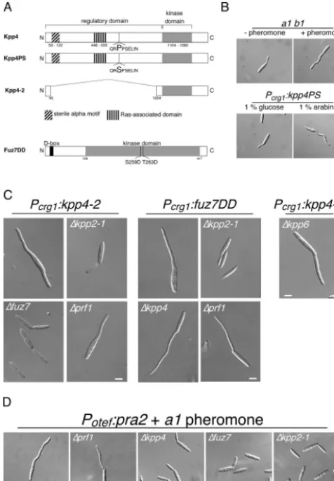
activity to STE11 of S. cerevisiae (15, 48). kpp4PS carries a
P681S substitution, and inkpp4-2the coding region for amino
acids 45 to 1055 of the presumed regulatory domain was de-leted (Fig. 2A). To generate a constitutively active allele of
fuz7(fuz7DD), we introduced two point mutations resulting in S259D and T263D substitutions, which likely mimic an acti-vated kinase. These alleles were placed under the control of
thecrg1promoter, which is repressed by glucose and induced
by arabinose (10), and were introduced in single copy into the
iplocus of FB1 (32). Under repressing conditions, strains
har-boringkpp4PS,kpp4-2, orfuz7DDwere morphologically indis-tinguishable from wild-type strains (Fig. 2B and data not shown). However, 4 h after transfer to arabinose-containing
medium, cells expressing either kpp4PS, kpp4-2, or fuz7DD
developed irregular filaments at one or both poles of the cell (Fig. 2B, lower panel, and C). These filamentous structures appeared to be curved and resembled conjugation tubes (Fig.
2B, upper panel). Moreover, in cells expressing kpp4PS,
kpp4-2, or fuz7DD, we could detect only one nucleus, which localized to the growing filament (not shown). These results show thatkpp4PS,kpp4-2, andfuz7DDinduce the formation of conjugation tube-like structures.
As shown in Fig. 2C, the MAPK cascade componentsfuz7
and kpp2 are essential for the morphological transition
in-duced bykpp4-2. The ability offuz7DDto trigger tube
forma-tion required kpp2 but was independent of kpp4 (Fig. 2C).
These results demonstrate thatfuz7acts downstream ofkpp4
and upstream ofkpp2. Unexpectedly, deletion ofprf1did not
abolish the morphological transition induced by eitherkpp4-2
orfuz7DD(Fig. 2C).
Prf1 is dispensable for conjugation tube formation. We
wondered whether a genetically activated MAPK cascade is
equivalent to a pheromone stimulus, since deletion of prf1
impairs conjugation tube formation (Table 2) but appeared to be dispensable for the morphological transition triggered by an activated cascade. Sinceprf1is required for the basal
transcrip-tion of theaandbgenes (22),prf1deletion mutants lack the
pheromone receptor and are blind to pheromone. Therefore,
we overexpressed the pheromone receptorpra2in FB2⌬prf1
TABLE 3. Plant infection assays
Inoculum
No. of:
% Tumor formationb
Infected
plantsa Plants withtumors
FB1⫻FB2 40 36 90
FB1⌬kpp4⫻FB2⌬kpp4 155 0 0
SG200 40 30 75
SG200⌬kpp4 117 0 0
FB1kpp4WT⫻FB2kpp4WT 44 38 86
FB1kpp4RA⫻FB2kpp4RA 37 15 40
HA103 135 82 60
HA103⌬kpp4 156 0 0
FB1⫻FB2 18 14 93
FB1⌬fuz7⫻FB2⌬fuz7 61 0 0
SG200⌬fuz7 42 0 0
HA103 38 32 84
HA103⌬fuz7 75 0 0
FB1⫻FB2 40 35 87
SG200 64 58 91
FB1kpp2-GFP⫻FB2kpp2-GFP 77 60 78
FB1⌬kpp2-1⫻FB2⌬kpp2-1 77 16 20
FB1kpp2WT⫻FB2kpp2WT 40 38 95
SG200kpp2WT 40 37 80
FB1kpp2AEF⫻FB2 36 18 50
FB1kpp2AEF⫻FB2kpp2AEF 76 2c 3
SG200kpp2AEF 115 0 0
aAll infections were performed twice with two independently generated
mu-tants.
bPercentage of plants that developed at least one tumor on a stem or leaf. cTumors observed were found on leaves only and did not extend 2 mm in
diameter. This is significantly different from infections with wild-type strains, where tumors develop on all green parts of the plant and reach diameters of up
to 50 mm.
on September 8, 2020 by guest
http://ec.asm.org/
by introducing the gene under the control of the constitutive
strong otef promoter in single copy into the ip locus. Upon
stimulation with synthetic a1 pheromone, this strain
(FB2pra2⌬prf1) formed conjugation tubes, while the
progen-itor strain FB2⌬prf1 did not react (Fig. 2D and Table 2). This shows that the inability of⌬prf1strains to develop conjugation
tubes is due to insufficient expression of the receptor gene. In contrast, overexpression ofpra2inkpp4,fuz7, orkpp2deletion strains did not rescue impaired conjugation tube formation, indicating that signaling via this cascade is crucial for this morphological response (Fig. 2D).
kpp4-2andfuz7DD induce pheromone gene expression.In previous experiments, conjugation tube formation and phero-mone-dependent gene expression always appeared to be linked. The finding that conjugation tubes can be formed in the absence of Prf1 led us to reinvestigate theprf1requirement for
mfa1 gene induction. We performed Northern analysis with
RNA isolated from FB2pra2 as well as FB2pra2⌬prf1 before
and 5 h after pheromone stimulation and observed thatprf1
was essential for pheromone-induced mfa2 gene expression
(Fig. 3A).
Next we analyzed pheromone gene transcription in strains
harboring eitherkpp4-2orfuz7DDbefore and after induction
FIG. 2. Constitutively active alleles ofkpp4andfuz7induce conju-gation tube-like structures. (A) Schematic representation of thekpp4
andfuz7alleles used in this study. Kpp4PS harbors a P681S substitu-tion in a conserved region, the so-called catalytic binding domain of STE11p-like kinases. In Kpp4-2 the regulatory domain is deleted. In Fuz7DD (accession number Q99078, 435 amino acids) two amino acid substitutions, S259D and T263D, were introduced in the conserved phosphorylation motif of MAPKK as described for, e.g., MEK1 (26). The D box (amino acids 13 to 21), described to be essential for MAPKK-MAPK interaction, is located in the N terminus of Fuz7 (6). (B) Expression of constitutively activekpp4PSinduces structures that resemble conjugation tubes. FB1 cells were treated with a2 pheromone (upper right panel), and FB1Pcrg1:kpp4PS was incubated with arabi-nose for 4 h (lower right panel). All pictures were taken with the same magnification. Bar, 10 m. (C) Kpp4, Fuz7, and Kpp2 act in one cascade. FB1 derivatives harboring eitherkpp4-2orfuz7DDare indi-cated on top and with the indiindi-cated gene deletions were used. Cell morphology was scored at 5 h after growth in arabinose-containing medium. All pictures were taken with the same magnification. Bar, 5 m. (D) Overexpression of the pheromone receptor rescues conjuga-tion tube formaconjuga-tion inprf1deletion strains. FB2 and all FB2-derived strains expressedpra2constitutively, carried the indicated gene dele-tions, and were stimulated with a1 pheromone. All pictures were taken with the same magnification. Bar, 5m.
FIG. 3. Constitutively activekpp4-2andfuz7DDtrigger pheromone gene expression. (A) Overexpression ofpra2does not rescue phero-mone gene expression inprf1deletion strains. The strains indicated on the top were treated for 5 h with synthetic a1 pheromone dissolved in DMSO (⫹) or with the same volume of DMSO (⫺). RNA was iso-lated, and 10g of total RNA was loaded per lane. The blot was probed with mfa2 and with rRNA as loading control. (B and C) Activation of the MAPK cascade elevates pheromone gene expression. FB1 and the FB1-derived strains indicated on the top were grown with glucose (⫺) or arabinose (⫹) as a carbon source. RNA was isolated, 15 g of total RNA were loaded per lane and the same filters were hybridized in succession with the probes indicated on the right.
on September 8, 2020 by guest
http://ec.asm.org/
of these alleles. Expression of either kpp4-2 orfuz7DD ele-vated pheromone gene transcription (Fig. 3B and C).
Further-more,mfa1induction bykpp4-2required the downstream
com-ponents fuz7 and kpp2, while the response to fuz7DD was
independent ofkpp4but dependent onkpp2(Fig. 3B and C).
These results demonstrate that components of the cascade which regulate the morphological response to pheromone are also required to observe pheromone-dependent gene
expres-sion. Prf1 was essential formfa1gene expression induced by
either kpp4-2 or fuz7DD (Fig. 3B and C), while it was not
needed for the morphological transition. Thus, signaling through this MAPK cascade appears to branch downstream of Kpp2.
MAPK cascade mutants are affected in
pheromone-respon-sive gene expression. To investigate pheromone-responsive
gene expression in⌬kpp4strains, we used synthetic a2
phero-mone. As shown in Fig. 4A, pheromone stimulation of a wild-type strain led to induced expression of theaandbgenes (pra,
mfaandbE,bW), as was shown before with compatible strains
(54). Analysis of the mutant strains revealed that in ⌬kpp4
mutants pheromone-inducedbgene expression was completely
abolished, while the induction of mfa1and pra1 was not
af-fected (Fig. 4A, lanes 3 and 4). A comparable response was observed in⌬fuz7strains (Fig. 4A, lanes 5 and 6). However, in
⌬kpp2-1mutants, pheromone induction ofmfa1,pra1, andb
was severely attenuated (37) (Fig. 4A, lanes 7 and 8). This indicates that upon pheromone stimulation the entire MAPK
module is required forbgene expression. In contrast,
induc-tion of genes in thealocus requires Kpp2, but the upstream
components, Kpp4 and Fuz7, are not needed.
We wondered whether this regulation of thealocus genes
might rely on Kpp2 kinase activity. To test this, we generated
two differentkpp2alleles,kpp2AEFandkpp2K50R. Kpp2AEF
contains two amino acid substitutions in the conserved phos-phate acceptor site (T182A and Y184F) and is therefore an
unphosphorylatable MAPK. The kpp2K50Rallele should
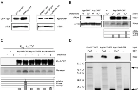
en-code a phosphorylatable but kinase-dead mutant protein due to a defect in ATP binding capacity. We assayed the kinase activity of Kpp2 and Kpp2K50R by mixing the respective pro-teins purified as GST fusions fromE.coliwith [␥-32P]ATP and MBP. These reactions were analyzed by SDS-PAGE for the presence of radioactively labeled MBP. As shown in Fig. 4B, GST-Kpp2 efficiently phosphorylated MBP, while the mutant
GST-Kpp2K50R did not show kinase activity. Thus,kpp2K50R
encodes a kinase-dead protein.
Next we constructed strains carrying kpp2AEF and
kpp2K50R as well as strains expressing GFP-tagged Kpp2,
Kpp2AEF, or Kpp2K50R by replacing the endogenous kpp2
allele in wild-type strains withkpp2AEF,kpp2K50R,
kpp2WT-GFP, kpp2AEF-GFP, or kpp2K50R-GFP (see Materials and
Methods). Western analysis revealed that kpp2WT-GFP,
kpp2AEF-GFP, andkpp2K50R-GFPare expressed to compa-rable levels (Fig. 5A, right panel). Moreover, the resulting
kpp2WT-GFPstrains showed no significant defect in mating or conjugation tube formation (not shown) or in pathogenic de-velopment (Table 3), demonstrating that the GFP moiety does not interfere with the function of Kpp2 in vivo.
Upon pheromone stimulation, FB1kpp2AEF and
FB1kpp2K50R failed to form conjugation tubes, indicating that phosphorylation as well as kinase activity of Kpp2 is es-sential for the morphological response (Table 2). When as-sayed for pheromone-induced gene expression, neither mutant
strain showed b gene expression (Fig. 4A, lanes 11 to 14).
However, the mutants differed with respect tomfa1induction:
while pheromone-responsive mfa1expression was attenuated
in FB1kpp2AEF (Fig. 4A, lanes 11 and 12), FB1kpp2K50R displayed a reduced basal level ofmfa1transcripts, while
pher-omone-inducedmfa1expression was comparable to the levels
seen in FB1 (Fig. 4A, lanes 13 and 14). These findings suggest
that the involvement of Kpp2 in the regulation ofalocus gene
expression is independent of its catalytic activity.
To elucidate whether Kpp2 kinase activity is required for
fuz7DD-induced pheromone gene expression, we introduced
Pcrg1:fuz7DDin FB1kpp2AEF and FB1kpp2K50R. After
ex-pression offuz7DD, both strains exhibited budding growth (not
shown) and did not show elevated expression ofaandblocus
genes (Fig. 4C). These results demonstrate that Fuz7DD can
trigger expression of the aandblocus genes, presumably by
increasing Kpp2 kinase activity.
FIG. 4. Pheromone signaling via the Kpp4/Fuz7/Kpp2 cascade af-fectsbgene expression. (A) FB1 and the FB1-derived strains indicated on the top were treated for 5 h with synthetic a2 pheromone dissolved in DMSO (⫹) or with DMSO (⫺). RNA was isolated, and 10g of total RNA was loaded per lane. Blots were hybridized with the probes indicated on the right. wt, wild type. (B)kpp2K50Rencodes a kinase-dead Kpp2 protein. The GST fusion proteins indicated at the top were purified fromE.coliand subjected to kinase assay with MBP as the substrate. The upper panel shows Coomassie blue staining, and the lower panel depicts the incorporated radioactive phosphate in MBP. (C) fuz7DD elevates b gene expression. FB1 and the FB1-derived strains indicated on the top were grown with glucose (⫺) or arabinose (⫹) as a carbon source. RNA was isolated, and 15g of total RNA was loaded per lane. The filter was hybridized in succession with the probes indicated on the right.
on September 8, 2020 by guest
http://ec.asm.org/
Taken together, our findings illustrate that genetic activation
of the MAPK module elevates transcription of genes in thea
locus as well as in theblocus. After pheromone stimulation,
this cascade is required only for pheromone-responsivebgene
expression and is dispensable for the induction of the genes located in thealocus.
Pheromone stimulation increases Kpp2 kinase activity.
MAPK activation results from phosphorylation on the threo-nine and the tyrosine residues in the TXY activation loop. With antibody raised against the phosphoepitope pTEpY of mammalian ERK1 and ERK2, we examined pheromone-stim-ulated phosphorylation of Kpp2. When protein extracts from
pheromone-treated kpp2WT strains were used, the
anti-pTEpY antibody reacted specifically with a protein of the size
expected for Kpp2 (41 kDa) (Fig. 5B, left panel), while no reaction was observed with proteins extracted from a strain expressing the unphosphorylatable derivative Kpp2AEF. This result suggested that Kpp2 is phosphorylated and activated upon pheromone stimulation. To assay the latter biochemi-cally, the catalytic activity of Kpp2 was monitored in an
immu-noprecipitation-kinase assay. Protein extracts from
FB2kpp2WT-GFP were prepared before and after treatment with pheromone. Kpp2WT-GFP was immunoprecipitated with anti-GFP antibody, and its ability to phosphorylate MBP was determined. In three independent experiments we observed a twofold induction of Kpp2 kinase activity after 40 min of stim-ulation (Fig. 5B). This indicates that Kpp2 becomes activated after pheromone stimulation.
FIG. 5. Pheromone as well asfuz7DDactivates Kpp2 kinase activity. (A) Expression levels ofkpp4andkpp2alleles constructed in this study. Left panel, Western analysis of protein extracts from SG200GFP-kpp4WT and SG200GFP-kpp4RA grown in CM-Glc by using anti-GFP antibody. In the lower panel the Western blot was stained with anti-␣-Tub antibody as a loading control. Right panel, protein extracts from FB1kpp2WT-GFP, FB1kpp2AEF-FB1kpp2WT-GFP, and FB1kpp2K50R-GFP grown in CM-Glc were analyzed with anti-GFP antibody. In the lower panel the Western blot was stained with anti-␣-Tub antibody as loading control. (B) Left panel, before and after pheromone stimulation, phosphorylation of Kpp2 was monitored with anti-pTEpY antibody. Extracts were prepared from FB1kpp2WT and FB1kpp2AEF 90 min after stimulation with synthetic a2 pheromone (90⬘) or after 90 min of DMSO treatment (0⬘). The upper panel shows a Western blot with anti-pTEpY antibody, and the lower panel shows a Western blot with anti-␣-Tub antibody as a loading control. Right panel, Kpp2 kinase activity in FB2 (wild type [wt]) and FB2kpp2WT-GFP was assayed by MBP phosphorylation at 0, 20, and 40 min after pheromone addition. The upper panel shows precipitated Kpp2WT-FB2kpp2WT-GFP detected with anti-GFP antibody. The middle panel shows MBP phosphorylation, and the lower panel summarizes relative kinase activity measured by quantification of incorporated phosphate in three independent experiments. Error bars indicate standard deviations. (C) FB1 and the FB1-derived strains indicated on the top were shifted to arabinose-containing medium. Extracts were prepared prior to (⫺) and 90 min after (⫹) the shift. The upper panel shows precipitated Kpp2-GFP derivatives detected with anti-GFP antibody. The middle panel illustrates MBP phosphorylation, and the lower panel depicts relative kinase activity measured by quantification of incorporated phosphate in three independent experiments. (D) To demonstrate Kpp2 interaction with Fuz7 in vitro, protein extracts were prepared from strains expressing eitherkpp2-GFP,
kpp2AEF-GFP, orkpp2K50R. These extracts were incubated with either GST-Fuz7 (Fuz7) or GST (⫺) bound to glutathione-Sepharose. The upper panel shows precipitated Kpp2-GFP (67 kDa) detected with anti-GFP antibody, and the lower panel illustrates GST fusion proteins (GST-Fuz7 [76 kDa] and GST [27 kDa]) bound to glutathione-Sepharose as detected by Coomassie blue staining. Experiments were performed twice with similar results.
on September 8, 2020 by guest
http://ec.asm.org/
Kpp2 is activated after expression offuz7DDand interacts
with Fuz7.To verify the activation of Kpp2 by constitutively
active fuz7DD, we replaced the endogenous kpp2 gene in
FB1Pcrg1:fuz7DD with eitherkpp2WT-GFP,kpp2AEF-GFP, or
kpp2K50R-GFP and performed immunoprecipitation kinase assays after 90 min of growth in arabinose-containing medium. As shown in Fig. 5C, the kinase activity of Kpp2WT-GFP
increased fourfold after expression offuz7DD(compare lanes
3 and 4). As expected, expression offuz7DDdid not enhance
the kinase activity of Kpp2AEF-GFP and Kpp2K50R (Fig. 5C, lanes 5 to 8). However, in all assays we detected more phos-phorylated MBP than in control experiments in which protein
extracts from FB1Pcrg1:fuz7DD lacking Kpp2-GFP were used
(Fig. 5C, compare lanes 1 and 2 with lanes 5 to 8). This might indicate that another kinase, whose activity was not altered by Fuz7DD, coimmunoprecipitated with Kpp2-GFP.
Interactions of MAPKs and regulatory proteins, e.g., MAPKKs or specific phosphatases, are known to be mediated by a so-called docking site (D box), which is defined by the consensus sequence (R/K)2-(x)2-6-L/I-x-L/I (19). Since such a D box is located in the N terminus of Fuz7 (Fig. 2A) (6), we tested the interaction of Fuz7 and Kpp2 with a GST pulldown assay. For this purpose, bacterially expressed GST and GST-Fuz7 fusion proteins were immobilized on glutathione-Sepharose beads and incubated with protein extracts prepared from either FB2kpp2WT-GFP, FB2kpp2AEF-GFP, or FB2kpp2K50R-GFP. After extensive washing, the beads were subjected to SDS-PAGE to examine for the presence of GST-Fuz7 and Kpp2 fusion proteins by Coomassie blue staining (Fig. 5D, lower panel) and by immunoblotting with anti-GFP antibody (Fig. 5D, upper panel), respectively. The results showed that Kpp2WT-GFP, Kpp2AEF-GFP, and Kpp2K50R-GFP inter-acted with GST-Fuz7 but not with GST alone (Fig. 5D).
Integrity of the MAPK module is essential for pathogenic
development.As described above, deletion ofkpp4abolishes
pathogenic development. In this respect⌬kpp4strains behave
like fuz7 deletion strains, which in our hands are
nonpatho-genic (Table 3). Furthermore, we did not observe disease
symptoms in corn plants infected with SG200⌬fuz7, which is
consistent with the described lack of tumor formation in dip-loid ⌬fuz7/⌬fuz7 strains (Table 3) (4). These results make it likely that Fuz7 and Kpp4 act in one cascade also during
pathogenic development. Since ⌬kpp4 and ⌬fuz7 strains are
impaired in bgene expression, we analyzed whether the
ob-served loss of tumor formation is simply due to insufficientb
gene expression. For this purpose, we introduced the deletion alleles into the haploid solopathogenic strain HA103 (a1 bcon), which expresses a bE1-bW2 heterodimer from constitutive promoters (22). HA103 as well as the resulting strains
HA103⌬kpp4 and HA103⌬fuz7 exhibited filamentous growth,
indicating functionality of the bE1-bW2 heterodimer (not
shown). However, plants infected with HA103⌬kpp4 or
HA103⌬fuz7 showed no disease symptoms, while the
progen-itor strain HA103 was able to induce plant tumors efficiently
(Table 3). This indicates that kpp4 and fuz7 are crucial for
pathogenic development and exert their function in parallel or downstream of the b heterodimer.
If Kpp2 acts downstream of Fuz7 and Kpp4 during
patho-genic development as it does during mating, kpp2 deletions
strains should be unable to cause disease. However,⌬kpp2-1
deletion strains are only reduced in pathogenicity (35, 37). This finding could reflect genetic redundancy on the level of the MAPK, as was observed in the yeast pheromone pathway. To analyze this possibility, we assayed the pathogenicities of
strains carrying either the inactive mutant allelekpp2AEFor
wild-type kpp2 as control. Interestingly, only 3% of plants
infected with mixtures of FB1kpp2AEF and FB2kpp2AEF showed small leaf tumors, while combinations of compatible
kpp2WT (FB1kpp2WT⫻ FB2kpp2WT) strains were able to induce tumors in 95% of the infected plants (Table 3). Since SG200kpp2AEF was also nonpathogenic, the pathogenicity
defect seen in mixtures of compatiblekpp2AEFmutant strains
cannot simply be a result of inefficient cell fusion (Table 3). In addition, mixtures of FB1kpp2AEF and FB2 induced tumor development in 50% of the infected plants, which makes it
unlikely thatkpp2AEFis a dominant-negative allele. Thus, the
unphosphorylatable allele of kpp2 (kpp2AEF) blocks disease
development much more efficiently than the deletion ofkpp2.
To address the question of which stage during pathogenesis
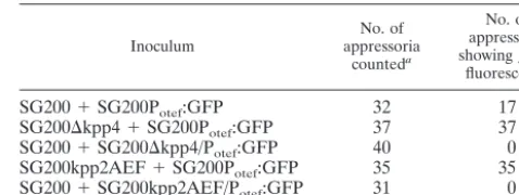
is affected in⌬kpp4, ⌬fuz7, orkpp2AEFmutants, we stained
fungal material on the plant surface with Calcofluor and Chlorazole Black E. The latter stain has recently been adapted for the visualization of infection structures ofU. maydis(11). On leaves prepared from plants infected with either SG200 or HA103, we observed vigorous filaments and formation of ap-pressoria (not shown). In contrast, after infections with either
SG200⌬kpp4, SG200kpp2AEF, HA103⌬kpp4, or HA103⌬fuz7,
only a small proportion of the inoculum developed filaments, and none of these formed appressoria (not shown). As we could not exclude the possibility that we might have overlooked some ap-pressoria, we directly examined development of appressoria by
coinfecting SG200 with either SG200⌬kpp4 or SG200kpp2AEF
(SG200 and derivatives cannot fuse and therefore allow for the simultaneous analysis of different strains). In order to distinguish between appressoria developed by SG200 and the mutant deriv-atives, we used combinations, in which one of the strains
ex-pressed GFP from the strong otefpromoter. While in control
infections with SG200 and SG200Potef:GFP approximately 50%
of Calcofluor-stained appressoria showed green fluorescence, in
combinations of SG200Potef:GFP and either SG200⌬kpp4 or
SG200kpp2AEF all stained infection structures derived from
SG200Potef:GFP (Table 4). When plants were infected with the
reciprocal combinations (SG200 with either SG200⌬kpp4/Potef:
GFP or SG200kpp2AEF/Potef:GFP), none of the appressoria
found showed GFP fluorescence (Table 4). These results show that integrity of the MAPK module is important for filament formation as well as for development of appressorial structures on the plant surface.
TABLE 4. Formation of appressoria
Inoculum appressoriaNo. of counteda
No. of appressoria showing green
fluorescence
SG200⫹SG200Potef:GFP 32 17
SG200⌬kpp4⫹SG200Potef:GFP 37 37 SG200⫹SG200⌬kpp4/Potef:GFP 40 0 SG200kpp2AEF⫹SG200Potef:GFP 35 35 SG200⫹SG200kpp2AEF/Potef:GFP 31 0
aAppressoria were counted on at least 10 different plants.
on September 8, 2020 by guest
http://ec.asm.org/
DISCUSSION
In this study we have demonstrated genetically as well as
biochemically that ubc4, fuz7, and ubc3/kpp2, which encode
components of a MAPK module, act in one cascade during mating and pathogenic development. This extends previous studies which demonstrated that the same three components are essential for the filamentous phenotype of mutants lacking adenylyl cyclase (1, 34, 35). The full characterization of the
MAPKKK gene kpp4shows that this gene is identical toubc4.
The deduced N terminus of Kpp4 contains a conserved protein-protein interaction domain termed the SAM domain (not present in Ubc4), which is a hallmark of other fungal MAPKKKs. In addition to the SAM domain (amino acids 59 to 122), Kpp4 harbors a putative RA domain (amino acids 446 to 555). These
domains are also present in the MAPKKK Byr2 ofS.pombeand
provide for a direct interaction with Ste4 and Ras1, respectively (52). Recently, twoU. maydisgenes,ras2andubc2, displaying homology to Ras1 and Ste4 ofS.pombe, respectively, were iden-tified. The respective deletion strains are impaired in conjugation tube formation and pathogenic growth, phenotypes that are also
associated with kpp4 deletion (30, 36). In this report we have
shown that strains expressingkpp4K481Eare attenuated in mating, conjugation tube formation, and pathogenic development. The
kpp4K481E allele carries a mutation analogous to Byr2FBR that prevents interaction with Ras1 and results in strongly reduced conjugation ofS.pombecells (52). Therefore, we consider it likely
that the regulation of Kpp4 is similar to Byr2 regulation in S.
pombeand involves interactions of Kpp4 with Ras2 via the RA domain and with Ubc2 via the respective SAM domains (Fig. 6).
Regulation of mating by the Kpp4/Fuz7/Kpp2 MAPK
mod-ule. Our data show that Kpp4, Fuz7, and Kpp2 act in one
module during mating. Structures resembling conjugation
tubes are induced by a constitutively active allele of kpp4
(kpp4-2), and this is dependent on the downstream
compo-nents Fuz7 and Kpp2. The same morphological switch is
trig-gered by a constitutively active allele of fuz7 (fuz7DD), and
here it requireskpp2but notkpp4. These genetic experiments
are supported by biochemical data. The kinase activity of Kpp2
is increased after expression of fuz7DD, and this enhanced
activity requires the conserved phosphate-acceptor sites (TEY) in Kpp2. In addition, Fuz7 interacts with Kpp2 in vitro as shown by a GST pulldown assay. On these grounds, we assume that Fuz7 activates Kpp2 in vivo (Fig. 6A).
Several observations suggest that the Kpp4/Fuz7/Kpp2 mod-ule is directly involved in transmitting the pheromone signal
(Fig. 6A). Disruption ofkpp4 attenuates mating and impairs
conjugation tube formation, phenotypes that were described before forfuz7andkpp2deletion strains (4, 35, 37). In none of these deletion strains does overexpression of the pheromone receptor rescue conjugation tube formation, indicating that all three components are required for transmitting the signal.
Activation of the cascade by eitherkpp4-2orfuz7DDinduces
conjugation tube-like structures, and concomitantly a and b
gene expression is increased. Finally, upon pheromone stimu-lation, Kpp2 was phosphorylated and its kinase activity was
shown to increase twofold. In comparable experiments withS.
cerevisiae, the MAPK activity of Kss1p as well as Fus3p was increased five- to sixfold (13, 42). It is presently not clear why
the increase of Kpp2 activity inU. maydisdoes not reach such
levels. One possibility might be the lower solubility or activity
of the lipopeptide pheromone applied toU. maydiscells
com-pared to the peptide pheromone used in the respective yeast experiments.
In line with the assertion that the Kpp4/Fuz7/Kpp2 module transmits the pheromone signal, we detected elevated levels of
bgene expression after expression offuz7DD, and this
induc-tion required the kinase activity of Kpp2. In the absence of
kpp4,fuz7, orkpp2, pheromone-inducedbgene expression was
prevented. However, with respect to pheromone-responsivea
gene expression the picture is different:mfa1induction is not
affected in kpp4 and fuz7 deletion strains (41) or in strains
FIG. 6. Proposed signaling processes during saprophytic growth and mating (A) as well as during the early steps of infection (B). The broken arrows indicate missing components or putative signaling events. See text for a detailed discussion.