PURIFICATION BY ION EXCHANGE CHROMATOGRAPHY AND ENZYME CHARACTERIZATION OF POTENTIAL DE-HAIRING ALKALINE PROTEASE FROM
BACILLUS CEREUS LS2B
Yendri Junaidi1, Ambar Partiwiningrum1, Yuny Erwanto1, Jamhari1, Lies Mira Yusiati1, Takashi
Hayakawa2, Tomoyuki Nakagawa2, Nanung Agus Fitriyanto1*
1Faculty of Animal Science, Gadjah Mada University, Jl. Fauna No. 3 Bulaksumur, Yogyakarta 55281,
Indonesia. 2Faculty of Applied Biological Sciences, Gifu University, 1-1 Yanagido, Gifu-Shi 501-1193,
Japan. E. mail: [email protected]
Article received 14.8.2017, Revised 3.6.2018, Accepted 9.6.2018
ABSTRACT
Objective: Effort to develop an environmental-friendly oriented method of animal leather tanning, especially de-hairing process, becomes a concern of this research. The aim was to purify the alkaline protease from Bacillus
cereus LS2B using a set of methods including ion exchange chromatography with DEAE Sepharose matrix. The
enzyme was considered for having a significant potential in the de-hairing process in tanning industries.
Method: The step of alkaline protease enzyme purifications including enzyme productions, ammonium precipitation, membrane dialysis and ion exchange chromatography was performed in this research. The total proteins and specific activity of every fractionation resulted from ion exchange chromatography were measured in this research. Identification of molecular weight by SDS-PAGE of each fraction and measurement of optimum pH and temperature of the purified enzyme was also described.
Result: The data showed that there was difference enzyme activity in every fraction obtained from the chromatography indicated the position of the enzyme. Alkaline protease from Bacillus cereus LS2B purification using ion exchange chromatography with DEAE Sepharose matrix has resulted in 35 fractions, with each fraction containing about 1.5 ml enzyme. The research was performed with a flow rate of 1.5 ml min-1. Each enzyme
fraction has different specific activity. The highest activity is shown at the 15th fraction that confirmed 64.4 U/mg.
The balance condition between protein concentration and specific activity was observed at the 21st fraction (45.5
U/mg). The 21st fraction was become interesting due to the total of enzyme protein was almost same as the total
enzyme activity. The correlation was considered becomes one of the indications of the effectiveness and efficiency of an enzyme purification. The result of SDS-PAGE determination showed that the 15th fraction has 3 bands of
protein enzyme with a molecular weight of 72 kDa, 20 kDa, and 13 kDa. The 21st fraction has smaller protein
bands, which are observed 20 kDa and 13 kDa.
Conclusion: Each fraction has dominant pure protein molecule around 20 kDa. Moreover, it is assumed that the
molecular weight of alkaline protease enzyme specific protein of Bacillus cereus LS2B is 20 kDa. Optimal
temperature and pH of the purified enzyme were 40°C and pH 8, respectively.
Keywords: Alkaline Protease, Bacillus cereus LS2B, Ion Exchange Chromatography, SDS-PAGE
INTRODUCTION
The leather industry faces challenges all over the world because of pollution-related problems, though it has been contributing significantly to economic development and employment and exp-ort potentials in many developing countries such as Indonesia. Leather processing involves four different unit operations: pre-tanning or beam house, tanning, post-tanning or wet finishing and finishing operations. The pollution is generated in various form from every unit operations, and the pre-tanning contribute the most in some total dissolved solids, chemical and biochemical
oxy-gen demand (Sundararajan et al., 2011; Bhat.,
2011). The main objective of liming is to remove hair and flesh. It also will open up the fiber struc-ture to get the excellent properties of the final lea-ther. Hence, liming is the most important pre-tanning operation in the leather processing. The conventional liming process involves the applicat-
ion of lime and sodium sulfide (Na2SO4) which results in the destruction of the skin hair and other proteinous materials impact in high pollution load regarding the biochemical oxygen demand (BOD) and chemical oxygen demand (COD). Moreover, sulfide is more toxic, and a significant amount of lime used to create a huge number of solid waste (Kanagaraj et al., 2006; Qureshi et al., 2009). Fur-thermore, to reduce the impact of tanning proce-sses on the environment, many researchers on this subject have been actively involved in the obser-vation and development of enzyme based de-hair-ing process; defleshde-hair-ing and fiber opende-hair-ing proces-ses to replace the conventional procesproces-ses using lime and sodium sulfide (George et al., 2014).
(Mothe and Sultanpuram., 2016). The critical role of the enzyme has a positive correlation with the level of enzyme production and catalysis ability of enzyme. Enzyme production, especially alkaline protease enzyme, can be performed by using the metabolism process of living things, such as bact-eria. Bacteria have a significant advantage comp-ared with animals, fungi, and plants in enzyme production. One of the bacteria types which can produce alkaline protease enzyme is B. cereus LS 2B ability is isolated from odorous farm soil at the tropical area (Fitriyanto et al., 2004). The enzyme
produced by Bacillus cereus LS2B must have
high catalysis ability if used as a commercial stan- dard. The catalysis ability of enzyme heavily depends on the level of enzyme purity. The level of catalysis power or specific enzyme activity is partly determined by the level of purity and char-acter of enzyme protein (Miyaji et al., 2005). The presence of a non-enzyme component in a crude enzyme strongly influences volume, purity, stabi-lity and catalysis power on the substrate. The usage of ion exchange chromatography method with sepharose matrix is a possible purification method.
Many researchers are able to purify enzyme using ion exchange chromatography method, incl-uding purification of the alkaline protease of Bacillus sp. HR-08 which was successfully puri-fied 20.2 times from crude enzyme using DEAE sepharose (Moradian et al., 2009). The ability of column ion exchange chromatography, an espec-ially with DEAE sepharose matrix in purifying enzyme can’t be separated from enzyme protein content and matrix content. The electrostatic inter-action of various types of charged ligand in a mat-rix with a group which can ionize in protein pro-duces to separation mechanism. Exchange of posi-tively charged anion binds an acid molecule, while the exchange of negatively charged cation produces separation mechanism for wet molecule (Wu et al., 2013). Based on various references on ion exchange chromatography with a column fil-led with sepharose matrix in purifying enzyme, the purified alkaline protease enzyme from B. cereus LS2B is expected to have increased acti-vity and show profiles of alkaline protease speci-fic protein bands from data of molecular weight confirmation of protein using SDS-PAGE.
MATERIALS AND METHODS
Culture condition and enzyme production: Enzyme production was performed using a modi-fied method from Moradian et al., 2009. The pre-liminary procedure was started by making the stock solution medium using 1 g of meat extract, 1
g of microbiological peptone, and 0.5 g NaCl with 70 ml of distilled water into a beaker glass, conti-nued by stirring into homogenous. The medium was then set to pH 7.2 followed by adjusted with distilled water to a volume of 100 ml. Solid agar medium is made from 1 ml of stock solution, 1.5 g agar and 99 ml of distilled water in 250 ml Erlen-meyer flask. Agar medium is heated with a hot plate stirrer, and then wait until quite cold and poured in a petri dish. Furthermore, when agar medium has hardened, by using ose, bacteria were streak and incubated for 24 hours. Once bacteria have grown, it stored in a refrigerator at 4°C. Liq-uid medium was made with a mixture of 90 ml of distilled water and 10 ml of stock solution contin-ued by sterilization by autoclaving at 121°C for 15 minutes, then cooled in the Laminar Air Flow (LAF). One ose of the pure isolate was grown on agar or cultivated by putting into 5 ml stock liquid medium and incubated on a shaker for 24 hours. The growth of bacteria was confirmed by observ-ing the turbidity of the medium (Moradian et al., 2009; Pouryafar et al., 2015)
To produce an alkaline protease enzyme, 3 ml of overnight-grown pre-culture bacteria were grown in a liquid medium having 100 ml of stock solution and 3 ml of skim milk. The pH of the medium was same as the pH of the stock solution. The inoculated medium was then incubated for 24 h at 30°C at 120 rpm in an orbital shaker. The crude enzyme was separated from bacterial cell by centrifugation at speed 3500 rpm for 15 min at 4 °C and continued by stored in refrigerator tempe-rature 4°C for further observation.
Precipitation using ammonium sulfate: The amount of 100 ml of cell-free extract was put into Erlenmeyer flask, and it was mixed with 80% ammonium sulfate slowly with stirrer and kept at 4°C condition for overnight. In order to separate the precipitated protein enzyme, it was centrifu- ged at 3500 rpm for 30 minutes at 4°C. The comp-onents separated during the centrifugation proc-ess, i.e. pellet and supernatant were separated. The pellet was considered as protein enzyme and sup-ernatant was discarded. Membrane dialysis pro-cess was performed to remove ammonium sulfate content in the component of protein enzyme (Moradian et al., 2009; Ghosh et al., 2008; Abidi et al., 2007).
that contained 0.58 g NaCl, 1.38 g NaH2PO4, and 1.78g Na2HPO4 in 1000 ml distilled water. Enz-yme sample was put inside dialysis membrane sack (cut off membrane <10 kDa) and diluted by dialysis buffer pH 8 for 5 times of dilution (enz-yme 1: 4 buffer). Dialysis process started by inserting dialysis membrane sack containing the enzyme and buffer into 1000 ml of dialysis buffer solution at 4°C while stirring using stirrer at low speed. In every 4 hours, buffer solution outside of membrane sack was replaced. It was replaced three times, and at the last replacement, the sam-ple was dialyzed overnight (Li et al., 2016; Papagianni and Sergelidis., 2014).
Molecular fractionation of enzyme protein using Ion exchange chromatography column: Alkaline protease from Bacillus cereus LS2B that has been dialysised was then purified using ion exchange chromatography column with DEAE. Sepharose matrix. The volume of the column was 25 ml, with dimension 14.5 cm in length and diameter 1.5 cm. The preliminary implementation stage was calibrating column by feeding it with 0.05 M pH 8 of phosphate buffer for one night. Enzyme purification was performed in three sta-ges. In stage 1, the column was fed to pH 8 equi-libration buffer with mixture (KH2PO4 0.1 N and NaOH 0.2 N) 4 times of column volume with 1.5 ml min-1 flow rate. The amount of fraction in one running was 52.5 ml, which was stored in 35 tu-bes. It was followed by feeding enzyme sample to a number of column volumes. After all enzyme samples had been saved in the column, it was fed with elution buffer, which consisted of a mixture of pH 8 equilibration buffer (KH2PO4 0.1 N and NaOH 0.2 N) and 4 times the column volume 0.5 N NaCl. Every released elution after being fed with elution buffer was stored in Eppendorf tube and labeled according to the order of elution (Mothe and Sultanpuram., 2016; Maruthiah el al., 2013; Joo and Chang 2005). The processing pro-duced 52.5 ml elution per running with a total of 35 fractions. Every elution fraction kept was sto-red in the freezer if not immediately used. The fractions were then measured for a specific acti-vity, total protein, and protein profile.
Measuring alkaline protease enzyme activity of every fraction: The activity of enzyme was meas-ured from each fraction using modified method of Bergmeyer, 1983 (Bergmeyer et al., 1983). The test stage included measurements of blank, stand-ard tyrosine and sample. Blank was measured by mixing 0.5 ml of pH 7 buffer added with 0.5 ml of casein and put into a test tube and incubated at 37°C for 10 minutes. 1.0ml of 10% TCA and 0.75 ml of enzyme were added, incubated for 10
minutes at room temperature and centrifuged for 15 min. 0.75 ml of supernatant was collected and added with 2.5 ml of Na2CO3 (0.5M), 0.5 ml of folin reagent and allowed to stand for 15 min at room temperature; then absorbance reading was performed at 578 nm. Standard tyrosine measure-ment was carried out by mixing 0.5 ml of pH 7 buffer, 0.5 ml of casein and 1 ml of tyrosine and inserting them into a test tube. It was incubated at 37°C for 10 minutes, then added with 1 ml of 10% TCA, incubated for 10 minutes at room tempera-ture and then centrifuged for 15 min. 0.75 ml of supernatant was collected, added with 2.5 ml of Na2CO3 (0.5 M), 0.5 ml of Folin reagent and allo-wed to stand for 15 minutes at room temperature then absorbance reading was measured at 578 nm. The sample was measured by mixing 0.5 ml of pH 7 buffer, and 0.5 ml of casein for each fraction and 1.0 ml of the enzyme from each fraction was put into test tube labeled according to fraction order. It was incubated at 37°C for 10 minutes, then added with 1.0ml of 10% TCA, incubated for 10 minutes at room temperature. Each fraction was then put into Eppendorf tube and centrifuged for 15 min. 0.75 ml of supernatant was collected, added with 2.5 ml of Na2CO3 (0.5 M), 0.5 ml of Folin reagent and allowed to stand for 15 min at room temperature. Absorbance reading was then performed at 578 nm.
Measuring total alkaline protease protein of each fraction: Total enzyme protein was
measu-red using the Lowry’s method (Lowry et al.,
1951) using BSA (Bovine Serum Albumin) as a standard protein. The total protein was measured using 0.2 ml of the enzyme from each fraction with 1.0 ml of biuret reagent in each enzyme frac-tion, then incubating it at room temperature for 10 minutes. Afterward, 0.1 ml of reagent E (1:1 of Folin to distilled water) was mixed until it was homogenous in each fraction and incubated at room temperature for 30 minutes. Then, the absor-bance level of each sample fraction was measured using spectrophotometry with wavelength λ = 750 nm, and then total protein was calculated.
Analysis of profile of enzyme protein of each fraction using SDS-PAGE: Sodium dedocyl sulfate polyacrylamide gel electrophoresis (SDS-PAGE) was performed according to the Laemmli method (1970). The relative molecular weight of the purified protein was estimated by comparing its mobility to standard protein size marker (Bio-Rad., 2014)
of enzyme incubation time. Maximum pH was determined by mixing enzyme with 0.2M phos-phate buffer. The pH ranges used were 6-12. Phosphate buffer was first made with pH 8. Low pH 6 and 7 were made by adding 0.1 M HCL, and high pH 9-12 were made by adding 0.1 M NaOH to phosphate buffer. The enzyme was mixed with a buffer of varying pH then stored at 37°C for 30 minutes, the activity of the enzyme was measured as reported modified method of Asker et al., (2013).
RESULT AND DISCUSSION
The result of ammonium precipitation and dia-lysis before purification with ion exchange chr-omatography column: Enzyme protein deposit-ion obtained considerable number of 10 ml with specific activity 0.5 (U/mg) than specific activity from the crude enzyme just with 0.4 (U/mg). For the release of salt residues from the ammonium sulfate was dialysis process with a semi-perme-able membrane (cellophane) pore 10 <kDa. An increase in specific activity was quite good from the dialysis is 1.8 (U/mg), so based on that protein enzymes successfully separated with residual salt and residues of other ions, thus the enzyme results of dialysis can be used for fractionation using ion exchange chromatography column.
Purification of alkaline protease enzyme resul-tedfrom ion exchange chromatography column with matrix filled with DEAE sepharose: Alka-line protease from Bacillus cereus LS2B is cate-gorized as an extracellular enzyme with very div-erse enzyme solution component. A non-protein component in enzyme indicates low enzyme spe-cific activity. Enzyme protein is a protein contain-ing anion or cation, so the enzyme protein should be purified using compound or matrix which can bond with the charge, such as DEAE sepharose. Purification using ion exchange chromatography exchanges anion and cation of DEAE Sepharose which binds negatively or positively charged protein based on the density of its protein content (Hosseini and Nasiri, 2015).
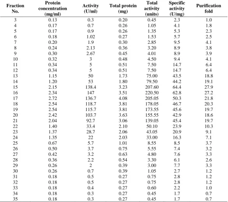
Based on the collected data (Table 1) alkaline
protease enzyme of Bacillus cereus LS2B was
successfully purified using ion exchange chroma-tography column with DEAE sepharose matrix. The results of ion exchange chromatography were 35 fractions where each enzyme fraction has diff-erent enzyme specific activity. Fractions 3-12 have low specific activity level from 2.3 (U/mg) to 14.7 (U/mg). Fractions 13-21 have very high specific activity level compared with other frac-tions, i.e. from 44.2 to 64.4 U/mg. Fractions 22-35 have significantly declined activity from 23.9 to
1.7 U/mg. The illustration of enzyme specific activity level shows the highest activity in the 15th fraction 64.4 U/mg with purity level up to 27.9 folds, the 16th fraction 62.8 U/mg with purity level up to 27.2 folds, and the 17th fraction 50.3 U/mg with purity level up to 21.8 folds. Increased purity to >20 folds was supported by data from various researchers such as Mothe and Sultanpuram (2016). Mothe and Sultanpuram (2016) who puri-fied alkaline protease enzyme of Bacillus caseiny-lticus using DEAE sepharose where the purifica-tion fold is 20.74. Purificapurifica-tion of alkaline protease
from Bacillus sp HR-08 using DEAE sepharose
resulted in 20.02 folds of purity level from crude enzyme (Moradian et al., 2009).
The data shows that there is great enzyme purification in the 15th fraction and the 21st fraction, where total protein in fraction 15 isn’t as high as fractions 16, 17, 18, 19 and 20 but has the highest specific activity of all fractions. It indicates that enzyme protein in the 15th fraction is pure alkaline protease protein which can perform catalysis process many times. Fractions 16-20 have the highest total protein but have specific activities below fraction 15. It’s interpreted that alkaline protease enzyme protein in the 16-20th fractions are still mixed with non-enzyme protein compounds or even bacterial cell protein, substr-ate protein and ammonium salt compound using in enzyme production, although dialysis has been performed. Non-enzyme protein compounds in enzyme component are a lipid, metal, phosphate and other apoenzyme parts, so the components support total enzyme protein captured by spectro-photometer wave so that the quantity of sample solution solid is higher (Zhang et al.,2016; Venka-tanagaraju and Divakar., 2015). The 21st fraction is kind of interesting to note because the total enzyme protein is almost the same as the total enzyme activity, so the correlation is considered an indication of effectiveness and efficiency of an enzyme of each protein unit in catalyzing a milli-gram of the substrate. This is supported by data of confirmation of molecular weight protein that the band formed in gel well row is fully clear.
Table 1: Summary of activity of alkaline protease enzyme of Bacillus cereus LS2B of each fraction
Confirmation of molecular weight of enzyme protein of each fraction using SDS-PAGE method: The SDS-PAGE result of alkaline pro-tease enzyme fraction of Bacillus cereus LS2B with ion exchange chromatography DEAE sepha-rose column separation shows different protein band profile. Band visualization of the first 11 fractions (Fig. 1) shows low protein density but with an entire protein band. Fractions 7 and 8 have 3 protein bands with molecular weights of 55 kDa, 20 kDa and 13 kDa, fractions 9-13 have 4
protein bands with molecular weight ranges of 72 kDa, 55 kDa, 20 kDa and 13 kDa. Visualization of protein bands of the second 11 fractions has a very high density (Fig. 2), dominated by protein bands with a molecular weight of 20 kDa. Fractions 14, 15, 16, 17, 18 19, 20 have protein with molecular weights of 72 kDa, 20 kDa and 13 kDa. Fractions 21-24 have declining whole enz-yme, protein band to 2 bands with molecular weights of 20 kDa and 13 kDa.
Fraction No.
Protein concentration
(mg/ml)
Activity (U/ml)
Total protein (mg)
Total activity
(units)
Specific activity (U/mg)
Purification fold
3 0.13 0.3 0.20 0.45 2.3 1.0
4 0.17 0.7 0.26 1.05 4.1 1.8
5 0.17 0.9 0.26 1.35 5.3 2.3
6 0.18 1.02 0.27 1.53 5.7 2.5
7 0.20 1.9 0.30 2.85 9.5 4.1
8 0.24 2.13 0.36 3.20 8.9 3.8
9 0.30 2.67 0.45 4.01 8.9 3.9
10 0.32 3 0.48 4.50 9.4 4.1
11 0.34 5 0.51 7.50 14.7 6.4
12 0.34 5 0.51 7.50 14.7 6.4
13 1.15 50 1.73 75.00 43.5 18.8
14 1.20 53 1.80 79.50 44.2 19.1
15 2.15 138.4 3.23 207.60 64.4 27.9
16 2.34 147 3.51 220.50 62.8 27.2
17 2.72 136.7 4.08 205.05 50.3 21.8
18 2.54 118.7 3.81 178.05 46.7 20.3
19 2.54 115.7 3.81 173.55 45.6 19.7
20 2.42 103.7 3.63 155.55 42.9 18.6
21 2.04 92.7 3.06 139.05 45.4 19.7
22 1.40 33.4 2.10 50.10 23.9 10.3
23 1.37 28.7 2.06 43.05 20.9 9.1
24 1.35 22 2.03 33.00 16.3 7.1
25 0.67 5.7 1.01 8.55 8.5 3.7
26 0.50 3.7 0.75 5.55 7.4 3.2
27 0.42 3.2 0.63 4.80 7.6 3.3
28 0.36 2.2 0.54 3.30 6.1 2.6
29 0.26 2 0.39 3.00 7.7 3.3
30 0.26 0.7 0.39 1.05 2.7 1.2
31 0.18 0.5 0.27 0.75 2.8 1.2
32 0.18 0.5 0.27 0.75 2.8 1.2
33 0.18 0.4 0.27 0.60 2.2 1.0
34 0.18 0.3 0.27 0.45 1.7 0.7
Figure 1: The profile of alkaline protease enzyme protein fraction from 3rd to the of ion exchange
chromatography. SDS-PAGE used 12% gel
concentration. M= Marker and number shows fractions
Figure 2: The profile of alkaline protease enzyme protein fractions from 14th to the 24th of ion exchange
chromatography. SDS-PAGE used 12% gel
concentration. M= Marker and number shows fractions
The 21st fraction is unique. The comparison of its enzyme specific activity and total enzyme pro-tein (Table 1) is equal, so the visualization of visi-ble protein band is also very clear, dominated by protein density with a molecular weight around 20 kDa. The statement is supported by Horikoshi (24) opinion that the efficiency of enzyme usage has correlation or equal proportion between enz-yme activity and enzenz-yme protein, so it can be assumed that every protein unit in enzyme has the same ability to catalyze one milligram of substrate compound. The visualization of protein bands of the last 10 fractions shows declining enzyme prot-ein band. There isn’t even any protprot-ein compound in enzyme solution, as shown in (Fig 3). In Fig. 5, the running enzyme sample is the 25th enzyme fraction. The lack of formed protein band indica-tes that there isn’t any protein compound.
Figure 3: The profile of alkaline protease enzyme protein fractions from of the 25th to the 35th of ion
exchange chromatography. SDS-PAGE used 12% gel
concentration. M= Marker and numbers shows
different fractions.
0 0.5 1 1.5 2 2.5 3 3.5 4 4.5
0 10 20 30 40 50 60 70
1 2 3 4 5 6 7 8 9 1011121314151617181920212223242526272829303132333435
Fractions
The protein band profile demonstrated by each fraction of fractions 3-35 shows that alkaline
protease enzyme of Bacillus cereus LS2B has
moderately good purification process using ion exchange chromatography column with DEAE sepharose matrix, where dominating protein band is found in molecular weight around 20 kDa. Miyaji et al., (2005) report that alkaline protease enzymes from Bacillus sp have the average mole-cular weight of 20-40 kDa. The resulting domina-ting protein band with molecular weight of 20 kDa is supported by the other study (Singh et al., 2012), who report that alkaline protease enzyme produced by Bacillus cereus SIU1 has a molecular weight of the protein of 22 kDa. Saxena and Singh (Saxena and Singh, 2015) report that the molecu-lar weight of alkaline protease enzyme protein
produced by Bacillus cereus B80 has a molecular
weight of 26 kDa. Prakash, Banik (Prakash et al., 2005) also report research data on the molecular
weight of alkaline protease enzyme from Bacillus
cereus, which is 28 kDa. The study of Venka-tanagaraju and Divakar (2015) finds the molecular weight of alkaline protease protein of Bacillus cereus GD55 which is a little different from the finding of this study. The molecular weight of alkaline protease protein of Bacillus cereus GD55 is 31 kDa.
Optimum temperature and pH of enzyme fraction with the highest activity resulted from ion exchange chromatography column: Temp-erature and pH are two of supporting factors of the catalysis ability of an enzyme, especially in the pure enzyme. At certain temperature and pH, the chemical reaction of the enzyme in catalyzing substrate can be very fast (high activity) and slow (low activity). The data of activity of alkaline pro-tease enzyme of pure Bacillus cereus LS2B at var-ious temperatures (Fig. 4) shows that the optimum temperature of alkaline protease enzyme of B. cereus LS2B is 40°C. It’s because the highest enz-yme activity is found at 40°C. The result is in line with the research result of Liu et al., (2010) who report that the optimum temperature of pure alka-line protease enzyme of Bacillus cereus MBL13 is 40°C. However, other researchers, such as Orhan et al., (2005), mention that the maximum tempe-rature of alkaline protease enzyme of Bacillus cereus is 60oC. However, alkaline protease enz-yme still has activity at 30, 50, 60 and 70°C although with a lower percentage than 40°C, so alkaline protease enzyme of Bacillus cereus LS2B can be called a thermostable enzyme. Orhan et al., (2005) state that the temperature range of alkaline protease enzyme produced by Bacillus cereus is
10-70°C. The optimum pH of alkaline protease enzyme of Bacillus cereus LS2B (Fig. 5) is pH 8. The result is in line with the research result of Orhan et al., (2005) who state that the optimum pH of pure alkaline protease enzyme of Bacillus cereus is pH 8.0. Liu et al., (2010) also report the same data, that the optimum pH of pure alkaline protease enzyme which can be isolated from Bacillus cereus MBL13 is pH8. Analysis of opti-mum pH of alkaline protease enzyme is also per-formed by Kim et al., (2001) who find that the optimum pH of alkaline protease enzyme of Bacillus cereus KCTC 3674 is pH 8.0. Although slightly different from the results of research (Margino, 2015) of the pure protease enzyme Bacillus sp. TBRSN- 1 with optimum pH of 7 and a temperature of 30°C.
Figure 4. The optimum temperature of purified
alkaline protease enzyme of Bacillus cereus LS2B
Figure 5. The optimum pH of purified alkaline
protease enzyme of Bacillus cereus LS2B
CONCLUSION
Alkaline protease from Bacillus cereus L2SB
has been purified through fractionation process using ion exchange chromatography column with
0 20 40 60 80 100 120 140
20 30 40 50 60 70
En
zy
m
e a
ctiv
ity
(U/m
l)
Teprature oC
0 20 40 60 80 100 120
6 7 8 9 10 11 12
En
zy
m
e a
ctiv
ity
(U/m
l)
DEAE sepharose matrix. The flow rate of the ion exchange chromatography tool is 1.5 ml min-1 with 35 fractions, each fraction containing 1.5 ml of enzyme. Dominating enzyme specific activity is found in the 15th fraction with a specific activity of 64.4 U/mg. The molecular weight of dominant protein and interpreted as alkaline protease protein of Bacillus cereus LS2B resulted from PAGE determination is 20kDa. The result of SDS-PAGE also shows that the cleanest enzyme prot-ein is the 21st fraction. The maximum temperature and pH of pure alkaline protease enzyme of Bacillus cereus LS2B are 40oC and pH 8.
ACKNOWLEDGEMENTS
This research was supported by Scholarship Indo-nesia Endowment Found for Education Ministry of Finance/ Lembaga Pengelola Dana Pendidikan (LPDP), with sponsor NO. S-1783/LPDP.3/2015 and Decree of awardees NO. 000699/Te/M/AF-2014. I gratefully thanks to the LPDP because for the full research fund.
REFERENCES
Abidi, F., Limam, F. and M.N. Marzouki, Purifi-cation and characterization of an alkaline protease prot 1 frombotrytis cinerea, Appl. Biochem. Biotechnol. 141: 361-376 (2007). Asker, M.M., Mahmoud, M.G., El Shebwy, K.
and M.S.A. el Aziz, Purification and charac-terization of two thermostable protease frac-tions from Bacillus megaterium, Journal of Genetic Engineering and Biotechnology 11: 103-109 (2013).
Bergmeyer, H., Bergmeyer, J. and M. Grassl, Enzymes 3: peptidases, proteinases and their inhibitors. Deerfield Beach: Verlag Chemie GmbH (1983)
Bhat, B.A., Cleaner Processing or Technology Vis-A-Vis Conventional Technologies, Jour-nal of Indian Leather Technologist Associa-tion, LXI, 729-747 (2011).
Bio-Rad. A guide to Polyacrylamide Gel Electro-phoresis and Detections, Bio Rad Labo-ratories Inc. Bulletin 6040 Rev B. Jerman (2014).
Fitriyanto, N.A., Oktaria, V., Erwanto, Y., Rusman, Hayakawa, T., Nakagawa, T. and K. Kawai, Isolation and Characterization of Prot-ease Producing Strain Bacillus cereus from Odorous Farm Soil in Tropical Area. in The 16th Asian Australasian Animal Production
Congress: The Asian-Australasian Associa-tion of Animal ProducAssocia-tion Societies (2004). George, N., Sondhi, S., Soni, S.K. and N.Gupta,
Lime and Sulphide-Free Dehairing of Animal Skin Using Collagenase-Free Alkaline
Prote-ase from Vibrio metschnikovii NG155, Indian J. Microbiol. 54: 139-42 (2014)
Ghosh, A., Chakrabarti, K. and D. Chattopadhyay, Degradation of raw feather by a novel high molecular weight extracellular protease from newly isolated Bacillus cereus DCUW, J. Ind. Microbiol. Biotechnol. 35: 825-834 (2008). Horikoshi, K., Alkaliphiles-genetic properties and
application of enzymes (2006).
Hosseini, K.M.and S. Nasiri, Preparation of factor VII concentrate using CNBr-activated Sepha-rose 4B immunoaffinity chromatography. Medical Journal of the Islamic Republic of Iran 29: 170 (2015).
Joo, H.S. and C.S. Chang, Oxidant and SDS‐sta-ble alkaline protease from a halo‐tolerant Bacillus clausii I‐52: enhanced production and simple purification. J. Appl. Microbiol. 98: 491-497 (2005).
Kanagaraj, J., Velappan, K.C., Babu, N.K.C. and S. Sadulla, Solid waste generation in leather industry and its utilization for cleaner envi-ronment-A review. Journal of Science & Industrial Research 65: 541-548 (2006). Kim, S., Kim, Y. and I.K. Rhee, Purification and
characterization of a novel extracellular prote-ase from Bacillus cereus KCTC 3674, Arch. Microbiol. 175: 458-461 (2001).
Laemmli U.K., Nature 227: 680-685. (1970) Li, F., Yang, L., Lv, X., Liu, D., Xia, H. and S.
Chen, Purification and characterization of a novel extracellular alkaline protease from Cellulomonas bogoriensis. Protein Expression Purif. 121: 125-132 (2016).
Liu, L., Ma, M., Cai, Z., Yang, X. and W. Wang, Purification and Properties of a Collagenolytic Protease Produced by Bacillus cereus MBL13 Strain, Food Technology & Biotechnology 48: (2010).
Lowry, O. H., Rosebrough, N. J., Farr, A. L. and R.J. Randall, Protein measurement with the Folin phenol reagent. J. Biol. Chem. 193: 265-275 (1951).
Margino, S., Purification and Characterization of Protease From Bacillus sp. TBRSN-1, Indone-sian Journal of Biotechnology 18: (2015) Maruthiah, T., Esakkiraj, P., Prabakaran, G.,
Palavesam, A. and G. Immanuel, Purification and characterization of moderately halophilic alkaline serine protease from marine Bacillus subtilis AP-MSU 6, Biocatalysis and Agricul-tural Biotechnology 2: 116-119 (2013). Miyaji, T., Otta, Y., Shibata, T., Mitsui, K.,
from Stenotrophomonas maltophilia strain S-1. Lett. Appl. Microbiol. 41: 253-7 (2005). Moradian, F., Khajeh, K., Naderi-Manesh, H. and
M. Sadeghizadeh, Isolation, purification and characterization of a surfactants, laundry dete-rgents-and organic solvents-resistant alkaline
protease from Bacillus sp. HR-08. Appl.
Biochem. Biotechnol. 159: 33-45 (2009). Mothe, T. and V.R. Sultanpuram, Production,
purification and characterization of a thermo-tolerant alkaline serine protease from a novel species Bacillus caseinilyticus. Biotech. 6: 1-10 (2016).
Orhan, E., Omay, D. and Y. Gvüenilir, Partial purification and characterization of protease enzyme from Bacillus subtilis and Bacillus cereus. Appl. Biochem. Biotechnol. 121: 183-194 (2005).
Papagianni, M. and D. Sergelidis, Purification and biochemical characterization of a novel alkal-ine protease produced by Penicillium nalgio-vense. Appl. Biochem. Biotechnol. 172: 3926-38 (2014).
Pouryafar, F., Najafpour, G.D. and M. Jahanshahi,
ComparativeStudies of Alkaline Protease
Pro-duction in Solid State Fermentation; Tray Biorector and Flask. Pak. J. Biotechnol. 12: 70-13 (2015)
Prakash, M., Banik, R.M. and C. Koch-Brandt, Purification and characterization of Bacillus cereus protease suitable for detergent indus-try, Appl. Biochem. Biotechnol. 127: 143-155 (2005).
Qureshi, S.A. and M.U. Dahot, Production of Pro-tease by Staphylococcus Epidermidis EFRL12 Using Cost Effective Substraten (Molasses) as
A Carbon Source. Pak. J. Biotechnol. 6: 55-50 (2009)
Saxena, R. and R. Singh, MALDI-TOF MS and CD spectral analysis for identification and structure prediction of a purified, novel, organic solvent stable, fibrinolytic
metallo-protease from Bacillus cereus B80. BioMed
Research International (2015).
Sattayasai, N., Protein purification, Chemical Bio-logy. Prof. Deniz Ekinci (Ed.) Pp.3-18 (2012). Singh, S. K., Singh, S. K., Tripathi, V.R. and S.K. Garg, Purification, characterization and seco-ndary structure elucidation of a detergent stable, halotolerant, thermoalkaline protease from Bacillus cereus SIU1. Process Biochem. 47: 1479-1487 (2012).
Sundararajan, S., Kannan, C.N. and S. Chittibabu,
Alkaline protease from Bacillus cereus
VITSN04: Potential application as a dehairing agent. J. Biosci. Bioeng. 111: 128-133 (2011). Venkatanagaraju, E. and G. Divakar, Production and Purification of Alkaline Fibrinolytic Enz-yme from Bacillus cereus GD55 under Solid State Fermentation and Screening for Indus-trial Applications, Current Trends in Biotech-nology and Pharmacy 9: 348-356 (2015). Wu, J., Gao, J., Chen, H., Liu, X., Cheng, W., Ma,
X. and P. Tong, Purification and characte-rization of polyphenol oxidase from Agaricus bisporus. Int. J. Food Prop. 16: 1483-1493 (2013).