Implementation of Fuzzy Cognitive Map and Support
Vector Machine for Classification of Oral Cancers
Anuradha.K
1,
Uma K P
21Associate Professor, Department of MCA, Karpagam College of Engineering, Coimbatore, Tamilnadu, India,
2 Professorand Head, Department of Mathematics, Hindusthan College of Engineering and Technology, Coimbatore,
Tamilnadu, India, [email protected]
Abstract
Objective: Tumors at any stage may progress into cancer. Till now, cancer classification is a challenging task for the researchers. Research on the cancer biology starts with the changes in the tissues. Oral cancer is a malignant cell growth in the oral cavity.
Method: The proposed work combines Fuzzy Cognitive map (FCM) with Support Vector Machine (SVM) for grading oral tumor. The histological features are used as concepts and the interrelationships between the concepts are identified. FCM acts as a classifier to distinguish between benign and malignant cases. Further, the extracted output from FCM is fed as an input to the Support Vector Machine (SVM) classifier. This will improve the prediction capabilities. Result: The classification accuracy obtained for the proposed model is 92.10% for malignant cases and 94.11% for benign cases.
Conclusion: The experimental results show that the combination of FCM with SVM obtained a good result when compared to the hybrid model using FCM.
Keywords: Oral cancer, Fuzzy Cognitive Map, Histological Features, Support Vector Machine
Received on 07 May 2018, accepted on 11 July 2018, published on 12 September 2018
Copyright © Anuradha.K et al., licensed to EAI. This is an open access article distributed under the terms of the Creative Commons Attribution licence (http://creativecommons.org/licenses/by/3.0/), which permits unlimited use, distribution and reproduction in any medium so long as the original work is properly cited.
3rd International Conference on Green, Intelligent Computing and Communication Systems - ICGICCS 2018, 18.5 - 19.5.2018, Hindusthan College of Engineering and Technology, India
doi: 10.4108/eai.12-9-2018.155559
1. Introduction
Cancer cells grow rapidly than normal cells. According to World Health Organization (WHO), grading system is classified to benign (tumor, low – grade) and malignant (cancer, high – grade). Among other cancers, oral cancer is the sixth commonest cancer that affects both men and women (Vigneshwaran et al, 2014). In most cases, oral tumors are identified using Morphological features. But some tumors does not cause harm to the humans. They do not need medication also and resolve by themselves.
Among the cancers present in the mouth, Oral Squamous Cell Carcinoma (OSCC) is the most frequent and harmful type of Oral cancer. It is one of the most important malignant neoplasms which arise from the mucosal epithelium of the oral cavity (Neville et al., 2005). Oral cancers are curable when detected at an earlier stage. There are various grading systems available to predict the clinical characteristics of OSCC. TNM (Tumor, Node, Metastatis) staging system is used to grade these cancers. For OSCC, the neoplasms were considered to identify stage I and II. The assessment of clinical staging and
on Energy Web and Information Technologies
Research Article
cytomorphology of the neoplasms serves as a good and accurate method for prediction. The features for this cancer are unusual and are not found in other carcinomas. So, it becomes complex to predict this type of cancer. For any cancer, tissue biopsy is the best method. Frederico Omar Gleber-Netto et al.,(2016) used saliva to predict OSCC.
Computer – based techniques have been used by experts to improve the diagnosis of cancers. These techniques grade the tumors into normal/abnormal and staging classifications.
The aim of this work is to develop a grading tool based on histological features of oral tumor which helps the experts to predict the disease. The eight histological features referred from Fischer grading system were taken as input (concepts) and a Fuzzy Cognitive Map is constructed. Fuzzy Cognitive Map is a model which represents the causal relationship between the concepts. From the map, the concepts that influence the others are taken and given as input to Support Vector Machine Classifier. This system is a combination of FCM and SVM. This model has been proposed by many researchers for medical decision making. The paper is structured as follows: Section 2 describes the background literature in oral cancer classification. Section 3 presents the methodology used for the proposed model. Discussion and results are presented in Section 4. Conclusion is discussed in section 5.
2. Background Literature
Previous efforts were made by researchers to classify oral cancers using various techniques. Many experts and researchers have worked on cancer classification (Anuradha and Sankaranarayanan, 2012). Few researchers (Papageorgiou et al 2006) used Fuzzy Cognitive map and support vector machines for the classification of bladder tumor grading. As several grading techniques are available, it becomes complex for oral tumor classifications.
Ankur Bhargava et al., (2010) reviewed the histopathological grading system of Oral Squamous Cell Carcinoma. Various grading systems were studied and compared (Ankur Bhargava et al., 2010).
Oral cancer can be graded using Lymph node metastatis. Akhter M et al., (2011) performed a detailed study on the histological grading of oral squamous cell carcinoma and its co-relationship with regional metastatis. They reviewed various grading systems and concluded that Anneroth's classification is less expensive and can be taken as a diagnostic approach for lymph node metastasis (Akhter et al., 2011).
A comparative study on four grading systems (Broder's, Jakobsson's, Anneroth and Hansen's, and Brynes) in oral squamous cell carcinoma was made by Jamadar.S et al., (2014). They evaluated the histopathological parameters to predict lymph node metastasis. They used 20 excisional biopsies for their work (Jamadar et al., 2014) and concluded that when the
whole tumor is considered, parameters like nuclear polymorphism (NP), and the pattern of invasion (POI) were sufficient to predict the Lymph Node Metastatis.
Few researchers used computer based techniques for oral cancer classification. Anuradha.K and Uma.K.P (2017) graded the tumors using histological features. They modelled Fuzzy Cognitive Map and represented the relationship between the concepts. Scores were calculated and graded for each tumor case. Fischer grading system was used for classification.
Papageorgiou et al., (2006) developed an advanced diagnostic tool to grade urinary tumors using Fuzzy Cognitive Map and Active Hebbian Learning algorithm.
3. Methodology
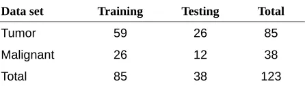
The proposed work uses 123 of oral tumor cases mostly in the age group 21 to 67. Using the current diagnostics tools available, 85 cases were diagnosed as normal and 38 cases were categorized as abnormal. Most of the abnormal cancer set was collected from Surya Dental Clinic, Coimbatore.
3.1. Fuzzy Cognitive Map
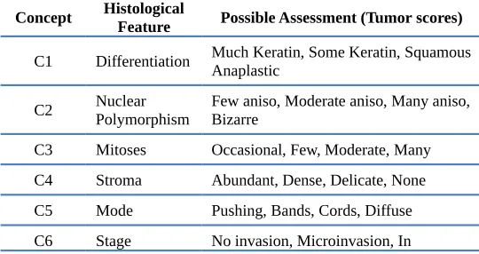
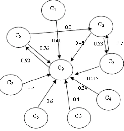
Fuzzy Cognitive Map was originally designed by Kosko, in 1986 (Kosko, 1986). A literature study reveals that a majority of FCM models were developed based on the experts knowledge (Aguilar, 2005). FCM can be applied to research and industrial areas for decision making. FCM is a symbolic representation which contains concepts, values and interrelationship between them. It is a combination of Fuzzy Logic and Neural Networks. Many researchers used Fuzzy Cognitive Maps for disease prediction and classification (Anuradha and Uma,2017; Papageorgiou et al., 2006; Nitin Bhatia and Sanheet Kumar, 2015; Papageorgiou et al., 2009; Papageorgiou et al., 2008; Papageorgiou et al.,2003) FCMs are used for future decision making if an initial state is given. Fischer's tumor grading system is used to construct the FCM (Fig 1). The grading system consists of eight concepts, C1 to C8 (Table 1):
Table 1. FCM for Oral Tumor Grading
Concept HistologicalFeature Possible Assessment (Tumor scores)
C1 Differentiation Much Keratin, Some Keratin, SquamousAnaplastic
connective tissue, Deep C7 Vascular None, Possible, Few, Many C8 Inflammatory response Marked, Moderate, Slight, None
C9 Degree of Tumor grade Low, high
The eight concepts represent the eight features of the tumor grading system. Concept C1 represents the differentiation, C2 represents Nuclear polymorphism, C3 represents Mitoses, C4 represents Stoma, C5 represents Mode, C6 represents Stage, C7 represents Vascular and C8 represents Inflammatory response. The ninth concept represents the degree of tumor grade. All the values are in the interval (0, 1). The threshold (0.5) decides which event is stimulated. The ninth concept C9 represents the degree of tumor grading (Anuradha and Uma, 2017). Histopathologists examined each tissue section and estimated the values of these variables. These values were converted between 0 and 1. These values were assigned to the corresponding concepts. The interrelationships among the FCM concepts indicate the cause and effect relationship. Consider Ci and Cj are the concepts with
values i,j= 1...8.
Figure 1. Fuzzy Cognitive Map for Oral Tumor grading
The degree of influence among the Concepts can be calculated using the IF...THEN conditions. The value (w) between the concepts indicates how strongly Ci influence
Cj.
If w > 0, there exists a positive causality between Ci and
Cj. That means, an increase in Ci will tend to increase the
value of Cj
When w< 0, it is a negative causality between Ci and Cj.
w=0, exhibits no relationships.
To develop a FCM, human expertise knowledge is required to decide the number of nodes and initial weights of FCM. The relationships between the concepts are described by experts using IF .... THEN rules. Here in Figure 1, there is no negative causality. But the increase in the concepts relies on the grading of the tumor (C9). If the value increases in C2 then there will be an increase in C3.
The value Ai of a Concept Ci, expresses a value
corresponding to its physical value allotted by the experts to a numerical value.
For each step, Ai of a Concepti is influenced by the values
of Concepts and the nodes connected to it.
The value for each concept is calculated using the equation given in (1).
Aj (t+1) = f (Aj(t) + Ai(t) ---(1)
where,
Aj (t+1) is value of concept Cj at step t+1,
Ai (t) is the value of concept Ci at step t, and Wij is the
weight of the arc from Concept Ci towards concept Cj and
f is a threshold function.
The initial weight matrix of the FCM is shown in Table 2:
Table 2. Initial Weight Calculation
Concept C1 C2 C3 C4 C5 C6 C7 C8 C9
C1 0 0 0 0 0 0 0 0 0.41 C2 0 0 0.53 0 0 0 0 0 0.48 C3 0 0.7 0 0 0 0 0 0 0.215 C4 0 0 0 0 0 0 0 0 0.34
C5 0 0 0 0 0 0 0 0 0.4
C6 0 0 0 0 0 0 0 0 0.6
C7 0 0 0 0 0 0 0 0 0.5
C8 0 0.3 0 0 0 0 0 0 0.76 C9 0 0 0 0 0 0 0 0.62 0
It is noted that Concept C8 (Table 2) influences more to grade the tumor.
3.2. Active Hebbian Learning
(2004) introduced Active Hebbian Learning algorithm to train FCM. Here this paper uses Active Hebbian Learning (AHL) algorithm which improves the result of FCM. AHL is a semi – automated learning algorithm, which requires the help from humans initially. The algorithm is based on Hebbian theory which can determine new FCM causal links. This will increase the classification capabilities of Support Vector Machines. The algorithms relate the values of concepts and values of weights in FCM and determine the sequence of activation. The sequence of activation contains more simulation steps. For each step, the active concepts influence other concepts till the iteration is over. After the implementation of AHL, the system starts to interact and the new adjusted weights are calculated. After few interactions, the value of Concept C9 for each case is calculated. The experts define the activation concepts, activated concepts, sequence of activation and the activation decision concept.
From Figure 1, it is seen that C8 is the activation concept; C2 and C3 are the activated concepts. All concepts together trigger the concept C9, which is the value to classify tumor/cancer. The activation value is calculated using (1).
3.3. Development of FCM and SVM
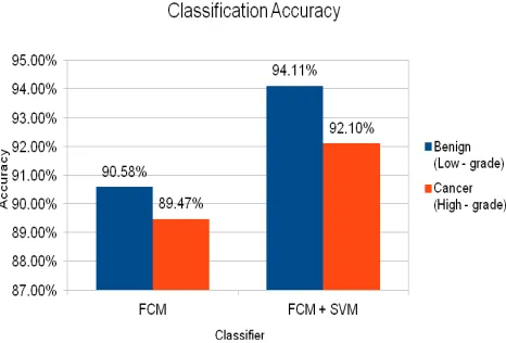
Support Vector Machine is a supervised learning algorithm used for classification. Papageorgiou. E et al., (2003) graded urinary bladder tumors using unsupervised learning algorithm for Fuzzy Cognitive Maps. They obtained a classification accuracy of 93.18% for high grade tumors and 90.59% for low grade tumors Anuradha.K and Uma.K.P (2017) developed a Fuzzy Cognitive Map to grade oral tumors. They achieved an accuracy of 90.58% for benign tumors and 89.47% for malignant cases. To improve further, another classifier SVM is used. The data set consists of 123 cases (85 tumor and 38 cancer).
For classification, this tool uses hold – out method. Two data sets (Training and Testing Set) are chosen randomly. As the data set is separated into two classes, Linear SVM is used.
Table 3. Data set for SVM classification
Data set Training Testing Total
Tumor 59 26 85
Malignant 26 12 38
Total 85 38 123
For training the SVM, 85 cases including 59 tumor cases and 26 cancer cases are used (Table 3). The remaining data are used for testing. The Hold – out method is used as it involves only a single run. So the time consumption is low.
Figure 2. FCM with SVM
As C9, C2 and C8 are more important for classification, they are used as input to Support Vector Machine. These features (concepts) are selected based on how they influence other concepts. Using the trained data and from the known class labels, the True Positives (TP), True Negatives (TN), False Positive (FP) and False Negative (FN) are obtained. The accuracy can be obtained using: Accuracy = Number of correct assessments / Total number of all assessments (2) i.e
AC = TN + TP / TN + TP + FP + FN (3)
where AC is the Classification Accuracy. Using the proposed method, the number of cancer cases correctly identified is 35 (out of 38) and normal cases are 80 (out of 85). In the previous work, [8] obtained an accuracy of 90.58% (low – grade) and 89.47% (high grade). Now to improve further, the combination of SVM and FCM is used.
4. Discussion and Results
Table 4. Classification Accuracy
Figure 3 shows the classification accuracy of the hybrid FCM model and the proposed FCM + SVM Model.
Figure 3. Classification Accuracy
5. Conclusion
In this paper, oral cancer is classified using Fuzzy Cognitive Map and Support Vector Machine. The data set consists of histological features more containing Oral Squamous Cell Carcinoma, Lichen Planus and Oral mucosa. The implementation of the tool is simple and can be used by the experts for classification. The Fuzzy Cognitive Map is constructed using the Histological features and the weights are calculated. To improve further, Active Hebbian Learning was used. Later, Support Vector Machine was used for classification. The classification rate obtained was 94.11% and 92.10% for low grade and high grade tumor respectively. The classification of oral cancer requires multiple iterations. In future, the model can be developed with minimum number of iterations. Also, Gray Level Co-occurrence Matrix features can be used to extract and classification can be obtained.
References
[1] Aguilar K. (2005) A survey about Fuzzy Cognitive Maps (Invited Paper), International Journal of Computational Cognition, 3(2): 27 – 33.
[2] Akhter M, Hossain S, Rahman QB, Molla MR. (2011) A study on histological grading of oral squamous cell
carcinoma and its co-relationship with regional metastasis. J Oral Maxillofac Pathol,15:168-176.
[3] Ankur Bhargava, Sonal Saigal, Monali Chalishazar. (2010). Histopathological Grading Systems In Oral Squamous Cell Carcinoma: A Review. Journal of International Oral Health, 2(4).
[4] Anuradha K. Sankaranarayanan K (2012). Identification of suspicious regions to detect oral cancers at an earlier stage – A literature survey,
International Journal of Advances in Engineering and Technology. 3(1): 84 – 91.
[5] Anuradha K, Uma KP. (2017). Histological grading of oral cancer using Fuzzy Cognitive Maps. Biomedical and Pharmacology Journal, 10(4). 1695 – 1700.
[6] Frederico Omar Gleber-Netto, Maha Yakob, Feng Li, Ziding Feng, Jianliang Dai, Huang-Kai Kao, Yu-Liang Chang, Kai-Ping Chang and David T.W. Wong.(2016). Salivary Biomarkers for Detection of Oral Squamous Cell Carcinoma in a Taiwanese Population, Clinical Cancer Research, 22(13), 3340 – 3347.
[7] Jamadar S, Narayan T V, Shreedhar B, Mohanty L, Shenoy S. (2014). Comparative study of various grading systems in oral squamous cell carcinoma and their value in predicting lymph node metastasis.
Indian J Dent Res, 25:357- 63.
[8] Kosko B.(1986). Fuzzy Cognitive Maps, Int. J. of Man – Machine studies, 24: 65 – 75.
[9] Neville, Damm, Allen, Bouqupt.(2005). Oral and Maxillofacial Pathol 2nd ed. India:Elsevier.
[10]Nitin Bhatia, Sanheet Kumar.(2015). Prediction of Severity of Diabetes Mellitus using Fuzzy Cognitive Maps, Advances in Life Sciences and Technology, 29:71 – 78.
[11]Papageorgiou EI, & Spyridonos P, Ravazoula P, & Stylios C, Groumpos P, Nikiforidis G.(2003). Grading Urinary Bladder Tumors Using Unsupervised Hebbian Algorithm for Fuzzy Cognitive Maps. Biomedical Soft Computing and Human Sciences, 9: 33-39.
[12]Papageorgiou EI, Stylios CD, Groumpos PP.(2004). Active Hebbian Learning algorithm to train Fuzzy Cognitive Maps, International Journal of Approximate Reasoning, 37: 219 – 249.
[13]Papageorgiou EI, Georgoulas G, Stylios CD, Nikiforidis G, Groumpos P.(2006) Combining Fuzzy Cognitive Maps with Support Vector Machines for Bladder Tumor Grading, Proceedings of the International Conference on Knowledge-Based and Intelligent Information and Engineering Systems, 515 – 523.
[14]Papageorgiou E I, Papandrianos N I, Apostolopoulos D J, Vassilakos P J. (2008).Fuzzy cognitive map based decision support system for thyroid diagnosis management, Proceedings of IEEE International Conference on Fuzzy Systems, 2008. FUZZ-IEEE (IEEE World Congress on Computational Intelligence). 1204-1211.
[15]Papageorgiou E I, Papandrianos N I, Karagianni G, Kyriazopoulos G C, Sfyras D A.(2009) fuzzy cognitive map based tool for prediction of infectious diseases, Proc. of IEEE International Conference on Fuzzy Systems. FUZZ-IEEE 2009. 2094-2099.
[16]Vigneshwaran N, Michelle D. Williams.(2014). Epidemiological Trends in Head and Neck Cancer and Aids in Diagnosis. Oral and Maxillofacial Surgery Clinics of North America. 26(2): 123-141.
Classification
method (Low - grade)Benign (High - grade)Cancer