Identification and Isolation of an Azoreductase from
Enterococcus faecium
*Corresponding author: Email: [email protected]
Horizon Scientific Press. http://www.horizonpress.com
Susan R. Macwana, Sumit Punj, John Cooper, Evan Schwenk and Gilbert H. John
Department of Microbiology and Molecular Genetics, Oklahoma State University, 418 LSE, Oklahoma State University Stillwater, OK 74075, USA
Abstract
Azo dyes are commonly used in many commercial industries. Some of the azo dyes can produce carcinogenic compounds after being metabolized by azoreductase. Several human intestinal microbiota possess azoreductase activity which plays an important role in the toxicity and mutagenicity of these azo dye compounds. The acpD gene product (AzoEf1) responsible for the azoreductase activity of Enterococcus faecium, an intestinal bacterium, was heterologously expressed, purified and characterized. The protein sequence shares 67% identity with the azoreductase from Enterococcus faecalis, AzoA. Although AzoEf1 possesses many commonalities with AzoA, there are differences in coenzyme preference, residues associated with FMN binding, substrate specificity, and specific activity. AzoEf1 utilized both NADH and NADPH for the reduction of azo dyes, and it contains a leucyl residue at position 104 and threonyl residue at position 19 which differ from AzoA at the active site. Its specific activity was 5095 µM/min/ mg and its catalytic efficiency for Methyl red reduction was lower than AzoA.
Introduction
An important function associated with intestinal bacteria is their ability to metabolize xenobiotics, such as azo dyes (Chung, KT. et al., 1978; Brown, JP. 1981; Cerniglia, CE. et al., 1982; Manning, BW., Cerniglia, CE. & Federle, TW., 1985). Azo dyes are characterized by the presence of one or more azo (R1-N=N-R2) bonds, and are widely used in the paper, textile, plastic, pharmaceutical, food, cosmetic, enamels, and drug industries (Collier et al., 1993; Dillon et al., 1994; Levine, 1991). The introduction of some of these dyes into the human body results in the reduction of their azo bonds and subsequent biotransformation to carcinogenic metabolites (Cerniglia et al., 1986; Bragger et al., 1997; Brown et al., 1983, Cerniglia, CE., 1982). Numerous in vivo and in vitro experiments support the toxicity of these metabolites (Morgan et al., 1984; Chung KT. 1983). Several intestinal bacteria are known to have azoreductase activity from culture studies (Chung K-T. et al., 1992b), thereby the isolation and characterization of azoreductase genes from these intestinal bacteria will provide information to their biochemical function within the cells and perhaps even allude to their evolutionary origin.
The azoreductase gene has been cloned, expressed and characterized from various intestinal organisms which include Enterococcus faecalis (Chen H. et al., 2004),
Escherichia coli (Nakanishi M. et al., 2001), Shigella (Ghosh, D.K. et al.), and Staphylococcus aureus (Chen, H. et al.). Interestingly, the properties of these azoreductases vary. For example, the azoreductase from E. coli is a 46 kDa homodimer, requires FMN as a cofactor, and uses NADH as an electron donor (Nakanishi M. et al., 2001); compared to Staphylococcus aureus which is a tetramer and utilizes NADPH for azo dye reduction (ref). In addition, the deduced protein sequences do not share significant sequence homology.
E. faecalis is a prominent organism found in the intestine, and the function of its azoreductase (AzoA) has been well characterized (Chen, H. et al., 2004, Punj, S. & John G.H., 2008b) including its structure, based on crystallization information (Liu, Z. & Chen, H. et al., 2007 add to reference). On the other hand, the azoreductase activity of E. faecium is not as well characterized. Although E. faecium is closely related to E. faecalis, there are some variations in the whole cell reduction of azo dyes (Punj, S. & John G.H. 2008a, Chung, K.T. et al. b), suggesting potential azoreductase differences in terms of catalytic and functional requirements. In addition, it has been shown that the orthologous azoreductases from several species of Bacillus possess different substrate specificities (Sugiura W, et al., 2006). Therefore, the present study focuses on characterizing the function of azoreductase from E. faecium, revealing catalytic differences between the closely related species, E. faecalis.
Methods
Bacterial strain, plasmids and culture conditions
Enterococcus faecium ATCC 6569 was grown in Brain Heart Infusion (BHI) broth supplemented with 0.05% (w/v) Vitamin K solution and 2% (w/v) yeast extract under anaerobic conditions at 37°C. The pCR2.1-TOPO (Invitrogen) plasmid was used for cloning the PCR fragment and pET-15b (Novagen) containing a hexa-histidine tag at the N-terminal was used for overexpression of the protein. For transformation, E. coli TOP10 (Invitrogen) competent cells and NovaBlue (DE3) (Novagen) cells were used. The E. coli strains were grown in Luria-Bertani (LB) medium containing 50μg ampicillin (Amp) ml-1 at 37°C.
3’contained the NdeI restriction site upstream of the start codon, and the reverse primer (AzoEf1r) 5 ’ C C C C A A A A A G A G G G AT C C G G G A A A ATAT C C 3’contained the BamHI site downstream of the stop codon. The PCR was performed in a Perkin Elmer DNA Thermal Cycler 480, and the conditions were 30 cycles: each cycle consisting of denaturing at 94ºC for 1 min, annealing at 55ºC for 1 min, extension at 72ºC for 1 min and a final extension at 72ºC for 7 mins. The PCR reaction mix contained sterile distilled water, 1X PCR buffer (Invitrogen), 1.5 mM Magnesium chloride (Invitrogen), 0.2 mM dNTP mix (Fischer Scientific), 10 ng genomic DNA, 50 pmol of forward and reverse primers, 2.5 Units Taq DNA polymerase (Invitrogen) in a final reaction volume of 100 μL. The amplified PCR product was run on a 0.8 % (w/v) agarose gel and the DNA eluted from the gel using the QIAquick® Gel Extraction kit (Qiagen).
Cloning and expression of AzoEf1
The amplified PCR product was directly cloned into the pCR2.1-TOPO vector which was subsequently transformed into E. coli TOP10 cells. The recombinant plasmid was extracted using the QIAprep® Spin Miniprep kit (Qiagen) and digested with the restriction enzymes NdeI and BamHI (Invitrogen) to release the insert. The released insert was eluted from a 0.8 % (w/v) agarose gel and the purified DNA was then ligated into the pET-15b vector, using a 1:6 vector to insert ratio. Ligation was carried out by T4 DNA ligase (Invitrogen) at 4ºC for 16 h. The recombinant plasmid, pAzoEf1 was transformed into NovaBlue (DE3) E. coli expression cells. The azoEf1 gene was sequenced from the pET-15b using the T7 promoter and terminator primers (Novagen).
Purification of the enzyme
NovaBlue (DE3) cells containing pAzoEf1 were inoculated in a LB/Amp (100 µg ml-1) medium and incubated overnight
at 37°C at 300 rpm. The cells were harvested, resuspended in 1 ml LB/Amp (100 µg ml-1) and transferred to 1 L of LB/
Amp (100 µg ml-1). The 1 L culture was incubated at 37°C
300 rpm. Upon reaching an OD600 = 0.6, the cells were
induced for 3 h with Isopropyl-1-thio-P-galactopyranoside (IPTG), final concentration of 1 mM. The induced culture was centrifuged at 6000 x g for 10 mins at 4°C to harvest the cells. The supernatant was discarded, and the pellet was frozen at -20°C.
The pellet was thawed, and resuspended in 10 ml lysis/ wash buffer (20% glycerol, 50 mM sodium monobasic phosphate, 750mM sodium chloride, 20mM Imidazole in double-distilled water pH, 8.0) Lysozyme (final concentration of 0.1 mg ml-1) was then added and the sample was incubated
on ice for 30 mins. The cells were disrupted by sonication using a Sonic 300 dismembrator with an intermediate tip and relative output of 60%. Sonication was performed 10 times for 5 sec on ice. The sample was centrifuged at 10,000 x g for 30 min at 4°C to remove cell debris. The supernatant was then subjected to batch purification using 50% Ni-NTA slurry (50% suspension in 30% ethanol, precharged with Ni2+).
four times and the protein was collected after each elution. All samples were stored at -20°C. The concentration of the protein was determined using a calibration curve using bovine serum albumin (BSA) as a standard curve.
SDS-PAGE
SDS-PAGE analysis was performed as previously described (Laemmli, 1970). A 1 ml culture (induced and noninduced) was collected and the cells were prepared for analysis by adding a final 1X concentration of SDS-PAGE loading buffer (4X=0.06 M Tris-HCL, pH 6.8, 10% (v/v) glycerol, 2% (w/v) SDS, 5% (w/v) 2β-mercaptoethanol, 0.0025% (w/v) bromphenol blue), boiling the sample for 15 min, and loading 20 µl onto a 12.5% SDS-PAGE gel to confirm the expression of the protein.
Enzyme assay
The assay was carried out in a polystyrene semi-micro cuvette (Sigma-Aldrich, St. Louis, MO) in a total reaction volume of 1 ml. The reaction mixture contained 25 mM potassium phosphate buffer (pH 7.0), 20 μM of azo dye and enzyme (0.5-1 μg). The reaction was initiated by the addition of 0.1 mM NAD(P)H. The extinction coefficient for Methyl Red at 430 nm was 23,360 M-1cm-1. The concentrations for the
remaining dyes in Table 1 were calculated from a calibration curve for each dye. The enzyme activity was measured by a decrease in absorbance over a 2 min period using a UV-visible spectrophotometer (UV-1601PC, Shimadzu). The enzyme activity was defined as the decrease in azo dye concentration (μM) per min ( For the control, the enzyme was denatured by boiling for 15 mins, followed by the addition of a few drops of hydrochloric acid (HCl).
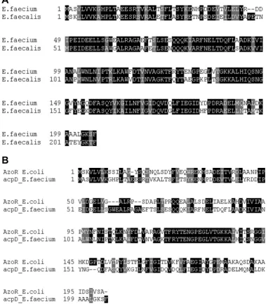
Fig. 1. Amino acid sequence homology. a) 67% sequence identity between E. faecium AcpD and E. faecalis AzoA, b) 42% sequence identity between E. coli AzoR and E. faecium AcpD.
A
Effect of pH and temperature on enzyme activity
To determine the effect of pH on the activity of the enzyme, a pH range from 4.0 to 9.0 was used. The three different buffer systems used for the experiments were: pH 4 and 5 sodium acetate (pKa 4.76), pH 6 and 7 potassium phosphate (pKa 6.86) and pH 8 and 9 Tris- HCl (pKa 8.06). For the temperature study, four different temperatures (12°C, 28°C, 38°C and 53°C) were tested. All reaction mixtures were preincubated at the selected pH and temperature for five minutes. The reaction was initiated by the addition of 0.1 mM NADH. Specific activities for each pH and temperature were determined as above.
Results
Identification of the E. faecium acpD gene
The AzoA protein sequence from E. faecalis and the AzoR from Escherichia coli were initially identified as an acyl carrier protein phosphodiesterase (AcpD) . Thus, the AzoA was used as the query in a BLAST search against the E. faecium (strain DO) database. The search identified a single AcpD protein that was 206 amino acids in length and showed 67% primary structure identity with AzoA (Fig. 1a). The amino acid residues involved in FMN binding, leu at position 104 and thr residue at position 19 differ from Se-Met-O and Ser in AzoA respectively (Liu ZJ et al, 2007. In
comparison with the AzoR from Escherichia coli, a 42% identity was observed (Fig. 1b).
Cloning, expression and purification of the acpD gene from E. faecium
Specific primers designed to amplify the acpD gene, renamed as azoEf1, successfully amplified the predicted 730 bp fragment from the genomic DNA of E. faecium The amplicon was directly cloned into the TOPO-TA vector and the sequencing results showed that the insert contained the complete open reading frame (ORF) for the gene. However, the ATCC 6569 strain contained 10 base pair differences resulting in two amino acid changes in the insert when compared to the sequence from E. faecium strain DO. The amino acid changes were at residues 88 and 174 (both Glutamic acid to Aspartic acid) . Reamplification and sequencing results revealed the same differences. The sequence was deposited into GenBank (accession number GQ479040). The pET-15b vector was used for overexpression and purification of the recombinant protein in DE3 E. coli.
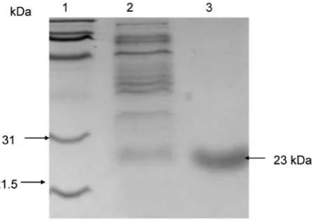
The crude induced DE3 cell extract exhibited azoreductase activity and yielded a yellow coloration (data not shown). The protein was purified using Ni-NTA resin, and the concentration of the protein was determined to be 12.5 mg ml-1. The SDS-PAGE gel showed the protein to be
approximately 23 kDa in size (Fig. 2). The purified protein also possessed a yellow coloration and the absorbance spectrum yielded a typical flavoprotein peak at 457 nm which is slightly shifted from the free flavins which have a peak at 445 nm (data not shown).
Substrate specificity
Table 1 shows that AzoEf1 utilizes either NADH or NADPH to reduce Methyl Red, Congo Red, Acid Red 88, and Direct Blue 15. However, Ponceau BS and Orange II were reduced in the presence of NADH only. The specific activity was highest for Methyl Red (5095 µM min-1 mg-1) when
NADH was used as the electron donor. The specific activity in the presence of NADPH for Methyl Red, Congo Red, Acid Red 88, were significantly lower. Interestingly, the specific activity for Direct Blue was the same for both NADH and NADPH (approximately 3.6 µM min-1 mg-1). No activity
was observed for Ponceau S, Tartrazine, and Amaranth in the presence of either electron donors. The opitimal pH was determined to be between 7.0 and 8.0 at 25°C for the reduction of Methyl Red using NADH as the electron donor, Fig. 2. SDS-PAGE showing the induced, purified and
and the optimum temperature was approximately 38°C (data not shown).
Apparent kinetic constants
The enzyme reactions were conducted by varying the concentration of one substrate, Methyl red or NADH, and maintaining a fix concentration of the other substrate. The Km and Vmax values were obtained from Linveweaver-Burk
plots. As shown in Table 2, the enzyme possesses greater activity and affinity for Methyl Red as indicated by its higher turnover number (kcat = 17) and catalytic efficiency (kcat/
Km =1) compared to NADH (kcat = 10, kcat/Km =.06) as a
substrate. No external addition of FMN was required for the assays which suggested that the FMN moieties already bound to the enzyme was sufficient for its activity.
Discussion
Knowledge of bacterial azoreductase is not fully understood. Azoreductase reductively cleaves a wide range of azo dyes and their activity is associated with numerous intestinal microorganisms, including the genera Enterococci (Chung K.T. et al., 1992 b,Walker R. et al., 1971). The isolation and characterization of azoreductase genes from these prominent Gram positive cocci is lacking, thereby their catalytic function and influence on human health is not fully understood. To date, only E. faecalis has been shown to possess an azoA gene, which encodes for an azoreductase that has broad substrate specificity.
The current study investigated the azoreductase activity from a related intestinal microorganism E. faecium which was shown to grow without inhibition in azo dyes (data not shown), as well as to reduce mono and bis-azo sulfonated azo dyes, indicating azoreductase was functionally expressed in the bacterium. Interestingly, the azoreductase genes from E. coli and E. faecalis were initially described as a putative acyl carrier protein phosphodiesterase (AcpD). Therefore, the protein BLAST search used the azoreductase AzoA from E. faecalis to identify an AcpD conserved domain protein from the E. faecium genomic database (DO strain). Interestingly, the ATCC 6569 culture strain had 10 base pair differences with the E. faecium DO strain, resulting in two amino acid changes at residues 88 (Glx>Asx) and 174 (Glx>Asx). The putative azoreductase identified (AzoEF1) showed 67% amino acid sequence identity with AzoA from E. faecalis. The purified AzoEf1 azoreductase is suggested to be associated with FMN as the absorption peaks were similar to a flavodoxin fingerprint motif (Chen.H., et al., 2004 and 2005). AzoEf1 reduced 6 of the 9 azo dyes tested (Table 1) and did not require any exogenous addition of FMN.
In contrast to AzoA (Chen.H et al., 2004), AzoEf1 did not reduce Amaranth and Ponceau S (Table I). This may be due to subtle differences in the active site of AzoEf1 compared to AzoA. AzoEf1 reduced the mono azo dye Methyl red in the presence of both cofactors NADH and NADPH but the specific activity for NADH was much greater. AzoA was also shown to prefer NADH over NADPH as the electron donor for the reduction of Methyl red with a similar Km but with
almost twice the catalytic efficiency for the dye (Punj S. & John GH, 2008b).
In conclusion, our results showed that although AzoEf1 shares many simalarities with AzoA, critical alterations in the active site of these enzymes can alter substrate specificity and catalytic rates. In spite of the close evolutionary link
between AzoA and AzoEf1, a further investigation of the differences in the active site of these two enzymes will provide a better understanding of the distinguishing factors involved in substrate specificity of azoreductase. Further, AzoEf1 has the ability to use both NADH and NADPH as an electron donor. The azo dye specificity showed differences as AzoEf1 was not specific for Panceau S and Amaranth, while AzoA has been reported to show activity for those azo dyes. Finally, the presence of an azoreductase in these two intestinal microorganisms is likely to play an important and varying role in azo dye metabolism in the intestine, and possibly impact human health.
References
Blumel, S., Knackmuss, H.J. and Stolz, A. (2002). Molecular cloning and characterization of the gene coding for the aerobic azoreductase from Xenophilus azovorans KF46F. Appl. Environ. Microbiol. 68, 3948-3955.
Bragger, J.L., Lloyd, A.W., Soozandehfar, S.H., Bloomfield, S.C., Marriott, C. and Martin, G.P. (1997). Investigations into the azo reducing activity of a common colonic microorganism. Int J Pharmaceut. 157, 61-71.
Brown, J.P. (1981). Reduction of Polymeric Azo and Nitro Dyes by Intestinal Bacteria. Appl Environ Microbiol 41, 1283-1286.
Brown, J.P., McGarraugh, G.V., Parkinson, T.M. and Wingard Jr., R.E. (1983). A polymeric Drug for treatment of inflammatory bowel disease. J Med Chem 26, 1300-1307.
Cerniglia, C.E., Freeman, J.P., Franklin, W. and Pack, L.D. (1982). Metabolism of azo dyes derived from benzidine, 3, 3'-Dimethylbenzidine and 3, 3'-dimethoxybenzidine to potentially Carcinogenic aromatic amines by intestinal bacteria. Carcinogenesis 3, 1255-1260.
Cerniglia, C.E., Zhou, Z., Manning, B.W., Federle, T.W. and Heflich, R.H. (1986). Mutagenic Activation of the benzidine-based dye Direct Black 38 by human intestinal microflora. Mutat Res 175, 11-16.
Chen, H., Wang, R.F. and Cerniglia, C.E. (2004). Molecular cloning, overexpression, purification, and characterization of an aerobic FMN-dependent azoreductase from Enterococcus faecalis. Protein Expression and Purification l34, 302-310.
Chen H, Hopper SL, Cerniglia CE (2005). Biochemical and molecular characterization of an azoreductase from Staphylococcus aureus, a tetrameric NADPH-dependent flavoprotein. Microbiology. 151, 1433-1441.
Chung, K.T., Fulk, G.E. and Egan, M. (1978). Reduction of azo dyes by intestinal anaerobes. Appl Environ Microbiol 35, 558-562.
Chung, K.T. (1983). The significance of azo-reduction in the mutagenesis and carcinogenesis of azo dyes. Mutat Res 114, 269-281.
Chung, K.T, Cerniglia, C.E. (1992a) Mutagenicity of Azo dyes, Structure-activity relationships. Mutat Res 277, 201-220.
Chung, K. T., Stevens, S. E., Jr., and Cerniglia, C. E. (1992b) The reduction of azo dyes by the intestinal microflora. Crit Rev Microbiol 18, 175-190.
Dillon, D., Combes, R. and Zeiger, E. (1994). Activation by caecal reduction of the azo dye D&C Red No. 9 to a bacterial mutagen. Mutagenesis 9, 295 -299.
Drasar, B.S. and Hill, M.J. (1972). Intestinal bacteria and cancer. Am J Clin Nutr 25, 1399-1404.
Ghosh DK, Mandal A, Chaudhuri J. (1992). Purification and partial characterization of two azoreductases from Shigella dysenteriae type 1. FEMS Microbiol Lett. 77, 229-233.
Hill, M.J. (1974). Bacteria and the etiology of colonic cancer. Cancer. 34, 815-818.
Levine, W.G. 1991. Metabolism of azo dyes, Implications for detoxification and activation. Drug Metab Rev 23, 253-309.
Laemmli, U.K. (1970). Cleavage of Structural Proteins during the Assembly of the Head of Bacteriophage T4. Nature 227, 680-685.
Liu ZJ, Chen H, Shaw N, Hopper SL, Chen L, Chen S, Cerniglia CE, Wang BC. (2007). Crystal structure of an aerobic FMN-dependent azoreductase (AzoA) from Enterococcus faecalis. Arch Biochem Biophys. 463, 68-77.
Manning, B.W., Cerniglia, C.E. and Federle, T.W. (1985). Metabolism of the benzidine-based azo dye Direct Black 38 by human intestinal microbiota. Appl Environ Microbiol 50, 10-15.
Morgan, D.L., Dunnick, J.K., Goehl, T., Jokinen, M.P., Matthews, H.B., Zeiger, E. and Mennear, J.H. (1994). Summary of the National Toxicology Program Benzidine Dye Initiative. Environ Health Perspect 102, 63-78.
Morrison, D., Woodford, N. and Cookson, B. (1997). Journal of Applied Microbiology Symposium Supplement 83, 89S-99S.
Murray, B.E. (1990). The life and times of the Enterococcus. Clin Microbiol Rev 3, 46-65.
Nakanishi, M., Yatome, C., Ishida, N. and Kitade, Y. (2001). Putative ACP phosphodiesterase gene (acpd) encodes an azoreductase. J Biol Chem 276, 46394-46399. Punj, S and John, G.H., (2008a). Physiological
characterization of Enterococcus faecalis during azoreductase activity. Microbiol Ecol Health Disease 20, 65-73.
Punj, S and John, G.H., (2008b). Purification and Identification of an FMN-dependent NAD(P)H Azoreductase from Enterococcus faecalis. Curr Issues Mol Biol. 11, 59-66. Sugiura, W., Yoda T., Matsuba T., Tanaka Y. and Suzuki
Y. (2006). Expression and Characterization of the Genes Encoding Azoreductases from Bacillus subtilis and Geobacillus stearothermophilus. Biosci Biotechnol Biochem 70, 1655-1665
Suzuki, Y., Yoda, T., Ruhul, A. and Sugiura, W. (2001). Molecular cloning and characterization of the gene coding for azoreductase from Bacillus sp. OY1-2 isolated from soil. J Biol Chem 276, 9059-9065.
Walker, R., Gingell, R., and Murrells, D. F. (1971). Mechanisms of azo reduction by Streptococcus faecalis. I. Optimization of assay conditions. Xenobiotica 1, 221-229.
• MALDI-TOF Mass Spectrometry in Microbiology
Edited by: M Kostrzewa, S Schubert (2016) www.caister.com/malditof
• Aspergillus and Penicillium in the Post-genomic Era
Edited by: RP Vries, IB Gelber, MR Andersen (2016) www.caister.com/aspergillus2
• The Bacteriocins: Current Knowledge and Future Prospects
Edited by: RL Dorit, SM Roy, MA Riley (2016) www.caister.com/bacteriocins
• Omics in Plant Disease Resistance
Edited by: V Bhadauria (2016) www.caister.com/opdr
• Acidophiles: Life in Extremely Acidic Environments
Edited by: R Quatrini, DB Johnson (2016) www.caister.com/acidophiles
• Climate Change and Microbial Ecology: Current Research and Future Trends
Edited by: J Marxsen (2016) www.caister.com/climate
• Biofilms in Bioremediation: Current Research and Emerging Technologies
Edited by: G Lear (2016) www.caister.com/biorem
• Microalgae: Current Research and Applications
Edited by: MN Tsaloglou (2016) www.caister.com/microalgae
• Gas Plasma Sterilization in Microbiology: Theory, Applications, Pitfalls and New Perspectives
Edited by: H Shintani, A Sakudo (2016) www.caister.com/gasplasma
• Virus Evolution: Current Research and Future Directions
Edited by: SC Weaver, M Denison, M Roossinck, et al. (2016) www.caister.com/virusevol
• Arboviruses: Molecular Biology, Evolution and Control
Edited by: N Vasilakis, DJ Gubler (2016) www.caister.com/arbo
• Shigella: Molecular and Cellular Biology
Edited by: WD Picking, WL Picking (2016) www.caister.com/shigella
• Aquatic Biofilms: Ecology, Water Quality and Wastewater Treatment
Edited by: AM Romaní, H Guasch, MD Balaguer (2016) www.caister.com/aquaticbiofilms
• Alphaviruses: Current Biology
Edited by: S Mahalingam, L Herrero, B Herring (2016) www.caister.com/alpha
• Thermophilic Microorganisms
Edited by: F Li (2015) www.caister.com/thermophile
• Flow Cytometry in Microbiology: Technology and Applications
Edited by: MG Wilkinson (2015) www.caister.com/flow
• Probiotics and Prebiotics: Current Research and Future Trends
Edited by: K Venema, AP Carmo (2015) www.caister.com/probiotics
• Epigenetics: Current Research and Emerging Trends
Edited by: BP Chadwick (2015) www.caister.com/epigenetics2015
• Corynebacterium glutamicum: From Systems Biology to Biotechnological Applications
Edited by: A Burkovski (2015) www.caister.com/cory2
• Advanced Vaccine Research Methods for the Decade of Vaccines
Edited by: F Bagnoli, R Rappuoli (2015) www.caister.com/vaccines
• Antifungals: From Genomics to Resistance and the Development of Novel Agents
Edited by: AT Coste, P Vandeputte (2015) www.caister.com/antifungals
• Bacteria-Plant Interactions: Advanced Research and Future Trends
Edited by: J Murillo, BA Vinatzer, RW Jackson, et al. (2015) www.caister.com/bacteria-plant
• Aeromonas
Edited by: J Graf (2015) www.caister.com/aeromonas
• Antibiotics: Current Innovations and Future Trends
Edited by: S Sánchez, AL Demain (2015) www.caister.com/antibiotics
• Leishmania: Current Biology and Control
Edited by: S Adak, R Datta (2015) www.caister.com/leish2
• Acanthamoeba: Biology and Pathogenesis (2nd edition)
Author: NA Khan (2015) www.caister.com/acanthamoeba2
• Microarrays: Current Technology, Innovations and Applications
Edited by: Z He (2014) www.caister.com/microarrays2
• Metagenomics of the Microbial Nitrogen Cycle: Theory, Methods and Applications
Edited by: D Marco (2014) www.caister.com/n2
Caister Academic Press is a leading academic publisher of advanced texts in microbiology, molecular biology and medical
research. Full details of all our publications at caister.com