R E V I E W
Open Access
Advances in understanding the cell types and
approaches used for generating induced
pluripotent stem cells
Jun Li
1†, Wei Song
1*†, Guangjin Pan
2and Jun Zhou
1Abstract
Successfully reprogramming somatic cells to a pluripotent state generates induced pluripotent stem (iPS) cells (or iPSCs), which have extensive self-renewal capacity like embryonic stem cells (ESCs). iPSCs can also generate daughter cells that can further undergo differentiation into various lineages or terminally differentiate to reach their final
functional state. The discovery of how to produce iPSCs opened a new field of stem cell research with both intellectual and therapeutic benefits. The huge potential implications of disease-specific or patient-specific iPSCs have impelled scientists to solve problems hindering their applications in clinical medicine, especially the issues of convenience and safety. To determine the range of tissue types amenable to reprogramming as well as their particular characteristics, cells from three embryonic germ layers have been assessed, and the advantages that some tissue origins have over fibroblast origins concerning efficiency and accessibility have been elucidated. To provide safe iPSCs in an efficient and convenient way, the delivery systems and combinations of inducing factors as well as the chemicals used to generate iPSCs have also been significantly improved in addition to the efforts on finding better donor cells. Currently, iPSCs can be generated without c-Myc and Klf4 oncogenes, and non-viral delivery integration-free chemically mediated
reprogramming methods have been successfully employed with relatively satisfactory efficiency. This paper will review recent advances in iPS technology by highlighting tissue origin and generation of iPSCs. The obstacles that need to be overcome for clinical applications of iPSCs are also discussed.
Keywords:Induced pluripotent stem cells, Tissue origin, Approach, Generation
At a glance
Induced pluripotent stem (iPS) cells (or iPSCs) have an extensive capacity for self-renewal, reproduction and dif-ferentiation, much like embryonic stem cells (ESCs).
Disease-specific or patient-specific iPSCs have both in-tellectual (e.g., disease modeling) and therapeutic bene-fits, and yet iPSCs face certain obstacles that hinder their applications in clinical medicine, especially the is-sues of convenience and safety.
Cells from three embryonic germ layers have been assessed to determine the range of tissue types amenable to reprogramming along with their particular characteris-tics. Some tissue origins have advantages over fibroblast
origins with respect to efficiency and accessibility. Human urine-derived cells can be an attractive choice from which to generate iPSCs.
The delivery systems and combinations of inducing factors as well as the chemicals used to generate iPSCs have also been significantly improved to provide safe iPSCs in an efficient and convenient way. Small mol-ecule strategy is much more promising because of its many advantages.
Extensive genetic screening and in vivo immunogen-icity testing should be standard procedure to ensure the safety of human iPSCs prior to their clinical use.
Introduction
Embryonic stem cells (ESCs), which are derived from the inner cell mass of mammalian blastocyst, have the ability to grow indefinitely while maintaining pluripo-tency and the ability to differentiate into cells of all three
* Correspondence:[email protected]
†Equal contributors 1
Department of Oncology, Shandong Provincial Hospital Affiliated to Shandong University, No. 324 Jingwu Weiqi Road, Jinan 250021, P.R. China Full list of author information is available at the end of the article
germ layers. Based on the hypothesis that factors that play important roles in the maintenance of ESC identity also play pivotal roles in the induction of pluripotency in somatic cells, in 2006, Takahashi et al. selected 24 candi-date genes for inducing pluripotency in somatic cells under ESC culture conditions [1]. They successfully con-verted mouse embryonic fibroblasts (MEFs) and adult tail-tip fibroblasts (TTFs) to an undifferentiated state similar to that of ESCs through retroviral transduction of four transcription factors: Oct 3/4 (O), Sox2 (S), Klf4 (K), and c-Myc (M). These cells were termed induced pluripotent stem (iPS) cells (iPSCs). Subsequently, they generated human iPSCs (hiPSCs) from human dermal fi-broblasts (HDFs) in the same way [2]. These hiPSCs were also generated by Yu et al. from fetal fibroblasts, newborn foreskin fibroblasts (BJ fibroblasts), and primary human fibroblast-like synoviocytes (HFLS) by introducing another four factors, namely, Oct3/4, Sox2, Nanog (N), and Lin28 (L), using lentiviral vectors [3].
Although the reprogramming process has been grad-ually revealed by intensive studies, the role of these tran-scription factors and the way they function in the acquisition and maintenance of pluripotency are still not fully understood. Simply speaking, the identity of som-atic cells is strictly protected by an epigenetic barrier, and these cells acquire pluripotency when the reprogram-ming factors break the epigenetic barrier [4]. Possible mechanisms include silencing of retroviral transgenes upon establishment of pluripotency [5], reactivation of en-dogenous pluripotency genes [1], establishment of bivalent chromatin domains in the promoters of developmentally regulated genes [6], global DNA hypomethylation and DNA hypermethylation of imprinted gene loci [7], reacti-vation of the inactive X chromosome in female iPSCs and reorganization of chromatin fibers [8].
iPSCs are found to be similar to ES in their morph-ology, proliferation, surface antigens, gene expression, epigenetic status of their pluripotent cell-specific genes, and telomerase activity. Furthermore, these cells can dif-ferentiate into cell types of the three germ layers in vitro as well as into teratomas in vivo. They are also capable of germline transmission [9]. Nevertheless, iPSCs are su-perior to ESCs in some aspects because they have the potential to bypass both the practical and ethical con-cerns associated with ESCs. Unlike ESCs, which require the use of embryos and are thus complicated by ethical controversies and possible immune rejection, iPSCs can be derived from directed reprogramming of somatic cells from individual patients. Because the lifespan of fully dif-ferentiated cells is usually short and because such cells cannot self-renew (with a few exceptions, e.g., hepato-cytes), iPS technology can provide them with an exten-sive ESC-like capacity to self-renew and the ability to generate daughter cells that can further differentiate into
various lineages or terminally differentiate into a func-tional state, thus opening a new field of stem cell re-search. These disease-specific or patient-specific stem cells provide a unique opportunity to understand novel disease mechanisms [10], to screen for more effective and safe drugs [11], to develop autologous cell therapies including in vitro expansion and differentiation into cells of the hematopoietic lineage [12,13], and to treat various diseases and injuries, such as Parkinson's disease [14] and cardiac damage [15].
Despite the potential implications of this technology, a number of questions about iPSC generation must be an-swered. There are several potential challenges to their pos-sible clinical application in humans: (i) virus-mediated delivery of reprogramming factors permanently integrates transgenes into the human genome, potentially altering genomic features and risking viral transgene reactivation [16]; (ii) the reprogramming factors Klf4 and c-Myc are oncogenic and can potentially lead to the development of certain cancers [17,18]; (iii) the low efficiency of iPS in-duction [1] and the lack of full and homogeneous iPS re-programming processes are both problematic.
A series of major breakthroughs have been achieved. To determine the range of tissue types amenable to re-programming, to decrease the tumorigenesis risk from transgene integration, and to increase the induction effi-ciency, investigators have extensively studied cells from three embryonic germ layers and identified some advan-tages of using tissues other than fibroblast. Delivery sys-tems and combinations of inducing factors, as well as the chemicals used to generate iPSCs have also been im-proved; for example, iPSCs can now be generated without the use of the oncogenes c-Myc and Klf4 [19], and non-viral integration-free delivery methods are being success-fully employed with reasonable efficiency. In this paper, we review recent advances in iPS technology, with a par-ticular focus on tissue origin and generation of iPSCs.
Tissue origins of iPSCs
Fibroblasts are the first cell type to have been success-fully reprogrammed into iPSCs. Takahashi et al. tried 24 candidate genes and found that the retroviral transduc-tion of OSKM reprogrammed MEFs and somatic TTFs into iPSCs [1]. These cells were also used to generate hiPSCs [2]. Fibroblasts are still widely used in iPSC tech-nology, and their reprogramming efficiency and differenti-ation ability are the gold standard to which new methods must be compared.
tissues from all three embryonic germ layers. iPSCs con-tribute to all tissue types, including the germ line, when injected into murine blastocysts. iPSCs from MEFs could generate “all-iPSC mice” following injection into tetra-ploid blastocysts, thereby satisfying the most stringent criterion of pluripotency [1]. The same procedures can-not be applied to hiPSCs, which must rely on embryonic bodies and teratomas as the most stringent tests. This paper will only focus on iPSCs of mouse and human origin.
Cells from the three embryonic germ layers have been studied by researchers in the iPS field to determine the range of tissue types amenable to reprogramming, to de-crease the tumorigenesis risk of transgene integration, and to increase the efficiency of induction. Malignant cell lines and primary cancer cells are also confirmed to be amenable to reprogramming by the iPS approach; however, this issue is not reviewed in this paper. To bet-ter understand the differences that result from the tissue origin of iPSCs, we will discuss them through two per-spectives: differences within the same tissue origin and differences resulting from different tissue origins.
Differences within the same tissue origin Efficiency
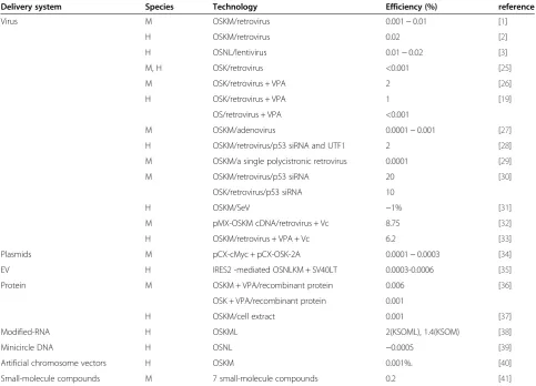
The efficiency of iPSC induction is defined as the per-centage of green fluorescent protein (GFP)-positive col-onies with clear iPS morphology with a certain amount of mother cells transfected with transgenes. The efficien-cies of iPSC induction from a single tissue of origin are different when using different technologies. It is hard to accurately correctly compare induction efficiency of dif-ferent methods given differences in their experimental settings. Furthermore, efficiency is indeed an important criterion for an induction technology, but it is not always the most important one. Some technologies were im-proved to reduce integration of transgenes while sacri-ficing efficiency. In general, there are a few lessons regarding the efficiencies of iPSC induction from a single tissue origin: (i) efficiency is higher in mice than in hu-man, successful induction using some technologies has not been reported in humans due to very low efficiency; (ii) decreasing the number of transgenes with the same methods would reduce the induction efficiency by orders of magnitude; (iii) non-integration methods are much less efficient than integration methods, with the excep-tion of the Sendai virus (SeV) versus retrovirus and lentivirus vectors; (iv) certain chemicals and microRNAs (miRNAs) can also markedly improve the efficiency. A detailed summary of the efficiencies of different induc-tion methods using fetal and somatic fibroblasts are listed chronologically in Table 1 according to the publi-cation date of descriptive papers, which straightfor-wardly represents the development of the technology.
Another example of different efficiencies in the same tissue origin in response to different induction technolo-gies is found in human adipose stem cells (hASCs). Sun et al. [42] reported that when hASCs are transduced with individual lentiviruses containing OSKM, the inci-dence of ESC-like colonies is 0.2%, whereas Vc or Vc + VPA with retroviral pMX vectors containing OSKM cDNAs reprogrammed hASCs with a much higher effi-ciency (up to 7.06%) [32], nevertheless, the reprogram-ming efficiency is much lower with minicircle DNA, which contains a single cassette of OSNL plus a GFP re-porter gene separated by self-cleavage peptide 2A se-quences. This system yields an overall reprogramming efficiency of ~0.005% [39].
Some small molecules can increase the efficiency of re-programming primary human fibroblasts to a pluripo-tent state [26]. When the same three-factor combination
(OSK) via retroviral transduction is used, the addition of VPA improves reprogramming efficiency by a factor of 1,000-fold. Furthermore, VPA could enable repro-gramming with only two factors (Oct4 and Sox2) with efficiency similar to that of three factors, suggesting that VPA treatment effectively dispenses the need for Klf4. Other small molecules that could obviate the need for certain exogenous factors will be reviewed below.
Somatic coding mutations
human Mendelian disorders suggests that the risk of dis-eases other than cancer should be evaluated as well for hiPS-cell-based therapeutic methods. Thus, although all hiPSC lines are extensively characterized for pluripo-tency and have normal karyotypes before DNA extrac-tion, pre-existing and new mutations occur during and after reprogramming. These mutations can produce gen-etic and epigengen-etic changes in the hiPSCs such that ex-tensive genetic screening should become a standard procedure to ensure the safety of hiPSCs before clinical use. One corollary is that if reprogramming efficiency is improved to a level such that no colony picking and clonal expansion is necessary, the resultant hiPSCs could potentially be free of mutations.
Copy number variations (CNVs)
Significantly more CNVs are present in early-passage hiPSCs than in intermediate passage hiPSCs established either by retroviral or piggyBac (PB) transposon delivery methods [44]. Fortunately, most CNVs render the af-fected cells at a selective disadvantage; thus remarkably,
the expansion of hiPSCs in culture selects rapidly against mutated cells, driving the lines toward a genetic state re-sembling human ESCs.
Differences resulting from different tissue origins Accessibility and universality
There is still no consensus regarding the preferred tissue from which to harvest donor cells for iPSC reprogram-ming. The ideal cell source should be easily accessible, susceptible, and universal (any age, sex, ethnic group, and body condition). Different cell sources are associated with distinct advantages and disadvantages.
Dermal fibroblasts are currently the most frequently used cells for reprogramming, but their obtainment re-quires biopsy, and candidates sometimes refuse to do-nate the necessary tissue. Additionally, the procedure is contraindicated in life-threatening skin diseases (e.g., se-vere epidermolysis bullosa) or burn cases. Keratinocytes have similar limitations [45]. They are keratin-dense epi-thelial cells and generate the outer protective epidermal barrier of the skin surface as well as appendages such as Table 1 Efficiency of different iPS induction methods in fibroblasts
Delivery system Species Technology Efficiency (%) reference
Virus M OSKM/retrovirus 0.001 ~ 0.01 [1]
H OSKM/retrovirus 0.02 [2]
H OSNL/lentivirus 0.01 ~ 0.02 [3]
M, H OSK/retrovirus <0.001 [25]
M OSK/retrovirus + VPA 2 [26]
H OSK/retrovirus + VPA 1 [19]
OS/retrovirus + VPA <0.001
M OSKM/adenovirus 0.0001 ~ 0.001 [27]
H OSKM/retrovirus/p53 siRNA and UTF1 2 [28]
M OSKM/a single polycistronic retrovirus 0.0001 [29]
M OSKM/retrovirus/p53 siRNA 20 [30]
OSK/retrovirus/p53 siRNA 10
H OSKM/SeV ~1% [31]
M pMX-OSKM cDNA/retrovirus + Vc 8.75 [32]
H OSKM/retrovirus + VPA + Vc 6.2 [33]
Plasmids M pCX-cMyc + pCX-OSK-2A 0.0001 ~ 0.0003 [34]
EV H IRES2 -mediated OSNLKM + SV40LT 0.0003-0.0006 [35]
Protein M OSKM + VPA/recombinant protein 0.006 [36]
OSK + VPA/recombinant protein 0.001
H OSKM/cell extract 0.001 [37]
Modified-RNA H OSKML 2(KSOML), 1.4(KSOM) [38]
Minicircle DNA H OSNL ~0.0005 [39]
Artificial chromosome vectors H OSKM 0.001%. [40]
Small-molecule compounds M 7 small-molecule compounds 0.2 [41]
Abbreviations:Mmouse,Hhuman,oriP/EBNA1Epstein-Barr nuclear antigen-1,EVepisomal vectors,IRES2internal ribosome entry site 2,SV40LTSV40 large T gene,
hair and nails. hASCs [42], a heterogeneous group of multipotent progenitor cells similar to the ones from fibroblast and the keratinocytic lineage can be readily and safely derived from adipose tissue of adult humans in very large quantities by lipoaspiration without ex-tended time for expansion, but lipoaspiration is invasive and sometimes harmful.
It is unclear if dermal papilla (DP) cells [46], which are specialized skin fibroblasts that are considered to in-struct hair follicle stem cells, are a good source for re-programming because the growth and quality of hair follicles depend on age, genotype, and medical condi-tions of the human donors, although they are repro-grammed more efficiently than skin and embryonic fibroblasts due to high endogenous expression of Sox2 and c-Myc.
Blood cells are among the most favorable cell types for iPSC induction, but their development has thus far been disappointing. They include stem/progenitor cells and terminal blood cells. Hematopoietic stem/progenitor cells are young cells that are expected to carry minimal somatic mutations and possess the immunological im-maturity of newborn cells. They are rare in peripheral blood but are naturally rich in the bone marrow (BM), umbilical cord blood (CB), and placenta. Harvesting mo-bilized CD34+ cells isolated from peripheral blood is a cumbersome, expensive, and time-consuming process [47] and granulocyte colony stimulating factor (G-CSF) mobilization is a procedure that is frequently associated with significant side effects, including bone pain, head-ache, fatigue, and nausea [48]. BM harvesting is an inva-sive procedure [49]. Other choices are the CB and the placenta [50], which are extra-embryonic tissues of par-ticular interest in regenerative medicine. They share an early developmental origin and are a source of vast amounts of cells with multilineage differentiation poten-tial. They are also poorly immunogenic and, unlike many other stem-cell sources, are not controversial. Moreover, these cells are likely exempt from incorporated muta-tions when compared with juvenile or adult donor cells such as skin fibroblasts or keratinocytes. Unfortunately, they are only available for a minority of individuals who have had their samples banked at birth.
Peripheral blood cells (PBCs) can be isolated by rou-tine venipuncture with minimal risk to the donor and can be obtained in sufficient numbers to enable repro-gramming without the need for prolonged expansion in culture. More importantly, PBCs provide convenient ac-cess to numerous patient samples stored in blood banks. Frozen blood samples, when reprogrammed to iPSCs, are of major interest because they would allow for retro-spective molecular analysis of rare diseases. However, some disadvantages associated with the blood specimens restrict their usage for generation of iPSCs. First, the
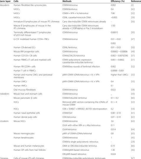
efficiency of this cell source is extremely low (0.0008 to 0.001%). Second, the main parent cells are mature T cells bearing specific T cell receptor (TCR) re-arrangements [51,52], which yield iPSCs with germ line IgH and TCR alleles, that are undesirable for some potential applica-tions in regenerative medicine. Moreover, in cases where blood infections are involved (e.g., hepatitis C virus, and HIV), giving blood is not exempt of concerns. Finally, reprogramming may be problematic in patients with blood diseases (e.g., hemophilia and leukemia).
Human urine -derived cells (HUCs) are another favor-able iPSC candidate [53,54], in particular because harvest-ing is completely non-invasive. In normal physiology, approximately 2000 to 7000 cells from an extensive net-work of tubules in the kidney and downstream parts of the urinary tract (the ureters, bladder, and urethra) be-come detached into the urine daily. They can be collected anywhere without medical assistance and are easily ex-panded. Their efficiency with retroviral pMX vectors con-taining the cDNAs of human OSKM is between 0.1 and 4%, while their efficiency with oriP/EBNA EVs carrying a combination of reprogramming factors encoded by OSK,
SV40LT, and the miRNA cluster MIR302–367 through
electroporation is approximately 0.2%. These are quite high efficiencies for the integration-free reprogramming method. Therefore, urine samples may be considered a preferred source for iPSC derivation. However, other re-ports indicate that the number of excreted cells range from 0 to 6500 showing a high degree of individual vari-ation, and cell proliferation is achieved in only approxi-mately 1/3 of the samples [55]. This raises the questions whether HUCs can be universally applied for generating iPSCs. Moreover, culturing HUCs usually requires long periods of time (3 ~ 6 days or more to form the HUC clones, 7 ~ 10 days or more for the clones to spread over the dish) prior to the introduction of reprogramming fac-tors into passage 2 ~ 4 HUCs [53]. Furthermore, although HUCs may produce iPSCs bearing fewer somatic cell mu-tations and copy number variations than iPSCs from the skin due to reduced direct exposure to radiation, iPSCs derived from this source have not yet been fully analyzed for the spectrum of mutations and other characteristics of iPSCs.
Cells of some tissue origins are excluded from easily-accessible cell resources because they are difficult to reach, with potential risks due to their positions in the body. These cell types include neural stem cells (NSCs), melanocytes, meningiocytes, hepatocytes, gastric epithe-lial cells, and pancreaticβcells.
Efficiency
found that the parental cells of different tissues of origin have different efficiencies. Some cell types are difficult to reprogram even with a virus-mediated method, let alone a non-integrating method. It is conceivable that epithe-lial cells are more amenable to reprogramming, perhaps because unlike fibroblasts, they need not undergo a mesenchymal-to-epithelial transition to produce iPSCs. Nevertheless, the high endogenous levels of transgenes required for iPSC generation in parent cells may also be associated with transcriptional and epigenetic states that are favorable to reprogramming. A detailed summary of the efficiency of various cell origins other than fibro-blasts is shown in Table 2.
Number of exogenous factors
Parent cells of different tissue origins require a different number of exogenous factors. Considering that the ectopic expression of c-Myc causes tumorigenicity in offspring and that retroviruses themselves can cause insertional mu-tagenesis, iPSCs generated with a minimal number of fac-tors were studied to identify cell types that can be more easily reprogrammed with fewer factors and with higher efficiency to increase the likelihood of ultimately replacing the remaining factors with small molecules.
Mouse and human fibroblasts can be reprogrammed into iPSCs in the absence of the c-Myc retrovirus, al-though their efficiency is as low as <0.001% [25]. Fur-thermore, it is believed that c-Myc plays a much smaller role in the generation of iPS-Hep and iPS-Stm cells than in iPS-fibroblasts [59]. This is because the omission of c-Myc decreased the colony numbers to 20 to 40% of those obtained by the four factors, in contrast to the generation of iPS-fibroblasts, which caused a 90% decrease in colony numbers in response to c-Myc omission.
Adult mouse NSCs express higher endogenous levels of Sox2 and c-Myc than ESCs. Exogenous Oct4 in com-bination with either Klf4 or c-Myc is found to be suffi-cient to generate iPSCs from NSCs [63], while Oct4 alone is confirmed to be both necessary and sufficient [64]. However, although this approach is potentially ap-plicable for generating hiPSCs, it is limited by the diffi-culty and potential risks of obtaining NSCs from the brains of patients.
Melanocytes are, like NSCs, of neuroectodermal origin and hence might require fewer factors for their conversion into iPSCs. Interestingly, with dox-inducible lentiviral vec-tors expressing OSKM, the efficiency of reprogramming melanocytes is more than 3-fold higher than that of fibro-blasts (0.19% versus 0.056%); this efficiency decreases to 0.03% with all three factors (with OKM) and to 0.02% in the absence of ectopic c-Myc, which is approximately 10-fold lower than the efficiency of melanocyte repro-gramming in the presence of c-Myc and fibroblast re-programming [63].
Epigenetic memory
iPSCs of different tissue origins are discovered to have different epigenetic memories when analyzed for DNA methylation. iPSCs derived from non-hematopoietic cells (neural progenitors and fibroblasts) are found to retain residual methylation at various loci where demethylation is required for a hematopoietic fate, as a result, they manifest a reduced blood-forming potential in vitro [68,69]. Epigenetic memory has a dual nature. For some applications, the epigenetic memory of the donor cell may be advantageous when the directed differentiation to spe-cific tissue fates is challenging; for others, epigenetic memory can restrict the use of the resultant iPSCs in re-generative medicine. For example, iPSCs generated from T cells from human PBMCs still contain TCR, restricting their broad application in regenerative medicine [52].
Differentiation potential
The tissue origin of iPSCs affects their differentiation potential [70]. For example, the teratoma-forming pro-pensity is different of secondary neurospheres generated from 36 mouse iPSC lines derived in 11 different ways. These iPSC lines can be characterized by (i) their origin (e.g., MEF, TTF, hepatocyte, or gastric epithelial cell); (ii) the presence or absence of c-Myc retroviral transduc-tion; (iii) the presence or absence of drug selection for Nanog or Fbxo15 expression. Retrovirus-derived iPSCs from adult TTFs and hepatocytes could contribute to chimeric mice; however, when the iPSCs are induced in vitro to differentiate into neural cells, they frequently form teratomas following cell transplantation. In con-trast, iPSCs prepared from MEFs show low teratoma for-mation, similar to ESCs.
Furthermore, the differentiation propensity and methy-lation profile of iPSCs can be reset. When blood-deficient neural progenitor-derived iPSCs are differentiated into blood cells, which are then reprogrammed into iPSCs, their blood-forming potential is markedly increased [69].
Susceptibility to tumorigenesis
epithelial cells [59] appear not only to be more easily re-programmed but also require fewer retroviral integra-tions than fibroblasts. No increased tumorigenicity was observed in chimera mice derived from iPS-Hep and iPS-Stm cells evaluated up to 30 weeks.
Somatic coding mutations of iPSCs have been con-firmed to be different even with the same cell origin of different fibroblast lines when different integrating and non-integrating methods are used [43]. However, no data exists regarding somatic coding mutations of iPSCs Table 2 The efficiency of cells of various non-fibroblast origins for generating iPSCs
Germ layer Cells Methods Efficiency (%) Reference
Mesoderm Human fibroblast-like synoviocytes OSKM/retrovirus 0.002 [2]
hASCs OSKM/lentivirus 0.4 [42]
hASCs OSKM + VPA + Vc/retrovirus 7.06 [32]
hASCs OSNL cassette/minicircle DNA ~0.005 [39]
Immature B lymphocytes of mouse iPS chimeras Carry dox-inducible OSKM retroviruses already - [56] Mature B lymphocytes of mouse in the
iPS chimeras
Carry dox-inducible OSKM retroviruses already + C/EBPalpha) or Pax 5 knockdown
1/30
Terminally differentiated T lymphocytes of p53-null mouse
OSKM/retrovirus 0.00015 [30]
G-CSF mobilized human CD34+ PBCs OSKM/retrovirus 0.01 ~ 0.02 [47] 0.002
Human CB-derived ECs OSNL/lentivirus 0.01 ~ 0.03 [50]
Mouse BW progenitor cells OSKM/retrovirus 0.00002 ~ 0.00006 [49] Human CD133+ CB cells OSKM,OSK,OS/retrovirus 0.002 ~ 0.007(OSK) [57] Human PBMCs (T cell and myeloid cell) OSKM polycistronic expression
cassette/dox-inducible lentivirus
0.001 ~ 0.0002 [51]
Human PB CD34+ cells OSKM/two rounds of lentiviral infection 0.002 [52]
Human T cell in PBMCs 0.0008 ~ 0.001
Human and murine CMCs and periosteal membrane
pMX-OSKM cDNA/retrovirus + Vc + VPA higher than UMCs [32]
Human UMCs pMX-OSKM cDNA/retrovirus + Vc + VPA 0.4 [33]
Human AMCs 0.1
Oral mucosa fibroblasts OSKM/retrovirus 0.022 [58]
Endoderm Mouse liver and stomach cells OSKM/retrovirus _ [59]
Mouse pancreaticβcells OSKM/inducible lentivirus 0.1 ~ 0.2 [60] HUCs Retroviral pMX vectors containing the cDNAs of
mouse OSKM
0.1 ~ 4 [53]
HUCs OSK + SV40LT + MIR302–367/EV electroporation 0.2 [54]
Human nasal epithelial cells OSKM/SeV 0.08 ~ 0.10 [61]
Human dental pulp cells OSK/retrovirus 0.01 ~ 0.10 [62]
Ectoderm Mouse NSCs OSKM/retrovirus 3.6 [63]
Oct4 with either Klf4 or c-Myc/retrovirus 0.11
Oct4/retrovirus 0.014 [64]
Mouse meningiocytes pMX of OSKM-cDNAs/retroviral 0.8 [65]
Human keratinocytes OSKM/retrovirus 1 [45]
OSKM/a single polycistronic retrovirus - [29] Mouse and human melanocytes OKM or OKS/dox-inducible lentivirus 0.19 [66] Human DP cells from hair follicles OSKM/pMX-based retrovirus 1.38 [46]
OK/pMX-based retrovirus 0.024
of different cell origin when the same non-viral, integration-free delivery methods are used.
Improvements in the approaches for iPSC generation
The currently available methods for iPSC induction can be divided into four categories based on their vector types: viruses, DNA and RNA (plasmid, episomal plas-mid, and transposon), cell penetrating peptides, and che-micals. Although retroviral and lentiviral vectors have a high reprogramming efficiency, the use of viruses encod-ing reprogrammencod-ing factors represents a major limitation of the technology because even low vector expression may alter the differentiation potential of iPSCs or induce malignant transformation, while viral transgenes can also be integrated into the somatic genome. Several groups have made considerable progress to overcome such technical problems. iPSCs can be generated without the oncogenes c-Myc and Klf4 [26]. Furthermore, genome-integrating but excisable systems have worked to remove the integrated transgenes [71,72]. Transient expression of reprogramming factors with adenoviral [27], SeV [31], and non-viral delivery integration-free methods such as plasmid [34], EV [35], proteins [36,37], and chemicals [41] have been successfully employed, albeit at a very low efficiency (with the exception of SeV). Nevertheless, further study should focus on whether there is any ference in the characteristics of iPSCs established by dif-ferent methods prior to their eventual use in medical applications.
Viral induction of iPSCs
According to the original method of iPS generation, cells were reprogrammed into an ESC-like state by viral trans-duction with defined combinations of factors. Thomson et al. [9] found that each iPS clone contained three to six retroviral integrations for each factor. Thus, each clone had more than 20 retroviral integration sites in total, which may increase the risk of tumorigenesis. In contrast, c-Myc is known as a proto-oncogene, and its reactivation may give rise to transgene-derived tumor formation [16]. Furthermore, although reprogramming has been achieved in the absence of c-Myc [25], the remaining integrated re-programming factors could also cause tumor formation [73]. To apply this technology to cell transplantation ther-apy, it is crucial to generate iPSCs with fewer viruses and transgenes.
Induction with varying numbers of transgenes
Transgene numbers can be decreased by changing the parent cell type. Considering that the ectopic expression of transgenes can cause tumorigenicity in offspring, the generation of iPSCs with a minimal number of factors has been extensively studied. Indeed, studies have shown that it is possible to generate iPSCs in the absence of
c-Myc with retroviruses from mouse and human fibro-blasts [25] and from mouse hepatocyte and stomach cells [59], to generate pluripotent stem cells with only oct4 from adult mouse NSCs [64], and to generate pluri-potency with dox-inducible lentiviral vectors expressing OSK or OKM from melanocytes [66].
Some small molecules could also replace certain ex-ogenous factors. When reprogramming primary human fibroblasts to a pluripotent state, VPA enables repro-gramming with OS alone with an efficiency similar to that of OSK, suggesting that VPA treatment effectively obviates the need for Klf4 [23]. Kenpaullone could activate Nanog expression in mouse fibroblasts and substitute for Klf4, although the underlying mechanism remains unclear [74]. A specific glycogen synthase kinase 3 (GSK-3) inhibi-tor, CHIR99021, can induce reprogramming of MEFs transduced with only OK. When combined with Parnate (also known as tranylcypromine), an inhibitor of lysine-specific demethylase 1, namely CHIR99021, human pri-mary keratinocytes can also be reprogrammed through transduction with OK [75]. The transient expression of OCT4 alone with the use of VPA and BIX01294, a histone methyltransferase G9a inhibitor, is sufficient to allow for reprogramming of NSCs [76]. In addition, the pan-Src family kinase (SFK) inhibitors Dasatinib and PP1 are able to replace Sox2 for the generation of iPSCs from MEF [77]. Hou et al. identified a specific chemical combination of four small molecules: VPA, tranylcypromine, CHIR99021, and 616452 (VC6T). These four molecules are sufficient to permit the reprogramming of mouse embryonic and adult fibroblasts in the presence of a single transcription factor, Oct4, which can replace SKM. Two years later, they generated iPSCs using only seven chemicals [41].
Drug-inducible transgenic systems
Induced pluripotency is complicated by the need for in-fection with high-titer retroviral vectors, which produce genetically heterogeneous cell populations. Wernig et al. generated iPSC chimeras and produced genetically homogeneous ‘secondary’somatic cells that carry repro-gramming factors defined as dox-inducible transgenes [67]. A drug-inducible transgenic system could directly reprogram multiple ‘secondary’somatic cell types with-out the need for a virus. These cells could be repro-grammed upon dox exposure without the need for viral infection with 25- to 50-fold greater efficiencies than those observed using direct infection and drug selection for pluripotency marker reactivation.
cells and convenient for screening out cells that are not transfected by transgenes.
Single polycistronic vector
The nucleofection of a single polycistronic vector to generate iPSCs can reduce transgene insertion into the genome. A major impediment to the use of iPSCs for therapeutic purposes has been the viral-based delivery of reprogramming factors; multiple proviral integrations pose a greater danger of insertional mutagenesis. On the other hand, the cellular and genetic heterogeneity of fi-broblasts randomly infected with a single transgene coded by a separate virus complicates the exploration of important molecular events occurring during repro-gramming and limits the scalability required for high-throughput analyses. Carey et al. reported an approach to deliver up to four reprogramming factors in a single virus using 2A “self-cleaving” peptides. These peptides support efficient polycistronic expression from a single promoter with a single proviral copy in both embryonic and adult somatic mouse cells as well as in human kera-tinocytes [29]. These iPSC lines show no evidence of transgene insertion into their genomes. Most of all, the single polycistronic vector could generate genetically identical cell populations amenable to reprogramming without any further genetic interference. Several similar
methods [78–80] have been reported in the same year,
and these methods are widely used to this day.
An integrating but excisable system
To derive hiPSCs that were free of proviruses, Soldner et al. used dox-inducible lentiviral vectors that could be excised after integration with Cre-recombinase [71]. Vectors transducing either OSKM or OSK are used to generate hiPSCs after being transiently cotransfected with plasmids for Cre-recombinase and EGFP and sub-sequently EGFP-positive and Cre-expressing cells are sorted. The excision of all reprogramming factors is con-firmed by Southern blot analysis with a different restric-tion digesrestric-tion. Furthermore, polymerase chain reacrestric-tion (PCR) of the genomic DNA with primers specific for Cre-recombinase confirms that none of the clones has stably integrated the electroporated plasmids. However, it should be noted that a number of technical hurdles complicate the use of Cre-mediated DNA excision from stem cells, potentially limiting the application of these methods to easily generate iPSCs free of floxed trans-genes. First, the delivery of Cre to ESCs or iPSCs is known to be inefficient. Second, screening methods to detect successful Cre-recombination may be cumber-some. Finally, the clumping of cells after the delivery of Cre may result in mosaic colonies containing some cells that have failed to undergo Cre-recombination.
Sommer et al. improved the method of Soldner et al. [72]. A novel version of their single polycistronic vector containing a reporter fluorochrome allows for the direct visualization of vector excision in living iPSCs in real time. Cre-recombination efficiency and the excision of reprogramming transgenes from the resulting iPSCs could be readily visualized and monitored in individual living cells and colonies in culture. In addition, through the excision of only a single vector copy, their approach minimizes the risk of chromosomal translocations, which is a significant advance over prior methods. Fur-thermore, a direct comparison of iPSC clones before and after excision reveals that the removal of the reprogram-ming vector markedly improves the developmental po-tential and differentiation capacity of the iPSCs. It should be emphasized that their approach leaves ap-proximately 200 base pairs (bp) of exogenous DNA, pri-marily viral long terminal repeat (LTR), behind in the iPSC genome. Although the viral LTR is inactivated in their studies and possesses no intrinsic promoter/enhan-cer activity, there is still a theoretical risk that insertional mutagenesis may arise from this genomically integrated exogenous DNA.
Papapetrou et al. further improved the above methods [81] and developed a strategy that includes additional steps for the mapping of the integration sites in the gen-ome. Their method also allows for the selection of clones with few or a single appropriate site for the inte-gration of the residual LTR.
Adenoviral vector
Adenoviral vectors allow for transient, high-level expres-sion of exogenous genes without the integration of such genes into the host genome. Stadtfeld et al. generated mouse iPSCs (adeno-iPSCs) from fibroblasts and liver cells [27] at extremely low efficiency of 0.0001% to 0.001%, which is significantly lower than that obtained with integrating viruses (~0.01 to 0.1%). This is most likely because many cells do not maintain viral expres-sion long enough to trigger entry into a state sustained by endogenous pluripotency factors.
PCR analysis of genomic DNA isolated from adeno-iPSC clones excludes the possibility of permanent viral integration. Southern blot analysis using the cDNAs of the four viral vectors carrying OSKM as probes con-firmed the PCR results and yielded no evidence for the continuous presence of adenoviral sequences in the adeno-iPSCs. Although highly unlikely, they cannot rule out the possibility that small pieces of adenoviral DNA have inserted into the genome of adeno-iPSCs but were not observed given the detection limits of Southern blot analysis.
adeno-iPSC lines were tetraploid, which is not observed in iPS cells produced with retroviral or lentiviral vectors.
SeV vector
SeV, a RNA virus carrying no risk of altering the host genome, is shown to be an efficient solution for generat-ing safe iPSCs in the work of Fusaki et al. [31]. SeV-derived transgenes are decreased during cell division; moreover, viruses could be easily removed by antibody-mediated negative selection utilizing the cell surface marker HN that is expressed on SeV-infected cells. SeV is shown to be an ideal vector for generating hiPSC. First, as emphasized repeatedly, the SeV vector allows the expression of transgenes without the risk of modify-ing the host genome. Second, the efficiency of iPSC gen-eration by gene transduction with SeV vectors is significantly higher (~1%) compared with other methods, especially without any transfection reagents or chemi-cals. Finally, it is easy to select iPSCs that deplete the viral genome from the cytoplasm.
Although the use of SeV requires stringent steps to purge the reprogrammed cells of replicating virus and the sensitivity of the viral RNA replicase to transgene se-quence content may limit the generality of this repro-gramming vehicle, this method is still used widely and can be used to efficiently deliver up to four exogenous genes into various mammalian cells, including primary tissue cells and human hematopoietic stem cells [82–86].
Non-viral induction of iPSCs
The development of novel approaches for generating integration-free iPSCs has eliminated concerns about in-tegrating virus-associated genotoxicity in clinical appli-cations. Among the virus-free method, PB transposons require a second step to remove the transgenes once re-programming has been achieved. Other one-step non-virus approaches make use of plasmids [34], minicircle DNAs [39], human artificial chromosome vectors (HACs) [40], protein transduction [36,37], and chemicals [41], but are very inefficient in generating integration-free iPSCs. In contrast, the use of modified mRNA [38] and oriP/ EBNA1-based EV [35] represent relatively efficient ap-proaches that have been readily reproduced in different la-boratories. The current most cost-effective approach is EV because it does not require the packaging of viral vectors and only needs simple infection instead of daily or mul-tiple additions of factors for successful reprogramming. However, small molecule strategy is much more promis-ing because of its many advantages.
PB transposons
DNA transposons are genetic elements that can relocate between genomic sites via a “cut and paste” mechanism. PB is originally isolated from Trichoplusia ni and
subsequently found to be a host-factor-independent method for efficient transposition in many different spe-cies. The PB transposon/transposase system requires only the inverted terminal repeats flanking a transgene and transient expression of the transposase to catalyze inser-tion or excision events. One important feature of the PB transposon is that it nearly always excises itself precisely, leaving no footprint behind [87].
Woltjen et al. demonstrated the successful and effi-cient reprogramming of murine and human embryonic fibroblasts using the dox-inducible OSKM transcription factor delivered by PB transposition [88]. By taking ad-vantage of the natural propensity of the PB system for seamless excision, the individual insertions can be re-moved without a trace from the established iPSC lines, providing an invaluable tool for discovery. The success-ful transposon-based reprogramming of fibroblasts to iPSCs represents a significant advance in the methods of transgene delivery. First, PB transposition permits technical simplification and improved accessibility of reprogramming methodology, making use of effortless plasmid DNA prep-aration and commercial transfection products for delivery. This eliminates the need for specialized biohazard contain-ment facilities and the production of high-titer, limited-lifetime viral stocks. Second, the range of somatic cell types that could be used for reprogramming is not limited by a decreased susceptibility to viral infection. Third, PB-mediated delivery will allow the option of xeno-free hiPSC production, contrary to current viral production protocols that use xenobiotic conditions. Finally, accurate transgene removal through transposase expression has been demon-strated in various cell lines. However, it is disadvantaged by labor intensive removal of multiple transposons.
Meanwhile, in the same journal, Kaji et al. reported that the non-viral transfection of a single multiprotein expression vector, the 2A-peptide-linked reprogramming cassette MKOS-ires-mOrange, could reprogram both mouse and human fibroblasts [89]. Moreover, the trans-gene could be removed once reprogramming had been achieved, when the single vector reprogramming system is combined with the PB transposon.
Although the PB transposon is thought to nearly always excise itself precisely, leaving behind no footprint, it is still found that significantly more CNVs are present in early-passage than in intermediate early-passage hiPSCs, as has been established by retroviral delivery methods [44]. Fortu-nately, most CNVs are a disadvantage to the affected cells. Remarkably, the expansion of hiPSCs in culture rapidly se-lects against mutated cells, driving the lines toward a gen-etic state resembling that of human ES cells.
Plasmid vector
(pCX-cMyc) and observed the highest efficiency of GFP-positive colony formation when the factors were in the order of OKS [34]. Interestingly, when they transfect pCX-OKS-2A on days 1 and 3, and pCX-cMyc on days 2 and 4, PCR analyses detected plasmid incorporation into the host gen-ome. However, after the transfection protocol was modi-fied to transfect pCX-OKS-2A and pCX-cMyc together on days 1, 3, 5, and 7, southern blot analyses did not detect any integration of the transgenes into the clones, while two of 11 GFP-positive clones were observed with amplifi-cation of the plasmid DNA. Nevertheless, the efficiency of the establishment of plasmid-iPSCs is very low; in 2010, the same group again reported that the efficiency is 0.0001 ~ 0.0003%, and this method is only applied to MEFs [90].
OriP/EBNA1 EV
Yu et al. reported that hiPSCs completely free of vector and transgene sequences could be derived from fibro-blasts by a single transfection with oriP/EBNA1-based EV [35]. Derived from the Epstein-Barr virus, oriP/ EBNA1 vectors are well suited for introducing repro-gramming factors into human somatic cells. These plas-mids can be transfected without the need for viral packaging and can be subsequently removed from cells by culturing in the absence of drug selection. One prob-lem is that the stable transfection efficiency of the oriP/ EBNA1-based vector is almost two orders of magnitude less than that of the lentiviral vector OSNL in human newborn fibroblasts (HNFs). To overcome this problem, they used internal ribosome entry site 2 (IRES2)-medi-ated expression of OSNL with oriP/EBNA1-based vec-tors, and the reprogramming efficiency is improved by approximately 10-fold (~0.1%). The addition of c-Myc and Klf4 further improves the reprogramming efficiency to over 1%. When they cloned all six reprogramming factors (OSNLKM) into an oriP/EBNA1 vector using IRES2 for coexpression, they initially failed to yield hiPSC colonies; possibly due to the toxic effects of high level c-Myc expression. They next included the SV40 large T gene (SV40LT) and produced iPSCs with a low efficiency (0.0003 ~ 0.0006).
With EV, exogenous DNA is not integrated into the hiPSC genome. Due to the gradual loss of cellular EV in the absence of drug selection, vector and transgene-free hiPSCs can be isolated through subcloning without fur-ther genetic manipulation. EV is now regarded as one of the most cost-effective and successful tools for repro-gramming of cells of different tissue origins from adult donors [91–99].
Modified mRNAs
Warren et al. described a simple, non-integrating strat-egy for reprogramming cells based on the administration
of a synthetic mRNA that is modified to overcome in-nate anti-viral responses [38,100,101]. They synthesized mRNAs incorporating modified ribonucleoside bases, encoding KSOM, and including a modified-RNA encod-ing LIN28 to form a five-factor cocktail (KMOSL) with the ability to reprogram four human fibroblasts. This system has an efficiency of over 2%, two orders of mag-nitude higher than those typically reported for virus-based derivations. Moreover, the resultant iPSC colonies emerge as early as 17 days, in contrast to virus-mediated iPSC derivations, which typically take four weeks. The efficiency of HNFs transfected with either KMOS-modified RNAs or transfected with the KMOS retrovi-ruses is 1.4% and 0.04%, respectively.
Minicircle DNA
Minicircle DNA is a novel compact vector free of bac-terial DNA and capable of sustaining high-level expres-sion within cells. Jia et al. [39] constructed a plasmid (P2PhiC31-LGNSO) containing a single cassette of OSNL plus a GFP reporter gene, each separated by self-cleaving peptide 2A sequences. This system yields an overall repro-gramming efficiency of ~0.005% in hASCs with minicircle DNA [102]. This efficiency is higher than that of previous plasmid-based transfection reprogramming methods, al-though this may be in part due to differences in donor cell types (HNFs vs. hASCs) and the number of reprogram-ming factors used. They then transfected HNFs with a minicircle vector and attained 10-fold less efficiency.
Although its efficiency is very low, minicircle DNA has its advantages. Compared with other iPSC derivations that are free of foreign or chemical elements, minicircle DNA is a simple method from adult donor sources, re-quiring only a single vector without the need for subse-quent drug selection, vector excision, or the inclusion of oncogenes such as SV40. Finally, minicircle DNA is already FDA approved, giving this novel method the po-tential for significant clinical translation.
HACs
HACs possess several characteristics that are required for gene therapy, including stable episomal maintenance and the capacity for large gene inserts. HACs can also carry genomic loci with regulatory elements, thus allow-ing for the expression of transgenes in a genetic environ-ment similar to a chromosome [103].
(e.g., tumor formation) occur. In addition, their HAC vec-tors encode EGFP. Because HACs are lost spontaneously at a low frequency, HAC-free cells can be isolated from re-programmed iPS populations by identifying EGFP-negative cells. The overall efficiency of reprogramming achieved with the HAC strategy was approximately 0.001%.
Protein delivery systems
Zhou et al. introduced a method using recombinant pro-teins to induce iPSC formation [36]. To generate recom-binant proteins that can penetrate the plasma membrane of somatic cells, they designed and fused a poly-arginine (i.e., 11R) protein transduction domain to the C-terminus of OSKM. Four repeated protein transductions of gramming proteins, with the help of VPA, could repro-gram the OG2/Oct4-GFP reporter MEF cells into iPSCs with an efficiency of 0.006%; for comparison, this effi-ciency is only 0.001% with proteins OSK.
This protein transduction method represents a signifi-cant advancement in generating iPSCs and has several major advantages over previous methods. First, it rules out any risk that the target cell genome will be modified by transgenes and consequently offers a method to gen-erate safer iPSCs. Second, the protein transduction method provides a substantially simpler and faster approach than the genetic method, which requires time-consuming sequential selection of potentially integration-free iPSCs. Finally, given the robustness and wide availability of large-scale recombinant protein production, this completely chemically defined reprogramming regi-men could potentially enable broader and more econom-ical application of reprogramming methodologies.
Kim et al. reported the generation of stable iPSCs from human fibroblasts by directly delivering proteins OSKM fused with a cell-penetrating peptide (CPP) in HEK293 cell extracts [37]. When the HNFs are treated with cell extracts, efficient intracellular translocation of each re-combinant protein was observed within eight hours. Re-peated protein treatment cycles (with 16 hours of protein treatment followed by six days of incubation in ES media) could yield hiPSCs starting from the 6th cycle. Overall, the establishment of these hiPS-like col-onies took approximately eight weeks, approximately doubling the time required for viral transduction. At present, the efficiency of iPSC generation is significantly lower using this protein-based protocol (approximately 0.001%). In particular, the whole protein extracts used in the present study limited the concentrations of factors delivered into the target cells, suggesting that iPSCs may be more efficiently generated using purified reprogram-ming proteins.
Compared to the work of Zhou et al., who reported that mouse iPSCs were not generated when only recom-binant proteins were used, the system of Kim et al.
generated hiPSCs with the direct delivery of reprogram-ming proteins in the absence of any chemical treatment. One possible explanation for these differences is that Kim et al. used reprogramming proteins expressed in mammalian cells, whereas Zhou et al. used refolded pro-teins after their expression in E. coli.
In general, protein-based iPSC generation requires ei-ther chemical treatment (e.g., VPA) or greater than four rounds of treatment. Furthermore, protein-based methods necessitate expertise in protein chemistry and handling, which are skills that many laboratories do not have. For these reasons, recombinant proteins are challenging to generate and purify in the quantities required. Lastly, low iPSC derivation efficiencies have been observed, presum-ably due to weak or non-constant expression of repro-gramming factors.
Small-molecule compounds
In their search for chemicals that could replace the transgenes needed to reprogram iPSCs, Hou et al. showed that pluripotent stem cells can be generated from mouse somatic cells at a frequency of up to 0.2% using a combination of just seven small-molecule com-pounds [41]. They confirmed for the first time that by
using small molecules, exogenous “master genes” are
dispensable for cell fate reprogramming. Small molecules have many advantages because they can be cell perme-able, non-immunogenic, more cost-effective, and more easily synthesized, preserved, and standardized. More-over, their effects on inhibiting and activating the func-tion of specific proteins are often reversible and can be finely tuned by varying their concentrations. This chem-ical reprogramming strategy has powerful potential for use in generating functional desirable cell types for fu-ture clinical applications. However, current reprogram-ming methods that do without transcriptional factors have relatively low and inconsistent efficiency. The tim-ing and dosage of specific small molecules need to be optimized. Moreover, the generation of iPSCs from hu-man somatic cells using this strategy has not yet been reported.
Perspectives
Successfully reprogramming somatic cells to a pluripo-tent state represents a significant advancement in stem cell research. To determine the range of tissue types amenable to reprogramming and their particular charac-teristics, to reduce tumorigenesis risk of transgene inte-gration, and to increase the efficiency of induction, many efforts have been put on the selection of ideal donor cells and optimization of iPSCs approaches. The
huge potential implications of disease-specific or
medicine. The development of iPS technology towards clinical application involves a thorough investigation of the most favorable tissue origin for iPSCs generation. Al-though fibroblasts are the first cell type from which iPSCs are developed and are still being widely used, they are not the ideal source. The procedure requires biopsy and is contraindicated in life-threatening skin diseases. To date, cells from all three embryonic germ layers have been analyzed by comparing with fibroblasts, and advan-tages of some tissue origins over fibroblasts were identi-fied. PBSs and HUCs are the most promising sources for
iPSCs, but each of them has its own limitations. The re-programming efficiency of PBSs is extremely low (0.0008 to 0.001%), and the mature T cells derived iPSCs harbor germ line IgH and TCR alleles; moreover, in some cases of infection and blood diseases, giving blood is not ex-empt of concerns. HUCs seem to be more promising than PBSs, they are non-invasive and can be repro-grammed with a high efficiency even with an integration-free reprogramming method (0.2%), However, other re-ports indicate a high degree of variability of excreted cell number in urine among individuals, and the proliferation
Donor body
From Ectoderm From mesoderm From endoderm
Fibroblastst Synoviocytes Liver cell
Neutral stem cell ASCs Stomach
Meningiocytes G-CSF mobilized human CD34+ PBCs P lls
Keratinocytes BW progenitor cells UCs
Melanocytes CD133+ CB cells Nasal epithelial cells
Dermal papilla cells PMBCs (T cell and myeloid cell) Dental pulp cells
Other cell sources Periosteal cell Other cell sources
UMCs
AMCs
CMCs
Other cell sources
Tissue origins of iPSCs
To evaluate by characteristics of tissue origin To evaluate by particular applications
To evaluate by accessibility Suitable is the best ?
Other cell sources > PMBCs UCs
To evaluate by efficiency
Other cell sources > UCs > PMBCs
To evaluate by safety of iPSCs generated
Other cell sources ? UCs ? PMBCs?
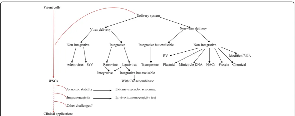
was achieved in only approximately 1/3 of samples [55]. Moreover, culturing HUCs usually requires long periods of time (at least two weeks) [53]. Furthermore, iPSCs de-rived from this source are not fully analyzed so far regard-ing genomic stability and differentiation potential. Thus, further efforts are still needed to identify the ideal tissue for iPS generation that would be effective, safe, and con-venient. A second major breakthrough is the elimination of the integration of the viral transgenes into the somatic genome. Current methods for iPSC induction can be di-vided into four categories based on their delivery system: viruses, DNA and RNA (plasmid, episomal plasmid, and transposon), cell-penetrating peptides, and chemicals. An-other classification is based on genome-integration: inte-gration, integration but excisable, and integration-free methods. Non-viral delivery integration-free methods, such as plasmid [34], EV [35], proteins [36,37], and chemi-cals [41], have been successfully employed to produce integration-free iPSCs, albeit at a lower efficiency than retroviral and lentiviral methods. The reasons for the low efficiency of iPSC induction are unclear, but it may be that the precise balance between transgenes and/or the expres-sion level of each transgene is important for reprogram-ming [18]. Generally, EV is now regarded to be one of the most cost effective, non-viral, integration-free vectors at present. It should be emphasized that the small molecule strategy is much more promising because of its outstand-ing advantages. Small molecule compounds can be cell permeable, non-immunogenic, and more easily synthe-sized, preserved, and standardized. Moreover, their ef-fects on the function of specific proteins are often reversible and cell fate can be manipulated in a controlled
microenvironment. This strategy could be more cost-effective if the efficiency of could be improved.
It is far too early to know for sure whether hiPSCs are safe for use in medical applications. Although hiPSC lines generated by integration-free methods are not found to have integrated transgenes by Southern blot analyses and PCR analyses, small pieces of vectors and transgenes could be inserted into the genome but not observed, given the detection limits of ordinary methods. Thus, because it is important to validate the quality of iPSCs, extensive genetic screening should become a standard procedure to ensure the safety of hiPSCs prior to their clinical use. Another pitfall that was discovered recently is potential immunogenicity against trans-planted iPSCs. Immune tolerance had been considered an advantage of autologous cell transplantation derived from patient iPSCs. However, it is found that some cer-tain but not all tissues derived from iPSC-derived cells can be immunogenic in syngenic hosts by using a tera-toma transplantation model. The reasons for this phenomenon are very complicated, and both genetic and epigenetic defects in iPSCs could contribute either dir-ectly or indirdir-ectly [104]. In vivo immunogenicity testing should be used as a screening platform to improve the reprogramming technology [105].
Conclusions and future directions
Generally speaking, researches to develop new methods for iPS cell generation for clinical applications are an on-going process. The range of tissue origins seems to have been extensively investigated, but we still have to find an ideal one for real practical application; rather, it is best
Parent cells
Delivery system
Virus delivery Non-virus delivery
Non-integrative Integrative Integrative but excisable Non-integrative
EV Modified RNA
Adenovirus SeV Retrovirus Lentivirus Transposons Plasmid Minicircle DNA HACs Protein Chemical
Integrative Integrative but excisable
iPSCs With Cre-recombinase
Genomic stability Extensive genetic screening
Immunogenicity In vivo immunogenicity test
Other challenges?
Clinical applications
to find one that most suit for a given application. For in-stance, PBSs which possess strong blood-forming poten-tial are attractive for hematologists whereas DP cells maybe a better cell source for dermatologists to grow hairs. More efforts should be put on the improvement of the culture conditions and technology (see Figure 1). As for the approaches used to generate iPSCs, although they have been improved from viral integration to integration-free, there are still many challenges down the road to achieve their clinical application in humans (see Figure 2). It counts on further improvement of current reprogram-ming technology to minimize the genetic and epigenetic difference between iPSCs and donors. In addition, a more comprehensive understanding of the reprogramming process will be crucial for the development of future ap-proaches for clinical applications of iPSCs.
Abbreviations
iPS cells or iPSCs:Induced pluripotent stem cells; ESCs: Embryonic stem cells; MEFs: Mouse embryonic fibroblasts; TTFs: Tail-tip fibroblasts; O: Oct 3/4; S: Sox2; K: Klf4: M, c-Myc; hiPSCs: Human iPSCs; HDFs: Human dermal fibroblasts; HFLS: Human fibroblast-like synoviocytes; N: Nanog; L: Lin28; SeV: Sendai virus; miRNAs: microRNAs; oriP/EBNA1: Epstein-Barr nuclear antigen-1; EV: Episomal vector; IRES2: Internal ribosome entry site 2; SV40LT: SV40 large T gene; VPA: Valproic acid; Vc: Vitamin C; siRNA: Small-interfering RNA; hASCs: Human adipose stem cells; GFP: Green fluorescent protein; mRNA: Messenger RNA; CNVs: Copy number variations; PB: piggyBac; DP cells: Dermal papilla cells; BM: Bone marrow; CB: Umbilical cord blood; G-CSF: Granulocyte colony stimulating factor; PBCs: Peripheral blood cells; TCR: T cell receptor; HUCs: Human urine-derived cells; NSCs: Neural stem cells; dox: Doxycycline; CMCs: Chorionic mesenchymal cells; UMCs: Mesenchymal-like cells from the umbilical cord matrix, AMCs, the amniotic membrane mesenchyme; GSK-3: Glycogen synthase kinase 3; SFK: pan-Src family kinase; PCR: Polymerase chain reaction; bp: Base pairs; LTR: Long terminal repeat; HACs: Human artificial chromosome vectors; HNFs: Human newborn fibroblasts; HSV-TK: Herpes simplex virus thymidine kinase; CPP: Cell-penetrating peptide.
Competing interests
The authors confirm that this article content has no conflicts of interest.
Authors’contributions
JL and WS carried out the primary literature search and drafted the manuscript. GP and JZ provided material input and helped in revising the manuscript. WS and GP conceived the study and provided field expertise. All authors read and approved the final manuscript.
Acknowledgments
This work was performed with support of the Department of Science and Technology of Shandong Province (2013GSF11811).
Author details
1Department of Oncology, Shandong Provincial Hospital Affiliated to
Shandong University, No. 324 Jingwu Weiqi Road, Jinan 250021, P.R. China.
2Guangzhou Institutes of Biomedicine and Health, Chinese Academy of
Science, 190 Kaiyuan Avenue, Science Park, Guangzhou 510530, P.R. China.
Received: 24 April 2014 Accepted: 1 July 2014 Published: 19 July 2014
References
1. Takahashi K, Yamanaka S:Induction of pluripotent stem cells from mouse embryonic and adult fibroblast cultures by defined factors.Cell2006, 126:663–676.
2. Takahashi K, Tanabe K, Ohnuki M, Narita M, Ichisaka T, Tomoda K, Yamanaka S: Induction of pluripotent stem cells from adult human fibroblasts by defined factors.Cell2007,131:861–872.
3. Yu J, Vodyanik MA, Smuga-Otto K, Antosiewicz-Bourget J, Frane JL, Tian S, Nie J, Jonsdottir GA, Ruotti V, Stewart R, Slukvin II, Thomson JA:Induced pluripotent stem cell lines derived from human somatic cells.Science 2007,318:1917–1920.
4. Watanabe A, Yamada Y, Yamanaka S:Epigenetic regulation in pluripotent stem cells: a key to breaking the epigenetic barrier.Phil Trans R Soc B 2012,368:2012292.
5. Jaenisch R, Young R:Stem cells, the molecular circuitry of pluripotency and nuclear reprogramming.Cell2008,132:567–582.
6. Spivakov M, Fisher AG:Epigenetic signatures of stem-cell identity.Nat Rev Genet2007,8:263–271.
7. Mikkelsen TS, Hanna J, Zhang X, Ku M, Wernig M, Schorderet P, Bernstein BE, Jaenisch R, Lander ES, Meissner A:Dissecting direct reprogramming through integrative genomic analysis.Nature2008,454:49–55.
8. Meshorer E, Misteli T:Chromatin in pluripotent embryonic stem cells and differentiation.Nat Rev Mol Cell Biol2006,7:540–546.
9. Thomson JA, Itskovitz-Eldor J, Shapiro SS, Waknitz MA, Swiergiel JJ, Marshall VS, Jones JM:Embryonic stem cell lines derived from human blastocysts. Science1998,282:1145–1147.
10. Imaizumi Y, Okano H:Modeling human neurological disorders with induced pluripotent stem cells.J Neurochem2014,129:388–399. 11. Gorba T, Conti L:Neural stem cells, as tools for drug discovery: novel
platforms and approaches.Expert Opin Drug Discov2013,8:1083–1094. 12. Xu D, TM O, Shartava A, Fowles TC, Yang J, Fink LM, Ward DC, Mihm MC,
Waner M, Ma Y:Isolation, characterization, and in vitro propagation of infantile hemangioma stem cells and an in vivo mouse model.J Hematol Oncol2011,6:54–64.
13. Yang J, Aguila JR, Alipio Z, Lai R, Fink LM, Ma Y:Enhanced self-renewal of hematopoietic stem/progenitor cells mediated by the stem cell gene Sall4.J Hematol Oncol.2011,6:38–51.
14. Takahashi J:Cell replacement therapy for Parkinson's disease using iPS cells.Rinsho Shinkeigaku2013,53:1009–1012.
15. Carpenter L, Carr C, Yang CT, Stuckey DJ, Clarke K, Watt SM:Efficient differentiation of human induced pluripotent stem cells generates cardiac cells that provide protection following myocardial infarction in the rat.Stem Cells Dev2012,21:977–986.
16. Okita K, Ichisaka T, Yamanaka S:Generation of germ-line competent induced pluripotent stem cells.Nature2007,448:313–317.
17. Yamamoto K, Okamura A, Sanada Y, Yakushijin K, Matsuoka H, Minami H: Micronuclei-associated MYC amplification in the form of double minute chromosomes in acute myeloid leukemia.Am J Hemato2013,88:717–718. 18. Okita K, Yamanaka S:Induced pluripotent stem cells: opportunities and
challenges.Philos Trans R Soc Lond B Biol Sci2011,366:2198–2207. 19. Huangfu D, Osafune K, Maehr R, Guo W, Eijkelenboom A, Chen S,
Muhlestein W, Melton DA:Induction of pluripotent stem cells from primary human fibroblasts with only Oct4 and Sox2.Nature Biotechnology 2008,26:1269–1275.
20. Liao J, Cui C, Chen S, Ren J, Chen J, Gao Y, Li H, Jia N, Cheng L, Xiao H, Xiao L: Generation of induced pluripotent stem cell lines from adult rat cells. Cell Stem Cell2009,4:11–15.
21. Liu H, Zhu F, Yong J, Zhang P, Hou P, Li H, Jiang W, Cai J, Liu M, Cui K, Qu X, Xiang T, Lu D, Chi X, Gao G, Ji W, Ding M, Deng H:Generation of induced pluripotent stem cells from adult rhesus monkey fibroblasts.Cell Stem Cell 2008,3:587–590.
22. Esteban MA, Xu J, Yang J, Peng M, Qin D, Li W, Jiang Z, Chen J, Deng K, Zhong M, Cai J, Lai L, Pei D:Generation of induced pluripotent stem cell lines from Tibetan miniature pig.J Biol Chem2009,284:17634–17640. 23. Shimada H, Nakada A, Hashimoto Y, Shigeno K, Shionoya Y, Nakamura T:
Generation of canine induced pluripotent stem cells by retroviral transduction and chemical inhibitors.Mol Reprod Dev2009,77:2. 24. Honda A, Hirose M, Hatori M, Matoba S, Miyoshi H, Inoue K, Ogura A:
Generation of induced pluripotent stem cells in rabbits: potential experimental models for human regenerative medicine.J Biol Chem2010, 285:31362–31369.