Developing Implant Esthetics in
Compromised Ridge
Rath SK,1 Ankit Gupta,2 Chavadar BK,3 Sandeep Tayal,4 Preeti Kaur5ABSTRACT:
Background: The article presents a case of anterior
compromised edentulous ridge managed with ridge augmentation utilizing guide bone regeneration (GBR) technique and subsequently restored by implant supported prosthesis to provide esthetics.
Methods: Prior to implant placement, the horizontal ridge
defect in the anterior maxilla was augmented utilizing guided bone regeneration technique with a mixture of alloplastic biomaterials, bioactive glass and medical grade calcium sulfate, covered with an absorbable bio-collagen membrane.
Results: After 6 months, upon exposure, it was observed that
the augmentation was achieved. This was confirmed during the exposure for implant placement, which was restored with satisfactory outcome.
Conclusion: Guided bone regeneration is a predictable
treatment modality for ridge deficiencies in the anterior maxilla which warrants high esthetic outcome. When combined with additional soft tissue augmentative procedures and prosthodontic rehabilitation, outcome often satisfies patient's esthetic expectations.
Key words: Maxillary anterior compromised ridge,
Guided-bone regeneration, Implant esthetics
doi: 10.5866/2015.7.10278
1Classified Spl (Periodontics)
Commanding Officer and Corps Dental Advisor 9 Corps Dental Unit, Yol Cantt
2,3Dental Officer
9 Corps Dental Unit, Yol Cantt 4Graded Spl (Prosthodontics)
5Graded Spl (Oral & Maxillofacial Surgery) 9 Corps Dental Unit, Yol Cantt
Article Info:
Received: October 10, 2015
Review Completed: November 8, 2015 Accepted: December 9, 2015
Available Online: March, 2016 (www.nacd.in) © NAD, 2015 - All rights reserved
Email for correspondence: [email protected]
Quick Response Code
INTRODUCTION
Traumatic tooth loss leads to alveolar resorption especially in sagittal direction, either due to direct loss of osseous structure because of injury or due to resorption of the alveolar crest that takes place after the loss of tooth. The alveolar process is often affected by the tooth loss, by a continuous resorptive process and has been seen to follow a predictable pattern, i.e., the labial aspect of the alveolar crest is the principal site of resorption, which first reduces in width and later in height.1, 2 Shortage of bone can
I
NDIANJ
OURNALOFD
ENTALA
DVANCEMENTSJ o u r n a l h o m e p a g e : w w w. n a c d . i n
prevent proper positioning of dental implants unless the volume of bone is increased before implantation. In the maxillary anterior area, this is also an issue of the esthetics. Several treatment modalities have been presented to augment the bone.
Guided bone regeneration (GBR) can maintain the bone volume and soft tissue contours. While this technique only requires a single surgical procedure, it is technically demanding.3 The GBR concept
teeth. On the other hand, guided tissue regeneration (GTR) was first developed in the early 1980s by Nyman et al.4 This concept is based on the principle
that specific cells contribute to the formation of specific tissues. Melcher described the concept of selective cell repopulation of defects to enhance healing.5
The present case report deals with a case of anterior compromised edentulous ridge managed with ridge augmentation using GBR technique and subsequently restored by implant supported prosthesis to provide esthetics as primary concern.
CASE REPORT
A medically and periodontally stable 25-year-old, non-smoking male reported to Department Of Periodontology, Army Dental Centre, Delhi Cantt and presented with a previously traumatic loss of tooth No. 11 (Figure 1A & 1B). The patient’s primary concern was esthetics and he also wanted rehabilitation with a implant supported prosthesis. Evaluation of the edentulous space with bone mapping revealed Siebert’s class III ridge defect i.e., loss in height and width of the alveolar ridge. The labio palatal width of the alveolar bone was measured to be just 3 mm at coronal crest which was quite inadequate for placement of implant. The Treatment Sequence followed included:
GBR at No. 11 site 6-month healing period
Implant placement in No.11site 3-month healing period
Guided soft-tissue growth
Final implant No 11 crown restoration
Routine haemogram and urine analysis was carried out to exclude any contraindication for surgery. Clinical findings were correlated with radiological findings of Orthopantomogram and Radio- visiography for confirmation of type of osseous defect and compare the post-operative gain of alveolar bone (Figure 1C).
Surgical phase:
Local anesthetic induction was achieved using
with a small right angle coronal modification was placed at the mesio-buccal line angle of No 12 and 21. A full-thickness flap was elevated past the mucogingival junction, and periodontal scoring took place near the base of the flap to facilitate the coronal advancement necessary for primary closure (Figure 2).
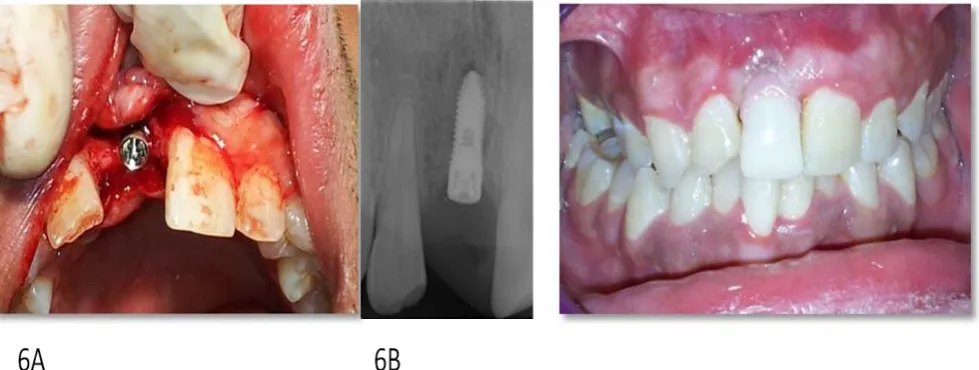
Decortication of the ridge was performed using a round bur to achieve bleeding points. Synthetic alloplast Bio active glass (PERIOGLASS) mixed with medical grade calcium sulfate was placed on the ridge to augment it buccally and coronally (Figure 3). An absorbable bio-collagen membrane (PROGUIDE) trimmed and used to cover the grafted area and secured with two sutures secured with soft tissue towards mucogingival junction at its apical aspect (Figure 4). The membrane was pulled down coronally to be secured with sutures with soft tissues from palatal side (Figure 5). After 6 months of uneventful healing, the available alveolar bone width was measured to be approximately 8 mm with a resultant gain of 6 mm. Sequential osteotomy was carried out till the implant length and width was achieved as per manufacturer’s instructions. IOPA radiograph was taken to confirm the angulation. A 2 stage endosseous implant measuring 3.25 X 9.5 mm was wrenched using appropriate torque (Figure 6A & 6B). After implant placement, flap was closed using simple interrupted 3-0 silk sutures. Suture removal was done after 10 days and post operative healing was uneventful.
Prosthetic phase:
Figure 1: A) & B) Pre-Operative intra-oral photograph of the patient; C) Pre-operative digital periapical radiograph
Figure 2: Recipient site exposure Figure 3: Defect coverage with a mixture of perioglass and medical grade calcium sulphate
Figure 6: A) & B) Two-piece implant placement (AB 3.25 x 9.5mm
months respectively. After 1year of clinical loading, the implant remained stable (Figure 10 A & 10B).
DISCUSSION
Among the different methods, guided bone regeneration (GBR) is the most popular and best documented for the treatment of localized bone defects in the jaws, probably due to its relative simplicity of use while allowing the placement of endosseous implants in areas of the jaw with bony defects and/or insufficient bone volume. Highly predictable success rates can be achieved using GBR; in fact, it has been shown that success rates of implants placed at GBR treated sites and sites without bone augmentation are comparable.6
Guided Bone Regeneration (GBR) has been used at the bone defect site to promote osseous regeneration and to preserve a large percent of grafted material which are placed and act by process of osteogenic, osteoconductive or osteoinductive activity.7-9 Prevention of soft tissue migration to the
grafted site is a necessary factor for creation and maintenance of space adequate enough for regeneration of bone.
GBR can be achieved by application of either resorbable or non resorbale membrane. Non resorbable barriers may provide predictable regeneration due to their inherent structural integrity but use of these membranes typically requires a second surgical procedure for their removal. Additionally, membrane exposure may necessitate premature removal, resulting in less than optimal results.10 It would be desirable
therefore, to use a resorbable membrane , but maintains its barrier function for as long as possible. Resorbable membranes with a rapid resorption profile may not exclude the connective tissue long enough for bone to completely fill the defect. In the present case, medical grade calcium sulphate has been moulded and applied as a membrane over the bone grafts and a resorbable bio collagen membrane used over it which was adequately covering the defect site.
Although it relieves supracrestal deficiencies, GBR best resolves space-making defects, which occur within an envelope of bone.11, 12 The intact
surrounding walls provide viable cells, blood supply, containment for graft particles, and a buttress to support the regenerative barrier and prevent collapse. Predictability of augmentation is directly proportional to the amount of bone that is enclosing the space. Ridge augmentation in the case under
discussion was not having intact walls. Therefore 100% regeneration and augmentation was neither expected nor achieved. But we could predictably achieve the bucco-palatal width adequate enough for placement of implant.
This clinical case presentation demonstrates the combination of grafting procedure by GBR followed by implant placement in one localized compromised case. The problem encountered in this case was long time period involvement and frequent visit by the patient
REFERENCES
1. Newman MG, Takei HH, Klokkevold PR, Carranza FA. Caranza’s Clinical periodontology, 10th ed. St Louis: Elsevier (Saunder’s); 2006: p. 301.
2. Lindhe J, Karing T, Lang NP. Clinical Periodontology and Implant Dentistry. 4th ed. New Delhi: Jaypee Brothers; 2003: p. 576.
3. Chen ST, Buser D. Clinical and esthetic outcomes of implants placed in postextraction sites. Int J Oral Maxillofac Implants 2009; 24:186-217.
4. Nyman S, Karring T, Lindhe J, Planten S. Healing following implantation of periodontitis-affected roots into gingival connective tissue. J Clin Periodontol 1980; 7(5):394-401.
5. Melcher AH. On the repair potential of periodontal tissues. J Periodontol 1976; 47(5):256-260.
6. Hammerle CH, Jung RE , Feloutzis A. A systematic review of the survival of implants in bone sites augmented with barrier membranes (guided bone regeneration) in partially edentulous patients. Journal of Clinical Periodontology 2002; 29:226-231.
7. Dahlin C, Gottlow J, Linde A, Nyman S. Healing of maxillary and mandibular defects using a membrane technique. An experimental study in monkeys. Scan J Plas and Recon Surg and Hand Surg 1990; 24:13-19.
8. Becker W, Dahlin C, Becker B, Lekholm U, van Steenberghe D, Higuchi K, Kultje C. The use of e-PTFE barrier membranes for bone promotion around titanium implants placed into extraction sockets: a prospective multicenter study. Int J Oral & Maxillofac Implants 1994; 9:31-40.
9. Mellonig J, Nevins M, Sanchez R. Evaluation of bioabsorbable physical barrier for guided bone regeneration. Part II. Material and a bone replacement graft. Int J Periodontics Restorative Dent 1998; 18:139-149.
10. Simion M, Trisi P, Maglione M, Piattelli A. A preliminary report on a method for studying the permeability of expanded polytetrafluoroethylene membrane to bacteria in vitro: A scanning electron microscopic and histological study. J Periodontol 1994; 65:775-781.
11. Tinti C, Parma-Benfenati S. Clinical classification of bone defects concerning the placement of dental implants. Int J Periodontics Restorative Dent 2003; 23(2):147-155.