Optimisation of qPCR-based methods for
Giardia lamblia and Cryptosporidium spp.
detection in human stool samples
Master’s Thesis
Rosa Mattila
Institute of Biomedical Technology (IBT)
University of Tampere
Acknowledgements
This thesis was carried out in Heikki Hyöty’s research group, Department of Virology, School of Medicine at the University of Tampere. I would like to thank him for this great opportunity to be a part of a truly interesting field of research - a linkage of autoimmune and allergic diseases to infections. I would also like to thank my supervisor, M.Sc. Sami Oikarinen, for guidance and for providing me with creative solutions to problems I encountered in my everyday labwork. A special thank you also for the laboratory technicians of the group for their great technical support and patience with me and my sometimes-weird requests and questions. Also the researchers M.Sc. Hanna Honkanen and M.Sc. Noora Nurminen deserve my thanks for their help and useful advice. Optimising of methods is not possible without samples known to be positive. Obtaining such samples when quite irregularly occurring parasites are in question is not always easy. As for these samples, I’d like to thank Kristin Elwin and Tapio Seiskari.
I would also like to thank my family for their always-reliable support and my friends for all the happy memories of my university years. Special thanks go to my great friends and grammar inspectors Julia Johansson and Jutta Laiho, without you this thesis would have quite a different outcome!
Lastly, but most importantly, I’d like to thank my fiancé Eero Juuti for always believing in me and putting up with my constantly changing moods during the making of this thesis.
This work was funded by EU 7th framework programme (DIABIMMUNE project, contract number HEALTH-F2-202063).
Tampere, May 2013 Rosa Mattila
PRO GRADU -TUTKIELMA
Paikka: TAMPEREEN YLIOPISTO
Biolääketieteellisen teknologian yksikkö (IBT) Tekijä: MATTILA, ROSA SOFIA
Otsikko: qPCR-pohjaisten seulontamenetelmien optimointi Giardia lamblian ja
Cryptosporidiumin toteamiseksi ihmisen ulostenäytteistä Sivumäärä: 47 sivua, 1 sivu liitteitä
Ohjaaja: FM Sami Oikarinen
Tarkastajat: Professorit Markku Kulomaa ja Heikki Hyöty Päiväys: Toukokuu 2013
________________________________________________________________________________
Tiivistelmä
Tutkimuksen tausta ja tavoitteet: Miljoonia ihmisiä kuolee joka vuosi suolistoloisten aiheuttamiin sairauksiin. Kaksi epidemiologisesti tärkeää loista, Giardia lamblia ja
Cryptosporidium, aiheuttavat esimerkiksi ripulia, oksentelua ja pahimmillaan imeytymishäiriöitä. Näiden loisten toteaminen on tähän asti tehty pääasiassa mikroskopoimalla, mutta kvantitatiivinen polymeraasiketjureaktio (qPCR) voisi olla menetelmänä tarkempi, herkempi sekä nopeampi. Menetelmällä voitaisiin myös tutkia useampia näytteitä yhtä aikaa. Tämän tutkimuksen tavoitteena oli optimoida menetelmät ja kehittää protokolla, jonka avulla voidaan todeta suolistoloiset Giardia lamblia ja Cryptosporidium (C. parvum ja C. hominis) kliinisistä ulostenäytteistä. Tavoitteena oli tehokas menetelmä, jota voidaan käyttää useiden näytteiden ja loisten yhtäaikaiseen tutkimiseen.
Tutkimusmenetelmät: Giardia lamblia- tai Cryptosporidium hominis -positiivisia ihmisen ulostenäytteitä käytettiin erilaisten esikäsittelyiden (konsentrointimenetelmät, lämpökäsittelyt, sulatus-jäädytyssyklit ja proteinaasikäsittelyt) sekä niiden yhdistelmien tutkimiseen, jotta löydettäisiin optimaalinen menetelmä mahdollisimman suuren DNA-määrän vapauttamiseksi loisten (oo)kystista. Kaikkiaan kokeiltiin 13 erilaista protokollaa herkimmän menetelmän löytämiseksi. Menetelmän kehittämiseen sisältyi myös kolmen kaupallisen DNA-eristysmenetelmän testaus sekä qPCR-reaktion aluke- ja koetinpitoisuuksien optimointi, jotka tehtiin ensin molemmille loisille erikseen, minkä jälkeen ne yhdistettiin samaan reaktioon. Menetelmän kehittämisen jälkeen DiabImmune-tutkimuksen näytteistä testattiin näitä loisia ja qPCR-reaktion toimivuus varmistettiin kliinisellä Cryptosporidium parvum –positiivisella
näytteellä.
Tutkimustulokset: Esikäsittely, joka koostui 10 minuutin lämpökäsittelystä (+98 °C) ja yön yli kestävästä proteinaasikäsittelystä (+56 °C), todettiin parhaaksi. Valinta tehtiin pääasiassa Giardia lamblia -näytteen tulosten perusteella, koska Cryptosporidium hominis –näyte monistui PCR-menetelmällä vain heikosti ja näin ollen tulokset eivät olleet luotettavasti toistettavia. Virus-RNA:n eristämiseen tarkoitettu eristysmenetelmä osoittautui herkimmäksi. Erot eri qPCR-aluke- ja
-koetinpitoisuuksien monistustehokkuuksien välillä olivat pieniä ja optimaalisiksi pitoisuuksiksi valittiin pienimmät konsentraatiot, jotka monistivat yhtä tehokkaasti kohdetemplaattia.
Cryptosporidium-qPCR:n todettiin havaitsevan sekä C. hominista että C. parvumia. Kaikki DiabImmune-tutkimuksessa otetut taustaväestöä edustavien lasten ulostenäytteet olivat negatiivisia.
Johtopäätökset: Tässä tutkimuksessa kehitetty protokolla on toistettava, herkkä ja useiden näytteiden yhtäaikaisen tutkimisen mahdollistava menetelmä, jolla voidaan todeta kyseisiä loisia ihmisten ulostenäytteistä. Koska Cryptosporidium hominis –näyte oli heikosti positiivinen, PCR-menetelmän toimivuus varmistettiin C. parvum-positiivisella näytteellä. Tässä tutkimuksessa optimoitu Giardia lamblian ja Cryptosporidiumin osoitukseen kehitetty protokolla on otettu käyttöön professori Heikki Hyödyn laboratoriossa.
MASTER’S THESIS
Place: UNIVERSITY OF TAMPERE
Institute of Biomedical Technology (IBT) Author: MATTILA, ROSA SOFIA
Title: Optimisation of qPCR-based methods for Giardia lamblia and
Cryptosporidium spp. detection in human stool samples Pages: 47 pages, 1 appendix page
Supervisor: M.Sc. Sami Oikarinen
Reviewers: Professors Markku Kulomaa and Heikki Hyöty
Date: May 2013
________________________________________________________________________________
Abstract
Background and aims: Millions of people die every year of intestinal parasite diseases. Two epidemiologically important parasites, Giardia lamblia and Cryptosporidium species (spp.) cause e.g. diarrhoea, vomiting and at worst malabsorption. Detection of these parasites has thus far been done by e.g. microscopy, but quantitative polymerase chain reaction (qPCR) would be a more accurate, sensitive and less time-consuming method and more samples could be tested at a time. The aim of this thesis was to optimise methods and to develop a protocol for simultaneous detection of human intestinal parasites Giardia lamblia and Cryptosporidium spp. (C. parvum and C. hominis) from clinical stool samples in a high-throughput format.
Methods: Human stool samples positive for Giardia lamblia or Cryptosporidium hominis were used to test different sample pretreatments (concentration methods, heat shocks, freeze-thaw cycles and proteinase treatments) and combinations to find the optimal one for maximal DNA release from the parasite (oo)cysts. Altogether 13 different protocols were tested. Three commercial DNA extraction kits were tested to find the best one for parasite DNA extraction. qPCR reaction was optimised first for primer and probe concentrations separately for both of the parasites and secondly these were combined into a multiplex reaction. DiabImmune study samples were tested for the two parasites after optimisation of methods and the functionality of qPCR reaction was confirmed for
Cryptosporidium parvum too.
Results: A pretreatment protocol consisting of a 10-minute heat shock at +98 °C and an overnight
proteinase treatment at +56 °C was considered the best. The choice was made mostly based on the
results of the Giardia lamblia sample, because the Cryptosporidium hominis sample showed constantly weak positivity. An extraction kit designed for viral RNA extraction was found the most effective. The differences in results obtained for different qPCR primer and probe concentrations were small and the concentrations were thus chosen cost-efficiently based on the lowest possible concentrations of the reagents. The Cryptosporidium spp. qPCR was found to detect both
Cryptosporidium hominis and parvum. All DiabImmune samples collected from children representing background population were negative
Conclusions: The protocol obtained as a result of this thesis is a reproducible, sensitive and high-throughput method for the detection of these two intestinal parasites in human stool samples. Even though the Cryptosporidium hominis sample used for optimisation was only weakly positive, the functionality of detection was confirmed with a C. parvum-positive sample. As a result of this thesis, professor Heikki Hyöty’s laboratory has started using the optimised protocol for detection of
Contents
Abbreviations... vii
1. Introduction ...8
2. Review of the literature...10
2.1 Giardia lamblia and giardiasis...10
2.2 Cryptosporidium spp. and cryptosporidiosis ...12
2.3 Proposed connection between intestinal parasite infections and allergic diseases...15
2.4 DiabImmune study description ...16
2.5 Theoretical background of the methods...17
2.5.1 Stool sample pretreatments ...17
2.5.2 DNA extraction ...19
2.5.3 qPCR and its use as a pathogen detection method...20
3. Main goals of the thesis...24
4. Materials and methods ...25
4.1 Samples ...25
4.2 Stool sample pretreatments ...25
4.2.1 Concentration methods...25 4.2.2 Freeze-thaw cycles ...26 4.2.3 Heat shocks ...26 4.2.4 Proteinase treatments ...26 4.3 DNA extraction...26 4.4 qPCR ...27
4.5 Testing of the optimised protocols ...29
4.6 Data analysis...29
5. Results ...30
5.1 Stool sample pretreatments ...30
5.2 DNA extraction...31
6. Discussion...36
6.1 Evaluation of the used methods...36
6.1.1 Stool sample pretreatments ...36
6.1.2 DNA extraction ...38 6.1.3 qPCR ...39 6.2 Future perspectives ...41 7. Conclusions ...43 8. References ...44 Appendix 1 ...48
Abbreviations
CRU UK Cryptosporidium Reference Unit CT threshold cycle
FAM fluorescein amidite
FRET fluorescence resonance energy transfer Fwd forward (in primers)
HLA human leukocyte antigen IBS irritable bowel syndrome LN liquid nitrogen
MGB minor groove binder
o/n overnight
qPCR quantitative polymerase chain reaction Rev reverse (in primers)
ROX 6-carboxy-X-rhodamine SD sample standard deviation
spp. species
SSU small subunit
8
1. Introduction
Diarrhoea was the fifth leading cause of death in low- and middle-income countries in 2008, accounting for approximately 5 % of deaths in total (WHO - mortality statistics, 2013). It was more common as a cause of death than HIV / AIDS in these countries. Certain viruses, bacteria or parasites cause these diarrhoeal diseases. In 2009, diarrhoea was reported the second leading cause of death in children under 5 years old, killing approximately 1,5 million children globally (WHO - diarrhoeal disease, 2013).
Giardia lamblia and Cryptosporidium species (spp.) are the two major intestinal parasites causing diarrhoea (Caccio et al., 2005). They share some common characteristics, e.g. their major transmission happens by faecal-oral route, either directly or indirectly (Caccio et al., 2005). Direct route can be due to insufficient hand hygiene or person-to-person contacts, whereas indirect routes include consuming contaminated water or food. Zoonotic transmission is also possible, being more common with Cryptosporidium than Giardia (Caccio et al., 2005). The infective form of these parasites is an (oo)cyst (oocyst for Cryptosporidium, cyst for Giardia lamblia).
These two parasites share the transmission mechanism and some other characteristics. Both have really low infectious dose: only 10 (oo)cysts might be enough to trigger an infection (Chen et al., 2002). The (oo)cysts are immediately infectious after their formation and very stable due to their hard walls (Caccio et al., 2005). It has been shown that Cryptosporidium oocysts are tougher than
Giardia lamblia cysts (Robertson and Gjerde, 2004). The (oo)cysts can survive in a latent form outside the body for months, taken that the location is damp enough (Sunnotel et al., 2006). They are resistant to virtually all traditional disinfectants and water purification systems. Furthermore, contaminated water requires filtration before use (Chen et al., 2002; Davies and Chalmers, 2009). On the other hand, some publications claim that filtration is not effective (Robertson and Gjerde, 2007), but filter pore size might also play a role. In addition, according to Sunnotel et al. (2006), conventional water purification systems are sufficient to get rid of Cryptosporidium oocysts. Evidently, this subject is still under debate and more research is required to draw definite conclusions.
Treatment options for both of these parasites are limited and ineffective: specific treatments are almost non-existent (Davies and Chalmers, 2009). Nitazoxanide, an anti-protozoal agent, is
sometimes used in immunocompromised Cryptosporidium patients to attenuate the symptoms and also for Giardia lamblia-infected adults in the USA (Davies and Chalmers, 2009; Sunnotel et al., 2006). Other medicines used usually have a general anti-parasitic activity (Davies and Chalmers, 2009). Some prospective new drugs, e.g. derivatives of benzimidazole are being studied for the treatment of Giardia lamblia, but these have so far been restricted to proof-of-principle and animal model studies (Tejman-Yarden and Eckmann, 2011).
2. Review of the literature
2.1 Giardia lamblia and giardiasis
Giardia lamblia is a eukaryotic, flagellated parasite, which taxonomically belongs to the phylum Metamonada, order Diplomonada and family Hexatimidae. It has seven genotypes (A-G), of which only the genotypes A and B cause disease in humans (Caccio et al., 2005). The different genotypes are phenotypically and genotypically quite heterogeneous, but morphologically almost identical within the species and thus impossible to distinguish by e.g. only microscopic examination (Caccio et al., 2005).
For historical reasons, Giardia lamblia is known by various names in the literature: Giardia duodenalis, Giardia intestinalis and Lamblia intestinalis are all synonyms for the name Giardia lamblia used in this thesis.
This parasite causes giardiasis, for which the main symptom is diarrhoea. The prevalence of the disease in developing countries is on average 20 % of the total population and on average 5 % in industrialised countries (Roxstrom-Lindquist et al., 2006). Risk factors for acquiring giardiasis include swimming in recreational pools (and ingesting the water), drinking contaminated tap water or eating food rinsed with it (Caccio et al., 2005). Zoonotic transmission is also possible, usually from dogs (Caccio et al., 2005). Symptoms vary between different countries (Cotton et al., 2011) and depend on several factors such as age, nutritional status, Giardia genotype, coinfections and host immune status (Roxstrom-Lindquist et al., 2006; Solaymani-Mohammadi and Singer, 2010). Immunocompetent individuals usually tend to be asymptomatic or have only mild symptoms, whereas immunocompromised individuals (e.g. HIV patients) are more likely to develop chronic diarrhoea (Roxstrom-Lindquist et al., 2006). Other symptoms include abdominal pain, vomiting and bloating.
Long-term effects of giardiasis can include malabsorption, allergies and growth problems in children (Caccio et al., 2005). Furthermore, giardiasis may cause symptoms in other parts of the body (e.g. joints, muscles, eyes and skin) even though the parasites stay exclusively in the gastrointestinal tract (Cotton et al., 2011). The reasons for these symptoms are not known. Giardiasis is also known to be able to trigger irritated bowel syndrome (IBS) (Cotton et al., 2011),
although it usually does not trigger mucosal inflammation, probably because the parasites do not invade the cells of the intestinal tract (Figure 1) (Roxstrom-Lindquist et al., 2006; Solaymani-Mohammadi and Singer, 2010).
Giardia lamblia’s lifecycle is extracellular at all phases (Roxstrom-Lindquist et al., 2006). The cysts have to be ingested, after which they travel to the upper small intestine (Roxstrom-Lindquist et al., 2006) where the thick walls are broken down presumably with the help of bile salts, proteases and lipases. This phase is called excystation (Figure 1). The released excyzoites differentiate into trophozoites which in turn start to replicate (Roxstrom-Lindquist et al., 2006). Once a certain threshold is surpassed, the symptoms are induced. The trophozoites adhere to the epithelia of the small intestine via an adherent disk (Cotton et al., 2011) and they have to constantly detach and reattach to avoid elimination by peristalsis (Roxstrom-Lindquist et al., 2006). In the next step, the trophozoites either disintegrate (Figure 1) or encyst to form new cysts, which are excreted with the faeces. Some trophozoites might also be excreted, but they are not viable outside the body.
The disease mechanism of giardiasis is still under debate. Shortening of the microvilli has been found in some patients, which could in turn explain the malabsorption symptoms often found in giardiasis (Cotton et al., 2011; Solaymani-Mohammadi and Singer, 2010). Increased transit rates, chloride hypersecretion and rearrangements of the host epithelial cell cytoskeletal actin network have also been reported (Cotton et al., 2011; Roxstrom-Lindquist et al., 2006). In addition, breakdown of the apical tight junctions between intestinal epithelial cells and increased apoptosis rates of these cells have been found (Cotton et al., 2011).
Some epidemiological studies have shown that previous Giardia lamblia infections are related to a reduced risk of re-infection and milder symptoms in such cases (Solaymani-Mohammadi and Singer, 2010). It has been established that the immune response against Giardia lamblia includes both innate and adaptive responses (Solaymani-Mohammadi and Singer, 2010), and that the variable surface proteins (VSPs) are the major antigens (Roxstrom-Lindquist et al., 2006), but the details of the aforementioned immune mechanisms are still not clear.
2.2 Cryptosporidium spp. and cryptosporidiosis
Cryptosporidium is a eukaryotic parasite that taxonomically belongs to the phylum Apicomplexa, order Eucoccidiorida and family Cryptosporidiidae. In humans, C. parvum and C. hominis are the most common forms (Caccio et al., 2005), but also C. muris, C. meleagridis and C. felis can infect humans (Chen et al., 2002). It seems that C. parvum has remained zoonotic and is often acquired from farm animals whereas C. hominis is quite strictly constricted to humans and is acquired usually via contaminated water or food (Davies and Chalmers, 2009; Robertson and Gjerde, 2007; Sunnotel et al., 2006). The prevalence of the different species varies geographically (Chen et al., 2002). However, the ones occurring more rarely could in fact be less pathogenic and asymptomatic and thus be frequently undiagnosed compared to the more pathogenic ones (Davies and Chalmers, 2009).
The Cryptosporidium spp. cause a disease called cryptosporidiosis. Its prevalence is on average 10 % of the total population in developing countries and on average 1-3 % in industrialised countries (Chen et al., 2002). As with giardiasis, cryptosporidiosis is more common in immunocompromised
Figure 1. Diagram of Giardia lamblia’s life cycle. (source: modified from http://www.idexx.com/view/xhtml/en_us/smallanimal/
education/reference-library/giardia.jsf; 3.5.2013)
individuals while infection is usually asymptomatic or mild in the immunocompetent ones. Zoonotic transmission is possible, usually from cattle (Caccio et al., 2005). What applies to Giardia lamblia, also applies to Cryptosporidium spp.: The parasites of the different species are phenotypically and genotypically heterogeneous, but morphologically almost identical (Caccio et al., 2005; Nichols et al., 2006).
Risk factors of cryptosporidiosis include age of the patient (very young or old being the most susceptible ones), travelling abroad and contacts with a person or a farm animal with cryptosporidiosis (Caccio et al., 2005; Chen et al., 2002). Symptoms are partly the same as in giardiasis: Diarrhoea is the main symptom, but vomiting and abdominal pain are also common (Chen et al., 2002). Apoptosis of neighbouring cells of the infected intestinal epithelial cell causes malabsorption and enhanced secretion; another suggested cause for the latter is an enterotoxin secreted by the parasite, but this is not widely verified yet (Chen et al., 2002). Symptoms of cryptosporidiosis can also be extraintestinal: Middle ear, pancreas and biliary tract can be infected, the latest being the most common one. How and why these aforementioned conditions occur is not known, but they are mostly observed in AIDS patients (Chen et al., 2002). Furthermore, lungs can be infected, because airborne transmission is possible for Cryptosporidium spp. (Caccio et al., 2005).
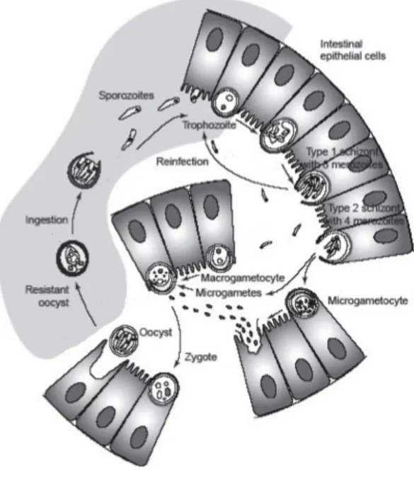
Cryptosporidium’s lifecycle is mainly intracellular but extracytoplasmic (Figure 2) (Chen et al., 2002). After the oocysts are ingested, they travel to the small intestine and excyst as a response to e.g. changed pH, producing infective sporozoites (Robertson and Gjerde, 2007). These attach to the epithelia. The epithelial cell in question forms a vacuole around the parasite, which however does not get totally endocytosed. In this vacuole, the parasite matures and undergoes asexual reproduction into type I meront that can either release merozoites to infect other cells or mature into a type II meront (Figure 2) (Chen et al., 2002). Type II meronts can in turn mature into gametocytes and start the sexual part of the life cycle. Gametocyte can either be a macro- or microgametocyte and the microgametes produced by the latter one fertilise the first mentioned (Figure 2) (Sunnotel et al., 2006). As a result, a zygote is formed and it undergoes encystation into an oocyst, which is excreted with the faeces.
Cryptosporidium has the ability to rearrange the cytoskeleton of its host by using actin-polymerising factors (Sunnotel et al., 2006). At the base of each vacuole, formed by these
cytoskeletal elements, is an electron-dense band which the parasite probably uses to take nutrients from the host (Chen et al., 2002).
Unlike Giardia lamblia, Cryptosporidium causes an inflammation reaction in the intestine (Chen et al., 2002). The inflammation affects only the most superficial epithelial surface of the mucosa. The mechanisms are not well known, however production of inflammatory cytokines can be involved in
Figure 2. Diagram of Cryptosporidium’s life cycle. (source: modified from Sunnotel et al. (2006)
the process. It is also possible to acquire resistance against re-infections of Cryptosporidium and interferon γ seems to have an important role in this phenomenon (Chen et al., 2002).
Cryptosporidium is also said to be able to trigger IBS, but this is still under debate (Davies and Chalmers, 2009).
2.3 Proposed connection between intestinal parasite infections and allergic diseases
Already from the very first months of human life, the immune system is broadly stimulated by different microorganisms (Olszak et al., 2012). The gastrointestinal tract and lungs are especially vulnerable, because they are the primary “landing sites” for the microbes. Furthermore, the gut and the barrier it forms are immature and quite permeable to different microbes to enter the body during infancy (Rautava et al., 2004). These early contacts (or the lack of them) may have a life-long influence on whether the person in question will get an allergic or an autoimmune disease (e.g. multiple sclerosis or type 1 diabetes) (Olszak et al., 2012).
Allergic diseases are an emerging problem in industrialised countries. In an allergic disease (e.g. asthma), seemingly innocuous antigens trigger a T-cell-mediated systemic inflammation (Romagnani, 2004). The causes are both genetic and environmental (Romagnani, 2004). Allergic diseases have been linked to decreased exposure to different pathogens and allergens, which in turn has raised a question about the possible role of the so-called hygiene hypothesis in the onset of these diseases (Ramsey and Celedon, 2005; Seiskari et al., 2007). The hygiene hypothesis suggests that exposure to different microorganisms during childhood protects from allergic diseases later in life due to the promoted maturation of the immune system (Yazdanbakhsh and Matricardi, 2004). One possible explanation for this is that early encounters with multiple microorganisms prevent e.g. the gastrointestinal tract from being colonised by harmful microbes. Alternatively, the exposure to diverse microorganisms might divert the immune system in such a direction that counteracts the development of allergic diseases (Ege et al., 2011).
Previously, these diseases along with several autoimmune diseases have been linked to gastrointestinal parasitic infections (Yazdanbakhsh and Matricardi, 2004). Some parasites (e.g.
Toxoplasma gondii, some helminths) have been found to be protective against these diseases whereas some (e.g. Campylobacter jejuni) are evidently predisposing (Ramsey and Celedon, 2005; Rautava et al., 2004; Seiskari et al., 2007). However, there are some inconsistencies between
different studies regarding these implications and sometimes other already existing diseases have an alternating effect on the results (Ramsey and Celedon, 2005).
2.4 DiabImmune study description
DiabImmune (complete title of the project: “Pathogenesis of type 1 diabetes: testing the hygiene hypothesis”) is a large, international collaboration project which aims to define the mechanisms of hygiene hypothesis in the pathogenesis of autoimmune and allergic diseases. Another objective is to assess the links between improved standard of hygiene and increased incidence of these diseases. At the border of Finland and Russian Karelia, there is a significant gradient in the standard of living. For example, the gross national product per capita of Russian Karelia is only one seventh of that of Finland. On the other hand, there are no significant differences in the mean temperatures or other physical factors, or in the background population regarding for example the distribution of the human leukocyte antigen (HLA) genotypes, which are used to assess predisposition to many autoimmune diseases. For these reasons, Finland and Russian Karelia represent a productive environment for studying gene-environment interactions in the pathogenesis of immune-mediated diseases. Estonia is included in the project because of its geographical location. It also represents a country that is undergoing a rapid transition. Furthermore, previous studies have shown that there are large differences in the occurrence of different microbes between these three areas (Viskari et al., 2004). For these reasons Estonia can be used as additional population of comparison.
In DiabImmune project, several different sample types (e.g. blood and stool samples) are collected from large cohorts of children in Finland, Russian Karelia and Estonia. One cohort comprises children that are assessed at the ages of 3 and 5 years. The second one is a birth cohort, in which the children are monitored from birth to the age of 3 years. The children are analysed for type 1 diabetes –associated autoantibodies and allergen-specific IgE-class antibodies, as well as for different infections, gene-expression profiles and nutritional factors. The different research groups participating in the study work in collaboration to bring new information about the connections between microbial exposure and allergic and autoimmune diseases.
The stool samples collected for DiabImmune study will be used in this thesis to test the optimised protocols for the possible presence of Cryptosporidium spp. and/or Giardia lamblia.
2.5 Theoretical background of the methods
Infections by Giardia lamblia and Cryptosporidium spp. are diagnosed by detecting (oo)cysts from stool samples. In most diagnostic laboratories around the world, stool sample microscopy is still the golden standard of detection for Giardia lamblia and Cryptosporidium spp. (Elwin et al., 2012; Schuurman et al., 2007). Usually multiple stool samples over time have to be tested, because (oo)cysts or trophozoites are not excreted constantly. However, microscopy as a technique has many drawbacks: It is time-consuming, labour-intensive, expensive and the results depend partly on the subjective observation (Schuurman et al., 2007; Verweij et al., 2003). Sensitivity of the technique is also a problem, because single (oo)cysts are difficult to detect.
ELISA, other immunoassays and qPCR can also be used for the detection of parasites (Schuurman et al., 2007; Verweij et al., 2003). However, antibody-based techniques can occasionally be non-specific due to cross-reactivity and have low sensitivity (Verweij et al., 2004). qPCR is thought to be more sensitive than microscopy, but it also has a cross-contamination risk. In addition, sensitivity depends on the system used: One Giardia lamblia cyst contains approximately 313 fg of chromosomal DNA (Schuurman et al., 2007) and not all qPCR systems detect such a small amount. The goal of this thesis was to develop new methods for the detection of these infections aiming at a high-throughput format, which would enable effective screening of large sample series. The practical aim was to use the same pretreatments and other assay steps for both Giardia lamblia and
Cryptosporidium spp., despite that in several previous publications different protocols have been used separately for different parasites.
2.5.1 Stool sample pretreatments
Stool samples usually require pretreating before they can be used to diagnose microbial agents, because they contain a lot of impurities (such as food degradation products and lipids) and inhibitors, which can affect downstream applications (Nantavisai et al., 2007). To overcome this problem, many publications have described stool sample pretreatments where so-called “spiked” stool samples have been used, meaning that purified (oo)cysts have been added to originally negative stool samples (Ng et al., 2005; Stroup et al., 2012). Alternatively, only prepurified (oo)cysts have been used to study wall disruption methods in several publications (Guy et al., 2003;
Minarovicova et al., 2009). Soil or sewage have also been used as sample material (Bertrand et al., 2004; Guy et al., 2003).
Since the amount of (oo)cysts in a stool sample of an infected individual is often very low (Chen et al., 2002) concentration step has been used in many protocols including simple centrifugation combined with pellet resuspension as well as PBS-ether-Percoll –method, which is based on sedimentation in discontinuous density gradient (Adamska et al., 2012; Haque et al., 2007)
In this thesis the focus is in different methods which are needed to disrupt the (oo)cyst wall. As mentioned before, the (oo)cyst wall is very tough, probably because of the cysteine- and disulfide-bond-rich VSP proteins that form it (Adamska et al., 2011; Roxstrom-Lindquist et al., 2006). The wall must be disrupted to access the DNA, which is used for downstream applications. Due to the vast selection of different methods, the present work focuses on those methods which are technically the most feasible for the aims of this study.
2.5.1.1 Freeze-thaw cycles
Freeze-thaw cycles with various lengths, repetition times and temperatures have been used in previous studies to disrupt the (oo)cysts’ wall. Their use is based on the idea that repeated temperature changes cause shearing forces on the (oo)cyst wall, thus leading to its destruction and consequently making the DNA accessible for extraction (Robertson and Gjerde, 2004). In most publications, the highest temperature varies from +95 °C to +100 °C and the lowest temperature is
usually achieved by liquid nitrogen (LN; -196 °C) (Adamska et al., 2011; Haque et al., 2007).
Adamska et al. (2011) noticed in their study that -70 °C was not cold enough in the freeze-thaw
cycling, because positive samples were negative in PCR detection. The length of the freeze-thaw cycling phases is usually 1-2 minutes (Fontaine and Guillot, 2002; Nichols et al., 2006) and the cycles are repeated 3 to 15 times (Adamska et al., 2011; Nichols et al., 2006). As can be seen from these examples, there is plenty of variation between published protocols. In addition, the execution varies; heating can for example be achieved with a water bath or a heat block (Adamska et al., 2011).
2.5.1.2 Heat shocks
Heat shocks of varying temperatures are one possible (oo)cyst wall disruption method. These usually only inactivate the (oo)cysts (Minarovicova et al., 2009) and for this reason they are not effective alone. Thus, heat shocks are usually combined with other steps, such as a proteinase treatment (see the next chapter) (Minarovicova et al., 2009; Verweij et al., 2003). In most publications heat shock refers to an incubation step at approximately +95 °C for 10-20 minutes,
though Sunnotel et al. (2006) claim that any temperature above +65 °C is enough to destroy the oocysts of Cryptosporidium.
2.5.1.3 Proteinase treatments
Proteinases have been used to disrupt parasite wall using different incubation periods (ranging from 1 h to overnight) at approximately +56 °C (Minarovicova et al., 2009). Proteinases are very
effective for Cryptosporidium spp., detaching the separate layers of the oocyst wall from each other (Adamska et al., 2011). Additional steps are often combined with the proteinase treatments, such as heat shocks (see chapter 2.5.1.2) or freeze-thaw cycles (see chapter 2.5.1.1), but the proteinase treatment has always been the last step of pretreatments (Nichols et al., 2006). Some publications also suggest that the proteinase reaction should be stopped by an incubation for approximately 15 minutes at +95 °C (Minarovicova et al., 2009) since the proteinase activity might inhibit some
downstream applications.
2.5.2 DNA extraction
Nowadays commercial kits are widely used for routine DNA and/or RNA extraction. Different kits usually follow the same principles: First the nucleic acids are bound to a porous filter of a column, the sample is washed and subsequently eluted. The differences are usually in the incubation times, the centrifugation parameters and in specific reagents used. In this thesis, different Qiagen (Hilden, Germany) kits were tested. These kits were chosen based on previous literature (Adamska et al., 2011; Guy et al., 2003; Haque et al., 2007) and the kits already in use in Heikki Hyöty’s laboratory.
2.5.3 qPCR and its use as a pathogen detection method
PCR is a method for DNA amplification. Conventional PCR has three phases including denaturation, annealing and extension (McPherson and Moller, 2000). In denaturation phase, the double-stranded template DNA is heated to a temperature where the two complementary strands separate. Next, in the annealing phase, the temperature is lowered so that the sequence-specific primers hybridise to their complementary target DNA sequences at the template and DNA polymerases start the extension from the 3’ end of the primers (McPherson and Moller, 2000). At the extension phase the temperature is optimal for the DNA polymerase to work and the extension continues. These three steps are repeated for a certain number of times, usually 35-45 cycles, depending on the application. Sometimes the annealing and extension steps can be combined, as is done in this thesis.
The PCR reaction requires different reagents. In this case, a ready-made master mix of the kit includes most of these reagents. It includes the thermostable Taq DNA polymerase that performs the actual DNA amplification. The DNA polymerase used in this thesis is of a modified form (HotStarTaq DNA polymerase) which is inactive in room temperature and has to be activated in the first step of the PCR reaction. This activation step reduces the amount of nonspecific PCR products. The master mix also includes buffer that is required to create optimal conditions for the reaction. This buffer is composed of NH4+ and K+ ions and MgCl2. The two ions promote specific primer
annealing and inhibit nonspecific binding and MgCl2 affects the stringency of the primer-template
interaction. Furthermore, the master mix includes a dNTP mix which has equivalent amounts of all four nucleotides that are used in the PCR reaction to synthesise the PCR products. Finally, this qPCR master mix includes a passive 6-carboxy-X-rhodamine (ROX) reference dye which is used as a baseline in the reaction.
qPCR is used both for DNA amplification (as conventional PCR) and for real-time monitoring of the amount of synthesized amplicons (McPherson and Moller, 2000). There are several ways to detect the amplification products. The conventional ones include for example SYBR Green or ethidium bromide, but they bind to all double-stranded DNA (dsDNA) present in the reaction tube, which excludes them from being used for diagnostic purposes (McPherson and Moller, 2000). Nowadays also fluorescence-based systems exist. The two most used ones are the TaqMan system and the two-probe system (fluorescence resonance energy transfer, FRET). Only the TaqMan system will be described here, for it is the one used in this thesis.
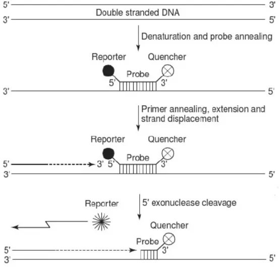
The TaqMan system uses, in addition to primers, an oligonucleotide probe that has a fluorophore reporter moiety at the 5’ end and a quencher moiety at the 3’ end (Figure 3). These moieties have an overlap in their emission and excitation spectra, thus making the fluorophore reporter unable to emit a detectable fluorescent signal at the presence of the quencher (McPherson and Moller, 2000). This probe is designed to hybridise to a complementary segment of the template DNA (downstream of the 5’ primer), but as mentioned before, as long as the quencher is bound to the other end of the same probe, the fluorophore moiety is not able to emit a fluorescent signal, regardless if it is bound to the target. During the extension phase of the PCR cycle, as the Taq DNA polymerase reaches the site where the probe is bound, it cleaves and thus liberates the 5’ fluorophore reporter moiety of the probe with its 5’ 3’ exonuclease activity. This leads to a detectable fluorescent signal (Figure 3) (McPherson and Moller, 2000). The amount of the specific PCR product is proportional to the amount of fluorescence.
Figure 3. Diagram of the principle of qPCR using the TaqMan system. (source: modified from McPherson and Moller (2000)
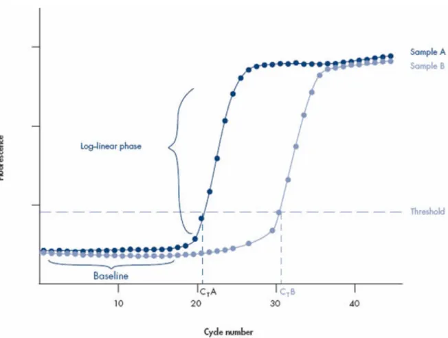
The ABI 7900HT qPCR machine does both the thermal cycling and fluorescence detection and also the measurements. The machine is connected to a computer with SDS software. It uses the ROX dye fluorescence to normalise the PCR product-related fluorescence signals. A baseline for the qPCR reaction is calculated based on the fluorescence acquired from the PCR products and it is reckoned the noise level of the early cycles when there is not yet detectable fluorescence from the amplification products (Figure 4) (QIAGEN - Data analysis 2013). The software produces a sigmoidal-shaped amplification plot (when using a linear scale, Figure 4), where the amount of detected fluorescence is plotted against the cycle number. The threshold is set to value above the baseline, but in the log-linear range of the plot. The threshold cycle (CT) values are determined
using this threshold: a CT value indicates the reaction cycle at which the amplification plot crosses
the set threshold (Figure 4) (QIAGEN - Data analysis 2013). The higher the CT value, the lower the
starting amount of the template: in Figure 4, sample A contains more starting template than sample B. CT values above 36 cycles are generally considered poorly reproducible and thus unreliable for
e.g. parasite detection (ten Hove et al., 2007). An increase of approximately 3,5 cycles indicates a 10-fold decrease in the amount of the template in the original sample.
qPCR is a powerful and worthy tool for parasite detection. It is based on the detection of the presence of parasite DNA in the samples. It is also possible to combine the detection of different parasites to one reaction in multiplex qPCR. Usually the combination of different primers in same PCR reaction tends to slightly inhibit each other, a phenomenon also known as “multiplex interference” (Hlousek et al., 2012). The benefit is still obvious, because working time and reagents can be saved and more analyses can be run with the machine. The main problem in using qPCR for parasite detection from stool samples is the presence of inhibitors that need to be removed beforehand (Abu Al-Soud and Radstrom, 2000). BSA is widely used for reduction of the inhibition in the PCR, but some publications claim it is useful only in the conventional PCR (Abu Al-Soud and Radstrom, 2000). Another problem is that often the stool samples are formalin-fixed. Formalin is used as sample preservative, but it is known to denaturate DNA, which partly inhibits e.g. qPCR reactions (Paglia and Visca, 2004; Srinivasan et al., 2002).
For Giardia lamblia, the small subunit (SSU) rRNA sequence is widely used in qPCR detection (Verweij et al., 2003) and it was also used in this thesis. For Cryptosporidium spp. detection, DNAJ-like gene was used (Bruijnesteijn van Coppenraet et al., 2009).
3. Main goals of the thesis
The main goals of this thesis were to optimise methods and to derive a protocol to detect human intestinal parasites Giardia lamblia and Cryptosporidium spp. (C. parvum and C. hominis) from clinical stool samples. These optimisations aimed at a protocol that can be used in a high-throughput format to screen large samples series in studies evaluating the possible links between allergic diseases and the aforementioned parasitic infections.
4. Materials and methods
4.1 Samples
Optimisation was carried out using clinical, formalin-fixed stool samples found positive for Giardia lamblia or Cryptosporidium spp. using traditional microscopic examination. Four Cryptosporidium -positive samples were obtained from the UK Cryptosporidium Reference Unit (CRU; Public Health Wales Microbiology ABM, Singleton Hospital, Swansea, UK). Three of them were positive for C. hominis and one for C. parvum. One of the C. hominis-positive samples was chosen for testing based on preliminary results (results not shown). The Giardia lamblia-positive sample was provided by the Fimlab Laboratories (Pirkanmaa Hospital District).
The stool samples were suspended into Hank’s buffer (Table S1, Appendix 1), which was supplemented with 4 % BSA (20 g of albumin (Sigma-Aldrich, Europe) in 500 ml of Minimum Essential Medium with Earle´s Salts, without L-Glutamine (PAA Laboratories, Pasching, Austria)). Stool samples were suspended to enable DNA extraction with Qiagen kits. Approximately 300 mg of stool was suspended into 3 ml of this buffer. The suspensions were stored in -20 °C.
4.2 Stool sample pretreatments
In the previuos literature, several different sample pretreatment techniques and their combinations have been tested to improve Cryptosporidium spp. and Giardia lamblia detection in human stool samples by PCR-based methods (Adamska et al., 2011; Haque et al., 2007). In this study, multiple different methods were tested including one with no pretreatments. In addition, sterile water samples were included as negative controls in every test. Summary of the methods and combinations is presented in Table 1.
4.2.1 Concentration methods
To test whether the (oo)cysts pellet during centrifugation, a stool suspension was centrifuged at 1118 g for 30 minutes at +4 °C (without braking). Another sample was treated without
aforementioned stool suspension solution (Table 1, protocol 1). Furthermore, sedimentation was tested as a gentler alternative to centrifuging. The stool suspension was left at the laminar to sedimentate for 1 hour and the supernatant was transferred to a new tube. This was repeated once and the final supernatant was used for downstream reactions (Table 1, protocols 1-7).
4.2.2 Freeze-thaw cycles
200 µl of stool suspension was incubated on a heat block of +100 °C for 2 minutes and
subsequently freezed in a closed container of LN for 2 minutes. These two stages were repeated for a different number of times: once, three times or ten times (Table 1, protocols 5-7 and 11-13).
4.2.3 Heat shocks
Heat shocks were carried out by incubating 200 µl of stool suspension on a heat block of +98 °C for
10 minutes. These heat shocks were used in combination with different proteinase treatments (Table 1, protocols 1-3 and 8-10).
4.2.4 Proteinase treatments
Proteinase treatments were executed with proteinase K (cat. no. 19133; Qiagen, Hilden, Germany). 15 µl of proteinase K was added to 200 µl of stool suspension, which was then incubated at +56 °C
on a heat block for 2 hours, 3 hours or overnight (o/n). The proteinase treatments were combined to a heat shock or freeze-thaw cycles (Table 1, protocols 1-13).
4.3 DNA extraction
The DNA extraction was carried out using commercial kits from Qiagen (Hilden, Germany): three different kits were tested. The kits were chosen based on literature and the laboratory’s own facilities: QIAamp DNA Blood Mini Kit (later on ‘blood kit’, cat. no. 51104) (Guy et al., 2003), QIAamp DNA Stool Mini Kit (later on ‘stool kit’, cat. no. 51504) (Babaei et al., 2011; Haque et al., 2007) and QIAamp Viral RNA Mini Kit (later on ‘viral kit’, cat. no. 52906).
The kits were tested by performing a sample elution series of 10-1 to 10-6 of a non-pretreated
Giardia lamblia sample. The most suitable kit was chosen based on sensitivity of the qPCR method. This testing was done in single tubes and columns and the optimised DNA extraction method was adapted onto 96-well plate system. The extracted DNA samples were either used immediately in the proceeding steps or stored in -20 °C.
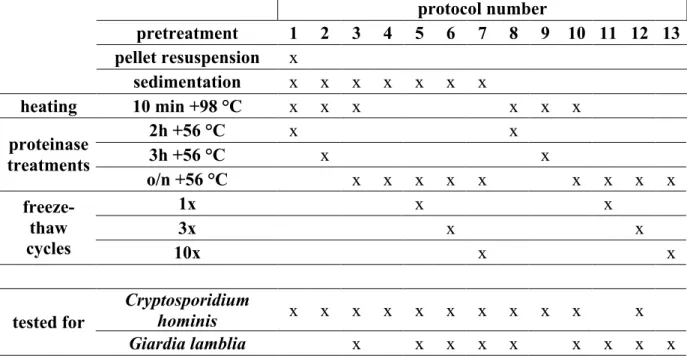
Table 1. Summary of the tested stool sample pretreatments. 'x' indicates that the step was included in the protocol. In the last two lines it indicates which protocols were implemented for each parasite.
protocol number pretreatment 1 2 3 4 5 6 7 8 9 10 11 12 13 pellet resuspension x sedimentation x x x x x x x heating 10 min +98 °C x x x x x x 2h +56 °C x x 3h +56 °C x x proteinase treatments o/n +56 °C x x x x x x x x x 1x x x 3x x x freeze- thaw cycles 10x x x Cryptosporidium hominis x x x x x x x x x x x tested for Giardia lamblia x x x x x x x x x
Some modifications were made on the kit protocols. The DNA extraction with viral kit was performed with combination of the viral kit reagents and RNEasy columns according to Heikki Hyöty’s laboratory’s routine practice. Also, the extraction with the stool kit was tested both with and without the InhibitEx tablets included in the kit.
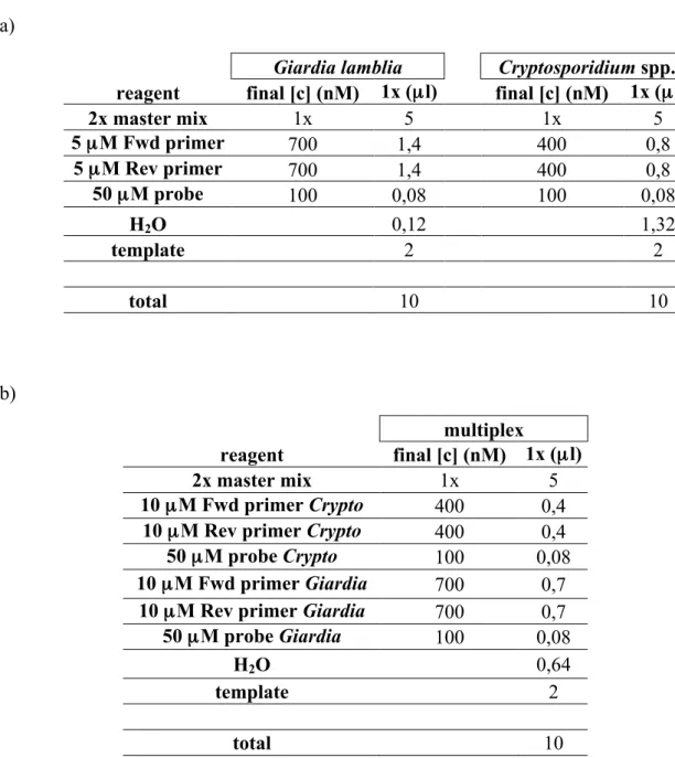
4.4 qPCR
The qPCR was performed with Applied Biosystems ABI 7900HT machine (LifeTech, Paisley, UK). The primer and probe sequences were selected from literature. These oligonucleotides had already been tested and shown to be specific and sensitive in previous publications. The Giardia lamblia -specific primers and probe were targeted to SSU rRNA gene of the parasite (GenBank accession no. M54878) to amplify and detect a 62-bp DNA fragment (Verweij et al., 2004). The Cryptosporidium
spp.-specific primers and probe were targeted to amplify and detect 70 bp fragment of the DNAJ-like protein gene (GenBank accession no. AF177278.1 / XM661034.1) (Bruijnesteijn van Coppenraet et al., 2009). The sequences of the primers and probes are shown in Table 2. The primers were ordered from Sigma-Aldrich (Europe) and the probes from AB Applied Biosystems (LifeTech, Paisley, UK). The primers and probes chosen for Cryptosporidium spp. recognise both
Cryptosporidium parvum and Cryptosporidium hominis, because it was not necessary to distinguish the two species. The qPCR was conducted with QuantiTect Probe PCR kit (cat. no. 204345; Qiagen, Hilden, Germany).
Table 2. Primers and probes used for qPCR. The abbreviations in the 5’ end and 3’ end of the probe sequences stand for the fluorescent dye and quencher moiety, respectively. ‘FAM’ stands for fluorescein amidite, ‘MGB’ stands for minor groove binder and ‘VIC’ is a proprietary name of Lifetech. The primers and probes for Giardia lamblia are from the publication (Verweij et al., 2004). The Cryptosporidium spp. oligonucleotides are from the publication (Bruijnesteijn van Coppenraet et al., 2009) with the exception that the fluorescent dye of the probe was changed from NED to VIC for technical reasons; ‘NED’ being a proprietary name of Lifetech.
The qPCR reactions were optimised for primer and probe concentrations according to the instructions of the qPCR kit and concentrations of 400-1000 µM for primers and 100-200 µM for
probes were tested. The total reaction volume was decreased from the directive 50 (25) µl to 10 µl,
because this reaction volume was already tested in the research group and it saves reagents. The volume of template sample was increased from the laboratory’s directive 0,8 µl to 2,0 µl (in 10 µl
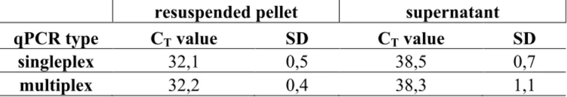
reaction) to ensure sufficient template amount in the reaction. Reactions for both parasites were first optimised separately as singleplex reactions and then combined to one, multiplex reaction.
Giardia lamblia
Forward primer GAC GGC TCA GGA CAA CGG TT Reverse primer TTG CCA GCG GTG TCC G
Probe FAM - CCC GCG GCG GTC CCT GCT AG - MGB
Cryptosporidium spp.
Forward primer CTT TTT ACC AAT CAC AGA ATC ATC AGA Reverse primer TGT GTT TGC CAA TGC ATA TGA A
The programme of the reaction (same for both singleplex and the multiplex reactions) is shown in Table 3, according to the instructions of the qPCR kit.
Table 3. The qPCR programme used for all qPCR reactions.
The threshold value used for the analysis of the qPCR data was the default value of 0,20 set by the software.
4.5 Testing of the optimised protocols
Clinical stool samples (n = 174) of the DiabImmune study were tested with the optimised protocols. The test was conducted in triplicate reactions. These samples were from Estonian and Finnish children representing background population and were all under two years of age. The samples had been stored in -70 °C until used. Sterile water samples were included as negative controls and samples positive for Giardia lamblia and Cryptosporidium hominis were included as positive controls.
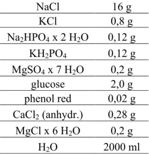
The Cryptosporidium spp. qPCR should detect both C. parvum and C. hominis and this was tested with purified DNA samples of the parasites provided by the CRU. The optimised protocol was tested with a positive sample that was acquired from HUSLAB (Helsinki and Uusimaa Hospital District), during an epidemic of Cryptosporidium parvum. This sample was tested from resuspended pellet and supernatant.
4.6 Data analysis
The results of the qPCR runs were obtained from the SDS software (version 2.2.2) and used to rank the pretreatments, alternative kits in DNA extraction and qPCR reagent concentrations. The results are presented with average C values and sample standard deviations (SD).
5. Results
The laboratory work of this thesis consisted of four major parts: Optimisations of stool sample pretreatments, DNA extraction and qPCR reaction steps and use of optimised methods for the detection of parasites in stools samples collected in DiabImmune project.
The optimisation of pretreatments and DNA extraction were tested using qPCR based on the qPCR kit’s basic protocol. Later the qPCR method was optimised to achieve the best possible sensitivity. All samples were tested by two or three parallel qPCR reactions. The qPCR results are presented in this thesis as average CT values of two or three tests and SD’s.
5.1 Stool sample pretreatments
There were clear efficacy differences between different stool sample pretreatments, especially when tested for the Giardia lamblia sample. The freeze-thaw cycles decreased the sensitivity of DNA extraction (Table 4; compare protocols 5-7 and 11-13 with protocols 3 and 10, respectively). Furthermore, the freeze-thaw cycles were very laborious. The 10-minute heat shock at +98 °C provided optimal results being a part of two of the tested protocols (Table 4, protocols 3 and 10). The proteinase K treatment at +56 °C was tested with incubation time of two hours, three hours and overnight. For the Giardia lamblia sample an overnight treatment (Table 4, protocols 3, 5-7 and 10-13) was more efficient than the two-hour treatment (Table 4, protocol 8) Sedimentation as a concentration step had a very small, if any effect on the Giardia lamblia results. Protocols 3 and 5-7 show only slightly stronger template amplification than protocols 10-13 (Table 4).
Testing of the Cryptosporidium hominis sample proved to be challenging, because the stool suspension contained only low concentration of Cryptosporidium hominis DNA. Due to that, only the most optimal pretreatments released enough target DNA for positive qPCR detection. Some of the pretreatment methods did not work or they were not effective enough (e.g. freeze-thaw cycles) and for these reasons negative results or very weak qPCR signals were obtained. Nevertheless, two protocols (Table 4, protocols 1 and 10) showed clearly detectable and reproducible template concentrations.
Concentration of original Cryptosporidium hominis sample was low, but centrifugation concentrated the oocysts and resuspension of the pellet increased the detection of Cryptosporidium hominis (results not shown). Using this pretreatment the sample was constantly positive in qPCR (Table 4, protocol 1). Sedimentation did not improve the results much, compared to non-sedimentated samples (Table 4; compare protocols 5-7 with protocols 11-13). Based on the results, the optimal protocol includes a 10-minute heating step at +98 °C and an overnight proteinase treatment at +56 °C (Table 4, protocol 10).
Stool samples with no pretreatments were tested with Giardia lamblia sample (Table 5). With the viral kit, the template amplification was the strongest and at approximately the same level as with the protocols 5-7 (Table 4).
5.2 DNA extraction
The viral kit was the most sensitive of the tested nucleic acid extraction methods. The same dilution series were extracted using viral, blood and stool kits and the qPCR amplification of extracted nucleic acid was strongest from extracts provided by the viral kit (Table 5). In addition, the viral kit already has a ready-made 96-well protocol, which supported its choice of being the most suitable kit for DNA extraction in professor Hyöty’s laboratory. Only this kit was used for the optimisation of the stool sample pretreatments and for the upcoming testing.
5.3 qPCR
Different primer and probe concentrations did not increase the sensitivity of either Giardia lamblia -specific or Cryptosporidium spp.-specific qPCR detection. The concentrations used in the subsequent reactions were thus chosen cost-efficiently based on the lowest possible concentrations of the reagents. The primer pair in concentrations of 700/700 nM (forward/reverse; Fwd/Rev) was selected for Giardia lamblia and the primer pair in concentrations of 400/400 nM (Fwd/Rev) for
Cryptosporidium spp. (Table 6). The tested probe concentrations showed similar sensitivities and the lowest concentration of probe (100 nM) was selected for both parasites (Table 7).
The final qPCR reaction mixes for both singleplex and multiplex reactions were based on the primer and probe optimisation (Tables 6 and 7). The mixes are presented in Table 8.
Table 5. Comparison of the DNA extraction kits. All samples were tested with three parallel qPCR reactions. The results are presented by average CT values and SD’s. ‘neg‘ indicates that the sample was negative. For negative samples the CT value of 45 was used for the calculations in the cases where some of the parallels were negative. The stool kit was tested both with (T) and without (NT) the InhibitEx tablets.
blood kit viral kit stool kit (T) stool kit (NT)
sample dilution
(log) CT value SD CT value SD CT value SD CT value SD
-1 27,8 0,2 26,7 0,6 32,1 0,3 35,0 0,3
-2 32,5 0,7 29,2 0,1 39,0 1,5 38,2 3,9
-3 37,7 0,1 34,1 0,8 neg neg
-4 neg 40,2 3,0 neg neg
-5 neg neg neg neg
-6 neg neg neg neg
Table 6. Results of the primer concentration optimisation. All samples were tested with two parallel qPCR reactions. The results are presented by average CT values and SD’s. The primer concentration pairs that were considered the most suitable are marked in green for both parasites.
Giardia lamblia Cryptosporidium spp. [c] (nM)
(Fwd/Rev) CT value SD CT value SD
400/400 35,7 1,3 34,5 0,1 400/700 35,1 0,1 35,0 0,4 400/1000 34,9 0,0 35,2 0,0 700/400 35,5 0,3 35,0 0,1 700/700 34,9 0,1 35,3 0,7 700/1000 35,1 0,3 34,7 0,2 1000/400 35,6 0,6 35,0 0,1 1000/700 35,1 0,3 34,8 0,1 1000/1000 34,3 0,1 34,6 0,4
Table 7. Results of the probe concentration optimisation. All samples were tested with two parallel qPCR reactions. The results are presented by average CT values and SD’s. The probe concentrations that were considered the most suitable are marked in green for both parasites.
Giardia lamblia Cryptosporidium spp.
[c] (nM) CT value SD CT value SD
100 33,7 0,1 34,0 0,3
150 34,3 0,9 33,9 0,7
200 33,7 0,9 34.9 0,0
Table 8. Final reaction mixes based on optimisation results. a) Reaction mixes for singleplex reactions for both parasites, b) reaction mix for multiplex reaction. ’Fwd’ stands for forward primer and ‘Rev’ stands for reverse primer.
a)
Giardia lamblia Cryptosporidium spp. reagent final [c] (nM) 1x (µl) final [c] (nM) 1x (µl)
2x master mix 1x 5 1x 5 5 µM Fwd primer 700 1,4 400 0,8 5 µM Rev primer 700 1,4 400 0,8 50 µM probe 100 0,08 100 0,08 H2O 0,12 1,32 template 2 2 total 10 10 b) multiplex reagent final [c] (nM) 1x (µl) 2x master mix 1x 5 10 µM Fwd primer Crypto 400 0,4
10 µM Rev primer Crypto 400 0,4
50 µM probe Crypto 100 0,08
10 µM Fwd primer Giardia 700 0,7
10 µM Rev primer Giardia 700 0,7
50 µM probe Giardia 100 0,08
H2O 0,64
template 2
5.4 Testing of the optimised protocols
As expected, the multiplex reaction showed slightly weaker template amplification for both parasites compared to the respective singleplex reactions (Table 9). The Cryptosporidium hominis
qPCR was negative in the multiplex reaction.
Table 9. Results of the multiplex qPCR testing. All samples were tested with three parallel qPCR reactions. The results are presented by average CT values and SD’s. In the table ‘neg‘ indicates that a negative result was obtained.
Giardia lamblia Cryptosporidium hominis
qPCR type CT value SD CT value SD
singleplex 20,6 0,3 35,8 0,2
multiplex 21,7 0,1 neg
The testing of the purified Cryptosporidium parvum and hominis DNA samples obtained from the CRU proved that the optimised protocols work and that the Cryptosporidium spp. qPCR detects both species. For C. parvum average CT value of three parallel qPCR reactions was 27,9 (SD 0,2)
and for C. hominis 30,5 (SD 0,1), respectively. The results acquired for the epidemic
Cryptosporidium parvum sample obtained from HUSLAB were positive for the pelleted and resuspended sample forms (Table 10).
Table 10. Results of the testing of the epidemic Cryptosporidium parvum sample. All samples were tested with three parallel qPCR reactions. The results are presented by average CT values and SD’s.
resuspended pellet supernatant
qPCR type CT value SD CT value SD
singleplex 32,1 0,5 38,5 0,7
multiplex 32,2 0,4 38,3 1,1
All of the tested (n = 174) DiabImmune study samples provided negative results. The reaction functioned properly, however, given that positive controls showed positive, reproducible signals.
6. Discussion
6.1 Evaluation of the used methods
The aim of this thesis was to optimise the following methods – stool sample pretreatments, DNA extraction and qPCR – to detect Giardia lamblia and Cryptosporidium spp. from clinical stool samples using PCR-based amplification of their nucleic acids. The sensitivity of the protocol depends on each of these steps, which are discussed in the following sections.
Stool sample pretreatments and their combinations in the previous literature were quite diverse. Furthermore, sample material types (stool, soil, sewage), DNA extraction methods and the qPCR amplification target genes differed considerably between publications. This made it difficult to choose the optimal methods, especially since their technical details were often poorly described.
6.1.1 Stool sample pretreatments
In this chapter, the different pretreatment protocols are discussed in terms of their execution and suitability for the aims of this thesis.
Stool samples are cumbersome starting material for DNA extraction due to the impurities and PCR inhibitors. Furthermore, sometimes processing of uncentrifuged stool suspensions requires a lot of manual work because of their residues.
Exactly similar pretreatment as in the optimised protocol no. 10 (a 10-minute heat shock at +98 °C
and an overnight proteinase treatment at +56 °C) has not been used in previous publications. Those
used in Verweij et al. (2003, 2004; Giardia lamblia and Cryptosporidium) and Minarovicova et al. (2009; Cryptosporidium) had most similarities with the method optimised in this thesis: The only difference was in the incubation time of the proteinase treatment. In some publications (Minarovicova et al., 2009) the proteinase K activity was removed with incubation of approximately 15 minutes in +95 °C. However, this step was not done to avoid an additional step to
the protocol. Furthermore, the proteinase K activity does not interfere with any of the downstream applications used in this thesis. In addition, such step is anyway included in the qPCR protocol.
Variable freeze-thaw cycles are widely used in the literature. It was hypothesised based on Yu et al. (2009) that three freeze-thaw cycles would be enough to release the maximal amount of DNA from the (oo)cysts. In this thesis one, three and ten freezing-thawing cycles were tested. Surprisingly, the results showed that the number of cycles did not have any effect on the outcome; for Giardia lamblia sample the CT values were equal for all the protocols that included freeze-thaw cycles
(Table 4, protocols 5-7 and 11-13). Effect was neither observed for the Cryptosporidium hominis
sample. In addition to having minimal effect on the outcome, the technical execution of freeze-thaw cycles is laborious and hard to apply in a high-throughput format. Overall, due to the lack of exact descriptions of the protocols used in previous publications, the assessment of the treatment effectivity is difficult.
In this thesis, the most sensitive method for Cryptosporidium hominis was protocol 1 which includes both pellet resuspension and sedimentation as concentration methods (Table 4). This means that the sample was first centrifuged and the pellet resuspended and the acquired sample was furthermore sedimentated. Thus, one explanation for the low positivity rate in the detection of this parasite could be very low concentration of the parasites in tested sample (it could only be detected after efficient concentration of oocysts). This could also explain why protocol 3 (in which sedimentation but no pellet resuspension was included) showed the second best results for the
Giardia lamblia sample, but with the Cryptosporidium sample this protocol provided negative results (Table 4). The chosen protocol (no. 10) does not include a separate concentration step, since it was considered important to develop a protocol that is applicable for both of the parasites. This is mainly due to the multiplex aspect of the qPCR detection, but it also enables the use of the high-throughput method. This requires compromises and the choice was based on the results of the optimisation carried out with the Giardia lamblia sample. The chosen protocol was eventually one of the two (in addition to protocol 1) that were the most sensitive for the Cryptosporidiumhominis
sample (Table 4).
However, in some publications describing multiplex reactions the different parasites were treated differently. For example in Haque et al. (2007), the Giardia lamblia-positive samples were washed with PBS, pelleted and treated with six free-thaw cycles, whereas the Cryptosporidium-positive samples were treated with the PBS-ether-Percoll –method and sonicated five times. This publication also included the parasite Entamoeba histolytica, the samples of which were treated the same way as the Giardia lamblia samples. Yet, no explanation was provided for the use of different