Cortically Coupled Image Computing
Zhengwei Wang
B.Eng., M.Sc.
A dissertation submitted in fulfilment of the requirements for the award of
Doctor of Philosophy (Ph.D.)
to the
Insight Centre for Data Analytics
School of Computing
Dublin City University
Advisors: Prof. Tom´as E. Ward and Dr. Graham Healy
September 2019
Declaration
I hereby certify that this material, which I now submit for assessment on the programme of study leading to the award of Doctor of Philosophy is entirely my own work, that I have exercised reasonable care to ensure that the work is original, and does not to the best of my knowledge breach any law of copyright, and has not been taken from the work of others save and to the extent that such work has been cited and acknowledged within the text of my work.
Acknowledgments
First and foremost, I would like to express my deepest gratitude to my advisors Prof. Tom´as E. Ward and Dr. Graham Healy. I am fortunate to have these two extraordinary advisors during my PhD. Thanks for your consistent support, understanding, technical guidance and encouragement through these four years. I met Tom´as when I was a final year undergraduate and he was my supervisor for the final year project. Given this chance, I had an opportunity to do this project in the area of neuroscience. He introduced lots of neuroscientific concepts to me, which I found quite hard to understand but very appealing to me at that time, and that was my first time hearing the terminology “brain-computer interface” in my life, which sowed a seed of doing research in this area in my mind. Thank you Tom´as for bringing me over to this exciting area. At the beginning of my research, I was overwhelmed by sophisticated explanation and concepts in neuroscience and everything was new to me when reading papers with my limited neuroscience background. Tom´as always sat down with me and we went through papers line by line together. Looking back my PhD, Tom´as constantly encouraged me when I was frustrated and was open-minded to the research that I chose to explore. He is my mentor but more like a friend, giving me encouragement when I was frustrate, providing me valuable opinion when my paper was rejected, and giving me the bravery to try new ideas. I am lucky to have Tom´as on board throughout years of my PhD. Graham has expertise in neuroscience field. He gave me tremendous help and guidance for my research through my PhD. When recording EEG signals from participants, I was inexperienced at scratching between electrodes and participants’ scalps as I was worried about hurting people. Graham taught me step by step and shared experience on doing this kind of scratch. While I was in my third year, I struggled to look for my PhD direction and Graham introduced generative adversarial networks to me, which brought me over to this exciting field. He suggested me to interact between deep neural networks and brain-computer interfaces. I had little experience on deep learning at that time and no one did research in that area before but I decided to have a try because of his encouragement and my own interest. We discussed lots of interesting problems, exchanged our ideas and inspired thinking to each other. I really enjoy working with him in the past four years. I appreciate everything that is given, taught and contributed by my two advisors for my PhD.
I also wish to thank Prof. Alan F. Smeaton for his numerous valuable suggestions on papers and rebuttals. I have known Dr. Qi She for over ten years since my secondary school and I am very excited that we have an opportunity to collaborate in research. I appreciate inspirations and knowledge in the area of machine learning and deep learning from Qi. He is not only my collaborator but also a soul mate.
I spent first two and half years of my PhD at Maynooth University. I would like to express my gratitude to all members in the Department of Electronic Engineering at Maynooth Univer-sity for producing a warm working atmosphere. In particular I would like to thank John Maloco for his technical help throughout the research and Joanne Bredin and Ann Dempsey for always being at the end of the email when needed.
Acknowledgments
University and Maynooth University as well as the entire Insight DCU family for their constant support. Jose Juan Dominguez Veiga, Damien kearney, Eoin Brophy, Lili Zhang and all of my friends, thanks for making a lovely atmosphere in which work, and fun, came easily. I really enjoy talking and discussing with you during my PhD.
In the last four years, I was supported by Insight Centre for Data Analytics which is spon-sored by Science Foundation Ireland under Grant Number SFI/12/RC/2289. Their generous support is also highly acknowledged.
My parents always encourage me to chase my dreams and give me endless support for my life and study. They cared about developing my hobbies and interests since I was a very young child. They gave me enough freedoms to learn anything I want. I was interested in Chinese Chess since I was very young and they let me learn it without any hesitation even the Chinese Chess is much less popular than Go in China. I have made great achievements in Chinese Chess and this hobby also develops my logic and strategic thinking skills, which has been utilized during my study. I appreciate their love and endless support throughout my life. I spent lots of my childhood with my grandparents and they taught me how to become a decent person. Thanks for your education and I will remember the words you have told me. To Anqi Hu, my girl friend, it is my pleasure to meet you in Ireland and thanks for your support, love and devotion throughout last few years.
Last but not the least, I would like to express my gratitude to those who I have met along the way and helped me in both study and life.
List of Publications
The following arejournal/book chapterpapers that have been submitted/published during the course of my PhD.
• Z. Wang, Q. She and T. E. Ward. “Generative Adversarial Networks: A Survey and Taxonomy,”IEEE Transactions on Emerging Topics in Computational Intelligence, June 2019. [Submitted]
• Z. Wang, Q. She, A. F. Smeaton, T. E. Ward and G. Healy. “Neuroscore: Using A Neuro-AI Interface for Evaluating Generative Adversarial Networks,” Neurocomputing, June 2019. [Submitted]
• Z. Wang, G. Healy, A. F. Smeaton and T. E. Ward. “Use of Neural Signals to Evaluate the Quality of Generative Adversarial Network Performance in Facial Image Generation,”
Cognitive Computation, Aug 2019.[Published]
• Z. Wang, G. Healy, A. F. Smeaton and T. E. Ward. “Spatial Filtering Pipeline Evaluation of Cortically Coupled Computer Vision System for Rapid Serial Visual Presentation,”
Brain-Computer Interfaces, vol. 5(4), pp. 132-145, Jan 2019.[Published]
• Z. Wang, G. Healy, A. F. Smeaton and T. E. Ward. “A Review of Feature Extraction and Classification Algorithms for Image RSVP based BCI,” in Signal Processing and Ma-chine Learning for Brain-maMa-chine Interfaces, pp. 243-270, The Institute of Engineering and Technology. Michael Faraday House, Six Hills Way, Stevenage, SG1 2AY, UK, 2018.
[Published]
The following areconferencepapers that have been published during the course of my PhD.
• E. Brophy, J. J. Dominguez, Z. Wang, A. F. Smeaton and T. E. Ward. “An Interpretable Machine Vision Approach to Human Activity Recognition using Photoplethysmograph Sensor Data”. Irish Conference on Artificial Intelligence and Cognitive Science (AICS), Ireland, 2018.
• E. Brophy, J. J. Dominguez, Z. Wang and T. E. Ward. “A Machine Vision Approach to Human Activity Recognition using Photoplethysmograph Sensor Data”. 29th Irish Signals and Systems Conference (ISSC), UK, 2018.
• Y. Wang, Z. Wang, W. Clifford, C. Markham, T. E. Ward and C. Deegan. “Validation of Low-cost Wireless EEG System for Measuring Event-related Potentials”. 29th Irish Signals and Systems Conference (ISSC), UK, 2018.
List of Publications
• Z. Wang, G. Healy, A. F. Smeaton and T. E. Ward. “An Investigation of Triggering Approaches for the Rapid Serial Visual Presentation Paradigm in Brain Computer Inter-facing”. 27th Irish Signals and Systems Conference (ISSC), UK, 2016.
The following areworkshop papers/manuscriptsthat have been published during the course of my PhD.
• E. Brophy, Z. Wang and T. E. Ward. “Quick and Easy Time Series Generation with Established Image-based GANs”. arXiv preprint arXiv:1902.05624, 2019.
• G. Healy, Z. Wang, C. Gurrin, T. E. Ward and A. F. Smeaton. “An EEG Image-search Dataset: A First-of-its-kind in IR/IIR. NAILS: Neurally Augmented Image Labelling Strategies”. In Proceedings of CHIIR Workshop on Challenges in Bringing Neuroscience to Research in Human-Information Interaction, Oslo, Norway, March 2017.
Contents
Declaration ii
Acknowledgments iv
List of Publications vi
List of Abbreviations xi
List of Figures xiv
List of Tables xvii
Abstract xviii
1 Introduction 1
1.1 Brain-computer Interfaces and Event-related Potentials . . . 2
1.2 Rapid Serial Visual Presentation Paradigm . . . 5
1.3 Cortically Coupled Computer Vision System . . . 7
1.4 Generative Adversarial Networks . . . 8
1.5 Neuro-AI Interfaces . . . 10
1.6 Research Motivation and Contributions . . . 12
1.7 Research Questions . . . 14
1.8 Overview and Organization of the Thesis . . . 16
2 Data Description 19 2.1 Introduction . . . 19
2.2 Neurally Augmented Image Labelling Strategies . . . 21
2.3 Neural Indices for Face Perception Analysis . . . 24
2.4 Conclusion . . . 26
3 Cortically Coupled Computer Vision Processing Methods 27 3.1 Introduction . . . 27
3.2 Overview of RSVP Experiments and EEG Data . . . 30
3.2.1 RSVP Experiment for EEG Data Acquisition . . . 30
3.2.2 Brief Introduction to RSVP-EEG Pattern . . . 31
3.2.3 RSVP-EEG Data Pre-processing and Properties . . . 33
3.2.4 Performance Evaluation Metrics . . . 35
3.3 Feature Extraction Methods Used in CCCV Research . . . 35
3.3.1 Spatial Filtering . . . 36
Contents
3.3.3 Other Feature Extraction Methods . . . 44
3.3.4 Summary . . . 44
3.4 Survey of Classifiers Used in CCCV Research . . . 44
3.4.1 Linear Classifiers . . . 45
3.4.2 Artificial Neural Networks . . . 49
3.5 Conclusion . . . 54
4 Spatial Filtering Pipelines for Cortically Coupled Image Classification 56 4.1 Introduction . . . 57
4.2 Methodology . . . 59
4.2.1 Pipeline Description . . . 59
4.2.2 Supervised Spatial Filtering . . . 60
4.2.3 Feature Generation . . . 65
4.2.4 Linear Classifiers . . . 65
4.2.5 Evaluation . . . 65
4.3 Results . . . 67
4.3.1 Impact of Number of Spatial Filters . . . 67
4.3.2 Performance Evaluation . . . 69
4.3.3 Source Reconstruction . . . 70
4.4 Discussion . . . 75
4.5 Conclusion . . . 79
5 Generative Adversarial Networks: A Survey and Taxonomy 81 5.1 Introduction . . . 82
5.2 Search Strategy and Results . . . 83
5.3 Previous Related Literature Reviews on GANs . . . 86
5.4 Generative Adversarial Networks . . . 86
5.5 Architecture-variant GANs . . . 87
5.5.1 Fully-connected GAN (FCGAN) . . . 87
5.5.2 Laplacian Pyramid of Adversarial Networks (LAPGAN) . . . 88
5.5.3 Deep Convolutional GAN (DCGAN) . . . 88
5.5.4 Boundary Equilibrium GAN (BEGAN) . . . 89
5.5.5 Progressive GAN (PROGAN) . . . 90
5.5.6 Self-attention GAN (SAGAN) . . . 91
5.5.7 BigGAN . . . 91
5.5.8 Summary . . . 92
5.6 Loss-variant GANs . . . 94
5.6.1 Wasserstein GAN (WGAN) . . . 97
5.6.2 WGAN-GP . . . 98
5.6.3 Least Square GAN (LSGAN) . . . 99
5.6.4 f-GAN . . . 101
5.6.5 Unrolled GAN (UGAN) . . . 101
5.6.6 Loss Sensitive GAN (LS-GAN) . . . 102
5.6.7 Mode Regularized GAN (MRGAN) . . . 104
5.6.8 Geometric GAN . . . 104
5.6.9 Relativistic GAN (RGAN) . . . 104
5.6.10 Spectral Normalization GAN (SN-GAN) . . . 106
Contents
5.7 Discussion . . . 111
5.7.1 Interconnections between Architecture and Loss . . . 112
5.7.2 Future Directions . . . 112
5.8 Conclusion . . . 112
6 Use of Neural Signals to Evaluate GANs 114 6.1 Introduction . . . 114 6.2 Related Work . . . 117 6.3 Methodology . . . 119 6.3.1 P300 Reconstruction . . . 119 6.3.2 Neuroscore . . . 120 6.4 Results . . . 120
6.4.1 Behavioral Task Performance . . . 120
6.4.2 Rapid Serial Visual Presentation Task Performance . . . 122
6.4.3 Comparison to Other Evaluation Metrics . . . 127
6.5 Discussion . . . 128
6.6 Conclusion . . . 130
7 Pseudo Neuroscore: Using A Neuro-AI Interface for Evaluating GANs 131 7.1 Introduction . . . 131 7.2 Related Work . . . 133 7.3 Methodology . . . 135 7.3.1 Neuro-AI Interface . . . 135 7.3.2 Training Details . . . 136 7.4 Results . . . 139
7.4.1 EEG Improves Model Performance . . . 139
7.4.2 Neuroscore Aligns with Human Perceptions . . . 142
7.4.3 Neuroscore Needs Much Smaller Samples . . . 142
7.4.4 Neuroscore Can Rank Images . . . 143
7.4.5 Generalization of Neuroscore . . . 144
7.5 Conclusion . . . 145
8 Conclusion 146 8.1 Summary . . . 146
8.2 Stepping into ERP Research . . . 148
8.3 Stepping into Computational Neuroscience . . . 149
8.4 Stepping into Neuro-AI Interface . . . 150
Bibliography 152 Appendix 176 A Investigation of Triggering Issue . . . 176
B Supplementary Tables . . . 182
C Notation on Chapter 5 . . . 183
C.1 Lipschitz Continuity . . . 183
C.2 Matrix Norm . . . 183
D Supplementary Figures . . . 184
E NAILS Ethic Approval . . . 187
List of Abbreviations
AI Artificial Intelligence
ANNs Artificial Neural Networks
AUC Area Under the Curve
BA Balanced Accuracy
BCIs Brain-computer Interfaces
BEGAN Boundary Equilibrium GAN
BLR Bayesian Linear Regression
BNNs Biological Neural Networks
CAR Common Average Reference
CCCV Cortically Coupled Computer Vision
CNNs Convolutional Neural Networks
CSP Common Spatial Pattern
DBN Deep Belief Nets
DCGAN Deep Convolutional GAN
DGMs Deep Generative Models
DNNs Deep Neural Networks
EEG Electroencephalography
ERPs Event-related Potentials
ERSP Event-related Spectral Perturbation
FCGAN Fully-connected GAN
FID Fr´echet Inception Distance
FPR False Positive Rate
GANs Generative Adversarial Networks
GLMs Generalized Linear Models
JS Jensen-Shannon Divergence
IC Independent Component
List of Abbreviations
IS Inception Score
ITC Inter-trial Coherence
KL Kullback-Leibler Divergence
LAPGAN Laplacian Pyramid of Adversarial Networks
LDA Linear Discriminant Analysis
LDR Light Diode Resistor
LDRCC Light Diode Resistor Comparator Circuit
LR Logistic Regression
LSGAN Least Square GAN
LS-GAN Loss Sensitive GAN
LSL Lab Streaming Layer
MLP Multi-layer Perception
MMD Kernel Maximum Mean Discrepancy
MRGAN Mode Regularized GAN
MTWLB Multiple Time Window LDA Beamformers
NAILS Neurally Augmented Image labelling Strategies
NIFPA Neural Indices For Face Perception Analysis
PCA Principle Component Analysis
PROGAN Progressive GAN
RBM Restricted Boltzmann Machine
RGAN Relativistic GAN
RNNs Recurrent Neural Networks
ROC Receiver Operating Characteristic
RSVP Rapid Serial Visual Presentation
SAGAN Self-attention GAN
SNAP Simulation and Neuroscience Application Platform
SN-GAN Spectral Normalization GAN
SNR Signal-to-noise Ratio
SSNR Signal-to-signal-plus Noise Ratio
STFT Short Time Fourier Transform
SVM Support Vector Machine
TPR True Positive Rate
UGAN Unrolled GAN
List of Abbreviations
WGAN Wasserstein GAN
List of Figures
1.1 An overview of a generic BCI system. . . 3
1.2 Three RSVP modes . . . 6
1.3 An example of CCCV system. . . 7
1.4 Architecture of a GAN. . . 9
1.5 Schematic of action potentials and postsynaptic potential. . . 10
1.6 Comparison between a biological neuron and an artificial neuron. . . 11
1.7 The word cloud of keywords presented in this thesis. . . 16
2.1 Electrode locations of 10-20 system used through this thesis. . . 20
2.2 Examples of target images in the NAILS task. . . 21
2.3 ERP butterfly plot example in the NAILS dataset. . . 22
2.4 ICs of the NAILS EEG dataset. . . 23
2.5 An example of eye-related artifacts present in EEG. . . 23
2.6 ERP butterfly plot example for BE task in the NIFPA dataset. . . 25
2.7 ERP butterfly plot example for RSVP task in the NIFPA dataset. . . 25
2.8 ICA components of the NIFPA EEG dataset. . . 26
3.1 RSVP paradigm protocol. . . 28
3.2 Block diagram of a typical BCI system. . . 29
3.3 RSVP experiment set up. . . 31
3.4 The P300 response example at the Pz channel. . . 32
3.5 Examples of ICA components (left) and ERP images (right). . . 40
3.6 Example of ERSP representation. . . 43
3.7 Projection of two different classes (with equal covariance) onto a line by LDA . 46 3.8 An example of a MLP architecture. . . 50
3.9 An example of CNN architecture for EEG classification. . . 51
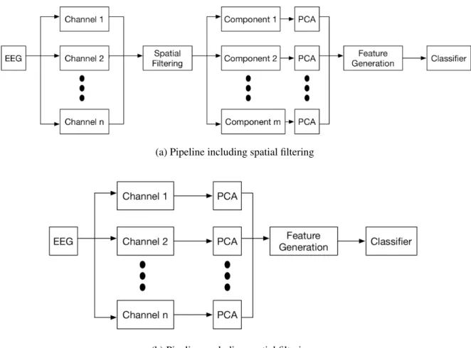
4.1 Two spatial filtering pipelines for RSVP-based EEG. . . 60
4.2 Spatial pattern estimation for LDA beamformer using whole EEG epoch via training data using CAR:Participant 2. . . 62
4.3 ERPs topography plot. . . 67
4.4 Example of estimated spatial patterns/filters for three spatial filtering approaches. 68 4.5 Spatial patterns topographical plots produced by xDAWN and MTWLB. . . 71
4.6 Time-course source N170 and P300 reconstructed by xDAWN and MTWLB. . 72
4.7 SNR for reconstructed N170 (top) and P300 (bottom). . . 73
4.8 AUC score of using the source signal (N170 top and P300 bottom) reconstructed by xDAWN and MTWLB. . . 74
List of Figures
5.1 Number of papers in each year from 2014 to 17th May 2019. . . 84
5.2 Categories of papers from 2014 to 17th May 2019. . . 84
5.3 Percentages of each category take account the total number of papers in each year. 85 5.4 Timeline of architecture-variant GANs. . . 87
5.5 Up-sampling process of generator in LAPGAN (from right to left). . . 88
5.6 Detail of DCGAN architecture for generator. . . 89
5.7 Illustration of BEGAN architecture. . . 89
5.8 Progressive growing step for PROGAN during the training process. . . 90
5.9 Self-attention mechanism architecture proposed in the paper. . . 91
5.10 Summary of recent architecture-variant GANs for solving the three challenges. 92 5.11 Illustration of training progress for a GAN. . . 95
5.12 JS divergence and gradient change with the distance betweenpr andpg. . . 96
5.13 Comparison of parameter distribution between WGAN and WGAN-GP. . . 99
5.14 Decision boundary illustration of original GAN and LSGAN. . . 100
5.15 An example of computation for an unrolled GAN with 3 unrolling steps. . . 102
5.16 Demonstration of the loss in equation (5.18) for LS-GAN. . . 103
5.17 SVM hyperplane used in Geometric GAN. . . 105
5.18 Doutput comparison between RGAN and original GAN. . . 106
5.19 Current loss-variants for solving the challenges. . . 107
5.20 Loss and gradient for the generator of different loss-variant GANs. . . 108
6.1 Schematic of the first type of neuro-AI interface. . . 116
6.2 Face image examples used in the experiment. . . 121
6.3 Reconstructed averaged (via LDA beamformer) P300 signal across 12 partici-pants. . . 123
6.4 Averaged P300 topography of each participant for each category. . . 124
6.5 Box plot of Neuroscore for each image category. . . 125
6.6 Correlation between Neuroscore and BE accuracy. . . 126
7.1 Schematic of different types of recorded neural signals. . . 133
7.2 Schematic of the second type of neuro-AI interface. . . 135
7.3 A neuro-AI interface and training details with adding EEG information. . . 136
7.4 Architecture of Shallow network used in this work. . . 137
7.5 Testing error of3models with and without EEG. . . 140
7.6 Scatter plot on the testing set of predicted and real Neuroscore of6models with and without EEG for training. . . 141
7.7 Neuroscore of different evaluated sample size for each type of GAN. . . 143
7.8 P300 amplitude predicted by proposed framework for each single image. . . 144
7.9 Generalization performance of the proposed framework for testing images. . . . 145
A.1 LDRCC architecture. . . 176
A.2 Captured hardware and software triggers. . . 177
A.3 Histograms of latencies derived from (paired) differences between hardware and software trigger timestamps. . . 179
A.4 Distribution of interval differences in timestamps for hardware triggers (in blue) and software triggers (in orange). . . 180
D.1 Correlation between Neuroscore and BE accuracy with normalization (includ-ing RFACE category). . . 184
List of Figures
D.3 Correlation between Neuroscore and BE accuracy without normalization (in-cluding RFACE category). . . 185 D.4 Two-stage training details for Chapter 7. . . 186
List of Tables
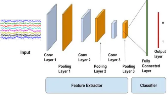
3.1 CNNs architectures in the literature. . . 53
4.1 Hyperparameter summary for each pipeline discussed in this chapter. . . 66
4.2 AUC score (%) for different pipelines across nine participants in testing session. 69 5.1 Summary of loss-variant for GANs. . . 109
6.1 Accuracy for face images generated from three GANs and real face images in the BE task. . . 122
6.2 Computed Neuroscore of each participant for each category. . . 125
6.3 Score comparison for each GAN category. . . 127
7.1 Comparison between Neuroscore and other metrics. . . 133
7.2 Number of trials for each stimulus type remaining after artifact rejection across each participant (ID) and different GAN categories. . . 138
7.3 Errors of9models for cross participants. . . 139
7.4 Performance of three conventional scores and Neuroscore. . . 142
A.1 Time-related latencies between image presentation in software and physical im-age presentation of different groups. . . 178
B.1 Details of SNR for Fig. 4.7 in Chapter 4. . . 182
Abstract
Cortically Coupled Image Computing
Zhengwei Wang
In the 1970s, researchers at the University of California started to investigate communication between humans and computers using neural signals, which lead to the emergence of brain-computer interfaces (BCIs). In the past 40 years, significant progress has been achieved in ap-plication areas such as neuroprosthetics and rehabilitation. BCIs have been recently applied to media analytics (e.g., image search and information retrieval) as we are surrounded by tremen-dous amounts of media information today. A cortically coupled computer vision (CCCV) sys-tem is a type of BCI that exposes users to high throughput image streams via the rapid serial visual presentation (RSVP) protocol. Media analytics has also been transformed through the enormous advances in artificial intelligence (AI) in recent times. Understanding and presenting the nature of the human-AI relationship will play an important role in our society in the future. This thesis explores two lines of research in the context of traditional BCIs and AI. Firstly, we study and investigate the fundamental processing methods such as feature extraction and clas-sification for CCCV systems. Secondly, we discuss the feasibility of interfacing neural systems with AI technology through CCCV, an area we identify as neuro-AI interfacing. We have made two electroencephalography (EEG) datasets available to the community that support our inves-tigation of these two research directions. These are the neurally augmented image labelling strategies (NAILS) dataset and the neural indices for face perception analysis (NIFPA) dataset, which are introduced in Chapter 2.
The first line of research focuses on studying and investigating fundamental processing methods for CCCV. In Chapter 3, we present a review on recent developments in processing methods for CCCV. This review introduces CCCV related components, specifically the RSVP experimental setup, RSVP-EEG phenomena such as the P300 and N170, evaluation metrics, feature extraction and classification. We then provide a detailed study and an analysis on spatial filtering pipelines in Chapter 4, which are the most widely used feature extraction and reduction methods in a CCCV system. In this context, we propose a spatial filtering technique named multiple time window LDA beamformers (MTWLB) and compare it to two other well-known techniques in the literature, namely xDAWN and common spatial patterns (CSP). Importantly, we demonstrate the efficacy of MTWLB for time-course source signal reconstruction compared to existing methods, which we then use as a source signal information extraction method to support a neuro-AI interface. This will be further discussed in this thesis i.e. Chapter 6 and Chapter 7.
The latter part of this thesis investigates the feasibility of neuro-AI interfaces. We present two research studies which contribute to this direction. Firstly, we explore the idea of neuro-AI interfaces based on stimulus and neural systems i.e., observation of the effects of stimuli produced by different AI systems on neural signals. We use generative adversarial networks
Abstract
(GANs) to produce image stimuli in this case as GANs are able to produce higher quality images compared to other deep generative models. Chapter 5 provides a review on GAN-variants in terms of loss functions and architectures. In Chapter 6, we design a comprehensive experiment to verify the effects of images produced by different GANs on participants’ EEG responses. In this we propose a biologically-produced metric called Neuroscore for evaluating GAN per-formance. We highlight the consistency between Neuroscore and human perceptual judgment, which is superior to conventional metrics (i.e., Inception Score (IS), Fr´echet Inception Distance (FID) and Kernel Maximum Mean Discrepancy (MMD) discussed in this thesis). Secondly, in order to generalize Neuroscore, we explore the use of a neuro-AI interface to help convolutional neural networks (CNNs) predict a Neuroscore with only an image as the input. In this scenario, we feed the reconstructed P300 source signals to the intermediate layer as supervisory informa-tion. We demonstrate that including biological neural information can improve the prediction performance for our proposed CNN models and the predicted Neuroscore is highly correlated with the real Neuroscore (as directly calculated from human neural signals).
Chapter 1
Introduction
Abstract: This chapter introduces basic concepts and presents the background information in related research areas to be discussed in this thesis. It also introduces the motivation and highlights the contributions in this thesis.
This thesis investigates the feasibility of deploying brain-computer interfaces (BCIs) in the area of media analytics. Research presented in this thesis spans fields including neuroscience, machine learning and deep learning. We discuss BCI research basically from two perspectives: (1) We investigate the imagery triaging ability of a traditional BCI system that is exposed to a rapid serial visual presentation (RSVP) protocol and we call this type of system as cortically coupled computer vision (CCCV) system. We explore and inspect the efficacy and performance of different spatial filtering pipelines for this type of system. Contents related to this type of research line will be introduced in Chapter 3 and Chapter 4; and (2) We deploy human neural responses to interface with AI systems. We demonstrate the concept of neuro-AI interfaces using two different frameworks. The first framework presents images produced by generative adversarial networks (GANs) to participants and uses participants’ neural feedback, electroen-cephalography (EEG) is used in this case, to assess the quality of images produced by GANs. This work will be demonstrated in Chapter 6. The second framework is to demonstrate that neural responses can be used as supervisory information to train a deep neural network (DNN), which can assist a DNN to accomplish some difficult tasks in the future i.e., evaluating im-age quality in our case. This work will be introduced in Chapter 7. Chapter 2 describes EEG datasets used in this thesis while Chapter 5 provides a review on generative adversarial networks (GANs) where GANs were used to produce the image stimuli in our experiment. Before start-ing to discuss the research work in this thesis, we first introduce some background knowledge
1.1. Brain-computer Interfaces and Event-related Potentials in related areas.
1.1
Brain-computer Interfaces and Event-related Potentials
Electroencephalography (EEG) is a non-invasive measurement of a human being’s brain waves that was firstly measured by the German psychiatrist, Hans Berger in 1924. EEG is the electric recording of the summed electric activity of populations of neurons, which is measured by us-ing electrodes placed on the scalp. It has been widely applied in clinical contexts. It is used in the evaluation of several types of brain disorders such as epilepsy detection [1, 2], lesions in the brain [3–5], sleep disorders [6, 7], Alzheimer’s disease [8, 9] and psychoses [10]. EEG is also able to provide indications for evaluating trauma [11], drug intoxication [12] and the extent of brain damage [13]. With successful deployment in clinical contexts, internal characteristics are also well researched in the literature. EEG comprises different waveforms, which can be gen-erally characterized by their frequencies, amplitudes, shapes as well as the locations i.e., sites on the scalp where they are recorded. Frequency is a key characteristic for classifying different types of EEG waveforms. Alpha rhythm [14] appears in the frequency ranging between 8 Hz and 13 Hz, which is activated during the relaxed wakefulness human brain and it is normally attenuated or abolished by visual attention and affected transiently by other sensory stimuli and by other mental alerting activities [15]. Theta rhythm appears in the frequency ranging between 4 Hz and 7 Hz, which is encountered in the front central regions and is usually related to drowsi-ness or heightened emotional states [16]. Alpha and theta reflect human cognitive and memory performance [17]. Delta rhythm [18] appears between 0.5 Hz and 4 Hz and is associated with sleep [19]. Beta rhythm appears between 14 Hz and 25 Hz, sometimes can be augmented by drugs [20] and increases with heavy breathing. Gamma appears over 30 Hz and is implicated in creating the unity of conscious perception [21]. Research related to those internal charac-teristics behind EEG provides the neurophysiological evidence that EEG is related to human behavior, brain function, external stimulus etc.From an engineering perspective, we are generally interested in leveraging patterns of ac-tivity in EEG for some real-world applications. BCIs enable such a way for engineering re-searchers to create a direct communication channel between the brain and computers. BCIs are basically divided into two types: (1) Non-invasive BCI where the sensors are placed on the head for the purposes of measuring signals related to brain activity e.g., EEG; and (2) Invasive BCIs,
1.1. Brain-computer Interfaces and Event-related Potentials
where the electrodes are placed directly into cortex by using surgical operation e.g., electrocor-ticography (ECoG) and local field potential (LFP). BCIs discussed in this thesis are EEG-based which all belong to the non-invasive category. A typical BCI comprises five parts [22]: (1) Data acquisition — the EEG (electroencephalogram) signals are recorded by an EEG amplifier; (2) Pre-processing — this step includes signal denoising e.g., filtering, artifact rejection, normal-ization and re-referencing. Good pre-processing yields EEG useful for subsequent analysis; (3) Feature extraction — this part aims to extract meaningful features that can be used for train-ing machine learntrain-ing algorithms later. The advantages of a suitable feature extraction method can include improving the signal-to-noise ratio (SNR) and allowing dimensionality reduction, which has effects on the later training part; (4) Classifiers and machine learning algorithms – in order to translate the features into device commands, it is necessary to apply machine learning techniques to the extracted features where both linear and nonlinear methods are often used in this step [23, 24]; and (5) Output device — the output device can be any type of electronic element that can receive the command in the last step e.g., a computer screen. Figure 1.1
il-Data acquisition Feature extraction Classification Signal processing Quadcopter control P300 speller Cortically coupled computer vision Motor imagery Neural spelling Neurally image computing Sensory feedback
Neural signals
Brain-computer Interfaces
Figure 1.1: An overview of a generic BCI system.
lustrates a visual overview of a generic BCI system. Three examples, specifically quadcopter control [25], P300 speller [26] and CCCV [27], are demonstrated. The end goal of a BCI system is to use brain signals (e.g., EEG is used in this work) to make the output device responsive to thoughts.
1.1. Brain-computer Interfaces and Event-related Potentials
brain structure in response to specific events or stimuli, which are time-locked to sensory, mo-tor or cognitive events [28]. Two types of ERPs are stated in the literature, namely exogenous ERPs and endogenous ERPs [29]. Exogenous (also referred to as sensory) components appear relatively early (50 ms to 250 ms) after stimulus presentation and depend on the physical pa-rameters of the stimulus e.g., shape and brightness [30]. Endogenous (or named as cognitive) components appear later (> 250 ms) after stimulus presentation and they are not sensitive to stimulus type. These components are typically related to cognition and information processing in the brain e.g., attention and memory. Each ERP is unique and is typically categorized using three properties: (1) Amplitude (10−6 V) is measured at the peak of ERPs. Polarity is used as the discriminant information; (2) Location spatial topography, where different ERPs can be recorded from different locations on the scalp; and (3) Latency refers to the time measured when the ERP’s peak appears after stimulus presentation. Latency is determined by the type of ERP. The early ERP components peaking at short latencies are normally related to exogenous components. The later ERP components peaking with a large latency usually belong to the family of endogenous components. This thesis is built up upon one of the endogenous ERPs — the P300 component, which reflects aspects of the attention process in humans.
The P300 component was first reported in 1965 [31] and can be elicited by an attention-driven task i.e., the P300 will appear when participants are paying attention for some specific stimuli to appear [32, 33]. The terminology “P300” indicates the amplitude and latency prop-erties mentioned before for the component. “P” refers to a positive-going peak of the ERPs waveform and “300” refers to the time window 300 ms – 600 ms that the P300 may appear in. The location of the P300 is distributed over the middle electrodes (Fz, Cz, Pz), which typically increases in amplitude from frontal to parietal electrode sites (it depends on the type of stim-uli) [34]. Moreover, the P300 comprises two subcomponents, which are known as P3a and P3b. The P3a can be elicited by an infrequent distractor stimulus randomly inserted in a presentation stream and it comes from frontal or central electrode sites on the scalp [35, 36]. The P3b is task-relevant, which is elicited during target stimulus processing [37,38] and presents parietally on the scalp [39]. This thesis mainly focuses on discussing the P3b because our experiments are designed upon target detection in this work.
The first P300-BCI was introduced in 1988 [40] and the P300 is the most widely used ERP component for a BCI system currently [41]. A P300-BCI benefits from its fast speed, a straightforward test (e.g., detect target) for participants and no requirement for training
par-1.2. Rapid Serial Visual Presentation Paradigm
ticipants. Despite the well known P300 speller system [42], recent research has shown that P300-BCIs have a wide range of different usages. For example, they can be used to assist the disabled [43–45]. In addition, new experimental paradigms related to P300-BCI applications have been demonstrated in the literature [26, 46, 47] and a P300-BCI was also demonstrated to improve human attention [48]. This thesis focuses on the use of a visual P300-BCI for searching and evaluating images.
1.2
Rapid Serial Visual Presentation Paradigm
Rapid serial visual presentation (RSVP) involves a series of images being rapidly presented to participants at a specific position on a screen. It was first introduced in [49]. It can be divided into text RSVP and image RSVP [50]. For the text RSVP, individual words are presented sequentially in a fixed place on a screen where it has applications such as high-speed in reading and assist reading for the disabled [50]. Similar to the text RSVP, the image RSVP involves rapid presentation of images to participants during the experiment. This thesis employs the image RSVP paradigm.
The concept of RSVP can be introduced using a familiar example, that of rapidly riffling through the pages of a book in order to locate a needed image [50]. In RSVP, a rapid succession of target and standard (non-target) images are presented to a participant on a display at a rate of 4 Hz – 10 Hz. The location of target images within the high-speed presentation is not known in advance by users and hence requires them to actively look out for targets i.e., to attend to target images. This paradigm where users are instructed to attend to target images amongst a larger proportion of standard images is known as an oddball paradigm and is commonly used to elicit ERPs such as the P300, a positive voltage deflection that typically occurs between 300 ms – 600 ms after the appearance of a rare visual target within a sequence of frequent irrelevant stimuli [39]. Since participants do not know when target images will appear in the presentation sequence, their occurrence causes an attentional-orientation response that is characterized by the presence of a P300 ERP.
RSVP was designed to explore the human visual processing system in the literature [51–55]. [51] showed how long a target image needs to be displayed so that it can be perceived by participants, implying an upper-bound in the display rate in a RSVP paradigm. An important finding during this procedure was that participants experienced a condition named attentional
1.2. Rapid Serial Visual Presentation Paradigm
blink [51]. This condition was characterized by failure to detect a target when it follows another target immediately presented within a stream of visual stimuli in rapid succession at the same spatial location on a screen. This happened when the second target appeared between 180 ms and 450 ms [52, 55] after the first. This result indicates an important point when designing a RSVP experiment, where the time interval between two targets cannot be too small otherwise the target interference may deteriorate the performance of the RSVP experiment [56].
There are three modes of RSVP paradigms (see Fig. 1.2): (1) Static mode, where images appear and disappear without moving; (2) Moving mode, where images appear and disappear sequentially; and (3) Multiple entries/exits, where images appear and disappear in many loca-tions or move along several paths [50]. Static mode has been shown to be more robust and
Figure 1.2: Three RSVP modes i.e., static mode, moving mode and multiple entries/exists mode (from left to right).
effective compared to other modes, where static mode is associated with a higher target recog-nition rate and a higher presentation rate (i.e., number of images presented per second) [57]. Multiple entries/exits mode is a more complex RSVP mode, which can be used for exploring gaze movement along the routes taken by images [58]. A good RSVP experiment should be designed with the following considerations: (1) Examine the tasks associated with the applica-tion to see what benefits can be provided by RSVP; (2) Select the mode that is influenced by availability of context presentation and manual control of rate and direction; (3) Presentation rates should be slower when a task has an increased complexity; and (4) Be aware of a potential link between eye-gaze movement and the success with which images may be recognized [50]. We follow previous researchers’ steps by using the static mode for designing our experiment in this thesis [59–61].
1.3. Cortically Coupled Computer Vision System
1.3
Cortically Coupled Computer Vision System
Cortically coupled computer vision (CCCV) is the integration of BCIs and the RSVP paradigm. CCCV uses neural signals to implement image computation (e.g., image detection/search) for a rapid image stream. Figure 1.3 illustrates a typical example of a CCCV system. A participant
Image Database Sample Output Labelling images Propagate labels EEG signals Target Target Standard
Figure 1.3: An example of CCCV system. The image stream sampled from the image database is presented to the participant and EEG signals are recorded simultaneously. Images are then triaged by using a participant’s EEG signal. The labels are then propagated back to the image database and output image of interest [62].
is asked to search for specific type of images (face images in particular in this case) in an image stream presented by a RSVP paradigm and EEG signals are recorded simultaneously. The P300 component would be elicited when participants see the appearance of target images. With proper machine learning techniques applied, images can then be labelled based on whether the P300 component is detected or not. Compared to current advanced computer vision systems, a CCCV system has some advantages: (1) Participants do not need to be trained regarding the
1.4. Generative Adversarial Networks
generation of P300; (2) This system is capable of being applied to those small image datasets (e.g., satellite image datasets [63]) whereas current deep learning approaches typically depend on a number of training samples; (3) This system has relatively light computation and literature has already demonstrated the possibility of real-time processing [27, 61, 64].
In this thesis, CCCV is explored with respect to searching for target images and evaluating images produced by generative adversarial networks (GANs). Regarding the first aspect, we explore and demonstrate efficient spatial filtering pipelines for a CCCV system. Moreover, we propose a spatial filtering approach called multiple time window LDA beamformer (MTWLB) for better reconstructing the time-course source signals for a CCCV system. In terms of the second aspect, we couple CCCV systems with two types of artificial intelligent (AI) systems, namely GANs and convolutional neural networks (CNNs), where we call this approach neuro-AI interfacing. We demonstrate the use of neuro-neuro-AI interfaces for (1) use in CCCV to produce a biologically neuro-produced metric called Neuroscore to evaluate the performance of GANs and (2) use in CCCV to provide biological neural information to train a CNN.
1.4
Generative Adversarial Networks
Generative adversarial networks (GANs) [65] are increasingly attracting attention in computer vision, natural language processing, speech synthesis and similar domains. Arguably the most striking results have been in the area of image synthesis. Figure 1.4 demonstrates the architec-ture of a typical GAN. The architecarchitec-ture comprises two components, one of which is a discrim-inator (D) distinguishing between real images and generated images while the other one is a generator (G) creating images to fool the discriminator. Given a distributionz∼ pz, generator defines a probability distributionpg as the distribution of the samplesG(z). The objective of a GAN is to learn the generator’s distributionpg that approximates the real data distribution pr. Optimization of a GAN is performed with respect to a joint loss function forDandG
min
G maxD Ex∼prlog[D(x)] +Ez∼pzlog [1−D(G(z))] (1.1) A GAN is a member of deep generative models (DGMs) family and was proposed to be trained by using a min-max game theory strategy between two players (a discriminatorDand generator
G) . The generator behaves like a “forger”, which aims to produce generated data to fool the discriminator. The discriminator behaves like a “detective”, which aims to distinguish the
1.4. Generative Adversarial Networks Real Face Generated Face Sampling Real or Fake ? Discriminator Generator Random noise
Figure 1.4: Architecture of a GAN. Two deep neural networks (discriminator (D) and generator G)) are synchronously trained during the learning stage. The discriminator is optimized in order to distin-guish between real images and generated images while the generator is trained for the sake of fooling discriminator from discerning between real images and generated images.
real data from the generated data. The optimal generator will produce the synthetic data that discriminator cannot tell is synthetic from real data. GANs have benefits compared to the traditional DGMs. First, the architectures in GANs are very flexible. Any type of architecture for a specific application can be used as the generator and the discriminator. Second, GANs are able to handle sharp density functions and produce high quality images. Third, they are able to produce diverse image samples. However, GANs suffer from problems such as being difficult to train and hard to evaluate. The first problem is more theoretic, which is discussed in Chapter 5. Regarding the second challenge, this thesis explores the use of human perception, which utilizes a CCCV pipeline to contribute that area.
There is limited research combining GANs and neuroscience in the literature. Current litera-ture focuses on using GANs to improve the spatial resolution of EEG signals or to produce syn-thetic EEG signals [66–68]. Research on exploring the use of outputs of GANs as experimental stimuli in the area of neuroscience is limited. This research direction has mutual benefits for both the deep learning and neuroscience domains. Firstly, the use of neurally-inspired metrics to evaluate the performance of GANs would improve the process of training GANs. Secondly, for the automatic generation of stimuli for a neuroscientific experiment. Traditional preparation of stimulus for a neuroscientific experiment is a time-consuming process. By using the modern deep learning techniques such as GANs, it saves lots of time and more importantly it enables experimental stimuli to be very flexible e.g., researchers can easily customize the type of image
1.5. Neuro-AI Interfaces
stimuli use for the experiment. We explore this research direction to bridge the gap in current literature by using CCCV systems with GANs.
1.5
Neuro-AI Interfaces
Biological Neural Networks In neuroscience, biological neural networks (BNNs) describe recognizable pathway of groups of interconnected neurons where these neurons communicate with each other by using electrical impulses. There are two main types of electrical activity associated with neurons, which are action potentials and postsynaptic potentials [69]. Action potentials are discrete voltages spikes that travel from the beginning of the axon at the cell body to the axon terminals (red flow as seen in Fig. 1.5), where neurotransmitters are released. A spike train is such a type of neural activity, where a neuron fires an action potential at a
Postsynaptic potentia
l
Figure 1.5: Schematic of action potentials and postsynaptic potential. Figure was constructed via [70] and [71].
sequence of recorded times [72]. Postsynaptic potentials (as seen in Fig. 1.5) are the voltages that arise when the neurotransmitters bind to receptors on the membrane of the postsynaptic
1.5. Neuro-AI Interfaces
cell, causing ion channels to open or close, leading to a graded change in the potential across the cell membrane [69]. ERPs discussed in this thesis are produced by postsynaptic potentials.
Artificial Neural Networks In general, an artificial neural network (ANN) comprises four components: (1) Neurons; (2) Weights; (3) Connectivity (topology of network e.g., fully-connected networks and CNNs); and (4) A learning rule [73]. An ANN is normally trained with weights carefully initialized in order to speed up convergence [74] and fixed connectivity. An optimization problem of ANNs is to map inputs to desired outputs, and weights in ANNs are updated through solving the optimization problem by using backpropagation [75].
Relationship between AI and Neuroscience It is well known that ANNs are inspired by BNNs. As seen in Fig. 1.6, an artificial neuron receives a number of inputs produced by
neu-Figure 1.6: Comparison between a biological neuron and an artificial neuron. Top figure describes a biological neuron while the bottom one demonstrates an artificial neuron. Figure from [76].
rons in previous layer and the neuron will be activated when the sum of input values are above the threshold, which is similar to that of a biological neuron that has dendrites to receive sig-nals from other neurons. A cell body in effect processes the inputs and via its axon(s) sigsig-nals with action potential fired by other neurons. However, BNNs have numerous differences to ANNs. Firstly, the initial state of BNNs are not random and are genetically influenced [77, 78].
1.6. Research Motivation and Contributions
Secondly, the connectivity and the weights are always changing over time and across differ-ent tasks. On the contrary, the connectivity of ANNs is fixed over time and the weights of ANNs are fixed after training. In the deep learning area, researchers focus on developing ANNs that are more biologically plausible in the past 30 years [79–81]. For instance, convolutional neural networks (CNNs) were developed by emulating human retina and brain visual system, which split images into tiny areas for object recognition [82–85]. Another example is recurrent neural networks, which use the neuron’s output as a feedback so it introduces memory func-tionality to the model [86]. This is also similar to BNNs as functional connectivity in BNNs, described by statistical dependencies such as transfer entropy, coherence, and correlations, are significantly similar in some respects [87–89]. Benefiting from the fundamental knowledge achieved in the areas such as neuroscience, deep neural networks (DNNs) and artificial intel-ligence (AI) have grown up and are revolutionizing our society today. From the other side, benefit by DNNs prevailing today, neuroscience research is pushed significantly forward by utilizing DNNs technology. For example, work [90] demonstrated that CNNs can be used to predict neural response in the V4 cortex when doing image recognition tasks. Other recent [91] showed that current ANNs are able to control the activity of neural populations, which has wide reaching therapeutic applications such as treatment for depressive disorder.
Both AI and neuroscientific research have achieved significant progress in their own fields, however, research on interconnections between these two areas are still limited and unexplored. Broadly speaking, it is under investigated what utility neural information can provide for AI systems. In this thesis, we explore this area by introducing the concept of “neuro-AI interfaces”, which creates a pathway between a human neural systems and AI systems. We introduce neuro-AI interfaces that use CCCV to interact with neuro-AI systems. We introduce neuro-neuro-AI interfaces via one of the fundamental problems in GANs: designing a proper evaluation metric to assess the quality of images produced by GANs, which is discussed in detail in Chapter 6 and Chapter 7.
1.6
Research Motivation and Contributions
Traditional CCCV research focuses on using a human’s neural signals (EEG) to find target images from a large database. A review of feature extraction and classification methods is pre-sented in Chapter 3, where the original content has been published as a book chapter in the
1.6. Research Motivation and Contributions
lots of research has been carried out to improve the classification performance for a CCCV system. In our work, we pay close attention to both classification performance and the neu-rophysiological interpretability when using a CCCV system. Motivated by this objective, we investigate the spatial filtering pipeline (i.e., the most widely used strategy in CCCV research) for a CCCV system in Chapter 4, which enables interpretation of EEG signals from both a spatial space and a temporal space. LDA beamformers based on multiple time windows are proposed for a CCCV system and we also demonstrate its efficacy on the reconstructed time-course source signals. This pipeline is then utilized to reconstruct a time-time-course source signal (the P300) for our neuro-AI interface in the later part of the thesis. Part of the content presented in Chapter 4 has been published in theJournal of Brain-Computer Interfaces[93].
GANs are attracting growing attention in the deep learning community and have been ap-plied to lots of different domains. Some reviews [94–98] on GANs have been published in the literature but discussion on the performance of GAN-variants is still lacking. In Chapter 5, we provide a survey on GAN-variants based on architectures and loss functions, where we discuss and analyze their performance (i.e., high quality image generation and stable training) in the context of the area of computer vision. We hope this review will help researchers both from and outside the deep learning community when deploying GANs in their research.
Lots of work has been carried out in regard to using DNNs for classifying the EEG re-sponses [99–101] where many approaches have achieved good classification performance for CCCV systems [102, 103]. With the blossoming of deep learning techniques today, the ex-ploration of the interconnections between areas of deep learning and biological neural systems deserves attention. Compared to traditional machine learning/deep learning systems, biological neural systems can be more robust to small perturbations on inputs, are more easily generalized to other tasks and directly reflect functionality in the brain system such as for cognition, atten-tion and memory. Studying the interconnecatten-tions between AI and neuroscience can be useful to enhance our understanding of how the brain works and improve the performance for AI systems through using informative knowledge derived from biological neural systems (or building more biologically plausible DNNs models). Thus investigation on this area has potential impact for both AI and neuroscience fields. In this thesis, we explore the interconnections between deep learning and neuroscience domains through CCCV technology. Firstly, we explore the feasibil-ity of using the P300 component, which is produced by the spatial filtering pipeline mentioned in the previous paragraph, to evaluate the performance of GANs in Chapter 6. We propose
1.7. Research Questions
Neuroscore as a biologically neurally-produced metric for scoring a GAN and demonstrate that Neuroscore is highly correlated with human perceptual judgment. To the best of our knowledge, this is the first research line that combines human perception with GANs. This chapter has been published in theCognitive Computation. Secondly, we consider if a DNN is able to learn useful information from neural signals and model the performance generated by human brain signals. In Chapter 7, we propose a CNN based neuro-AI interface to evaluate GANs, which synthesizes the Neuroscore. Importantly, we show that including neural responses during the training phase of the network significantly improves the prediction performance of the proposed model.
Finally, this thesis contributes two open EEG datasets: neurally augmented image labelling strategies (NAILS) and neural indices for face perception analysis (NIFPA). We hope these two datasets will be helpful to other researchers working in this field.
1.7
Research Questions
This thesis visits the areas of neuroscience, machine learning and deep learning. With respect to neuroscience, we aim to investigate human beings’ neural responses (i.e., ERPs in EEG signals) to image stimuli. We are interested in using neural responses in image target search and in image quality evaluation, in what we refer to as a CCCV system. From the machine learning aspect, we explore methods (i.e., feature extraction and classification) that efficiently performing classification and maintain neurophysiological interpretability. In terms of deep learning, we explore the possibility of using neural signals to evaluate image stimuli produced by GANs and the efficacy of adding neural responses as supervisory information to DNNs.
Below are three research questions that arise in these three areas (namely neuroscience, machine learning and deep learning), which shape the content of this thesis:
1. Can we improve on the extraction of discriminative ERP components while preserv-ing neurophysiological interpretability for a CCCV system?
This research question explores the effectiveness of different spatial filtering approaches in a CCCV system with respect to their classification performance and neurophysiologi-cal interpretability. Chapter 3 visits this area and we propose a spatial filtering approach named multiple time window LDA beamformer (MTWLB). Its neurophysiological inter-pretability is demonstrated and it is compared to existing approaches in the literature. An important problem for ERP research is that EEG signals suffer from a low SNR
is-1.7. Research Questions
sue, especially for CCCV systems. The ability to reconstruct ERP source signals is not only beneficial to the classification performance of a CCCV system but may also have positive effects on ERP research carried out using a RSVP protocol i.e., high SNR sig-nals (compared to the sigsig-nals recorded from one electrode) can be used for ERP analysis which gives more robust results in terms of different experimental conditions. We also investigate the efficacy of our proposed approach in this aspect — the performance of reconstructing the ERP source signals under the RSVP experimental protocol.
2. Can neural signals be used to provide indications on image quality that is consistent with human perceptual judgment and is it possible to use this as a biological score to evaluate generative models such as GANs?
AI has a significant impact on our society. Research into the interaction between humans and AI deserves further exploration and has only recently become a research focus. We explore the possible interface between generative models (i.e., GANs used in this case) and human neural systems. As a starting point, we investigate the feasibility of using neu-ral signals (EEG) to evaluate the performance of GANs. The main concern of evaluating the performance of GANs in the current literature is whether current evaluation metrics are consistent with human perceptual judgment. In Chapter 6, we address this question by introducing a neurally-produced metric called Neuroscore, which is produced by us-ing a CCCV system. A systematic comparison between Neuroscore, human perceptual judgment and other evaluation metrics is carried out.
3. Is it possible to interface biological neural systems and AI systems and if so, can biological neural signals provide any type of informative knowledge for helping AI systems to learn a difficult task?
The current literature has demonstrated the use of DNNs in emulating the encoding pro-cesses of the brain during an image object recognition task via invasive measurements, which indicates the encoding processes of the brain and of DNNs are similar to each other. Given this evidence, we are interested in understanding if biological neural information via a non-invasive measurement (EEG in this case) is transferable to DNNs to help DNNs accomplish difficult tasks. As a starting point, we introduce the concept of a neuro-AI in-terface, which uses the P300 signal as a source of supervisory information to help CNNs produce a Neuroscore for the purpose of evaluating the performance of GANs (Chapter 7).
1.8. Overview and Organization of the Thesis
1.8
Overview and Organization of the Thesis
The word cloud in Fig. 1.7 summarizes the key words in this thesis. This thesis discusses
Figure 1.7: The word cloud of keywords presented in this thesis.
CCCV systems from two perspectives. First, we investigate using CCCV systems for image search. From this perspective, we study the feature extraction and classification approaches that have been established in the literature. We also study the spatial filtering pipeline for using CCCV technology to search for target. We discuss the effective spatial filtering pipelines in the literature and propose a novel spatial filtering approach named multiple time window LDA beamformers (MTWLB) for CCCV systems. Second, we visit the area that interfaces CCCV systems with AI systems, where we call it neuro-AI interfaces. From this aspect, we first consider the use of human beings’ brain signals EEG to evaluate the performance of GANs, where we propose the Neuroscore as an evaluation metric for GANs. We then demonstrate a type of CNN based neuro-AI interface to generalize the use of Neuroscore, where the model was trained by using human’s brain signals.
The organization of this thesis is as follow:
• Chapter 2describes the two EEG datasets, namely the NAILS and the NIFPA, which are contributed by this thesis. EEG data in the NAILS was recorded to encompass a broad range of image search activities and coincident neural signals, which benchmarks the ma-chine learning strategies for RSVP-EEG (EEG signals recorded in the RSVP paradigm experiment) signals. The NIFPA is to explore the human being’s EEG response to
syn-1.8. Overview and Organization of the Thesis
thetic images produced by GANs along with the real images of the same type. Both N170 and P300 ERPs were elicited in this dataset. We introduce the properties of ERPs from the perspectives of time course and topographical distribution. Details of analysis and feature extraction of ERPs are introduced in Chapter 3.
• Chapter 3presents a comprehensive review on the processing methods for RSVP-EEG. We introduce the experimental set up for recording the RSVP-EEG data and study the feature extraction and classification algorithms applied for this type of data. In terms of the feature extraction, we mainly consider two most popular areas, spatial filtering and time-frequency representation, to extract informative features from ERPs. Regarding the classification methods, we study the traditional linear classification methods and the recent momentum deep learning methods.
• Chapter 4 analyzes and discusses the spatial filtering pipeline deployed in cortically coupled image classification. We propose a spatial filtering approach called multiple time window LDA beamformers (MTWLB), which uses LDA beamformers based on multiple time windows. The proposed MTWLB is compared with other two famous approaches in the literature, which are xDAWN and common spatial pattern (CSP). Moreover, we compare the performance between xDAWN and MTWLB in terms of reconstructing the time course of the source signals. The performance of three linear classifiers, linear dis-criminant analysis (LDA), Bayesian linear regression (BLR) and logistic regression (LR) are also discussed.
• Chapter 5 provides a review on GAN-variants from architecture and loss perspectives. We analyze the problems in the original GAN and study the GAN-variants that deal with those problems in the literature.
• Chapter 6 demonstrates a neuro-AI interface, which deploys a novel cortically coupled paradigm that uses neural signals to evaluate the performance of GANs. Neuroscore has been proposed as an evaluation metric for GANs, which closely mirrors behavioral human perception on the images produced by GANs. Neuroscore is compared to three conventional evaluation metrics in the literature.
• Chapter 7 extends the work in Chapter 6, in which a CNN based neuro-AI interface is proposed to produce the Neuroscore. We show that DNNs are able to learn informative
1.8. Overview and Organization of the Thesis
knowledge from human neural responses and successfully demonstrate a CNN trained by using neural responses to evaluate the performance for GANs.
• Chapter 8concludes the thesis. Future directions are also discussed with respect to the different research areas, which are ERP research, computational neuroscience and neuro-AI interfaces.
Chapter 2
Data Description
Abstract: This chapter describes electroencephalography (EEG) datasets that are used in this thesis. Two EEG datasets are introduced: (1) The neurally augmented image labelling strate-gies (NAILS) dataset is used to support a collaborative evaluation task in which participating researchers benchmarked machine learning strategies against each other. The experimental protocol used to capture the dataset was designed to encompass a broad range of image search activities and coincident neural signals; and (2) The neural indices for face perception analysis (NIFPA) dataset is to explore human being’s EEG responses to synthetic images generated by using generative adversarial networks (GANs) and real images of the same type. By doing so, we will be able to compare differences in neural responses that indicate whether images are perceived as being real or fake. Identifying and operationalizing such EEG responses would provide an alternative method for the evaluation of GANs and a feedback signal to improve their effectiveness of GAN. The NAILS dataset has already been released [104] and the NIFPA dataset will be released as well at some stage.
2.1
Introduction
A brain-computer interface (BCI) provides a communication pathway between the human brain and a computer system. This type of interface requires the development of algorithms for trans-lating the measured brain signals into computer commands. Brain signals can be measured invasively [105] and non-invasively [106]. Non-invasive BCIs are more commonly being re-searched in the BCI community [107]. Electroencephalography (EEG) signals, acquired via a non-invasive manner to enable BCIs, have been widely studied in the literature. Applications
2.1. Introduction
designed using such type of BCIs span broad areas e.g., information retrieval [108], rehabilita-tion engineering [109] and cortically coupled computer vision [59].
The P300, a type of event-related potential (ERP), is a well known EEG component that has been widely used to drive BCI systems (e.g. the P300 speller applications [110]). P300-BCIs for multimedia information retrieval have attracted growing interests in recent years [93, 103, 111]. Open EEG datasets related to this field are severely lacking. In this chapter, we provide de-tails of the two recorded EEG datasets used in this thesis: (1) The neurally augmented image labelling strategies (NAILS) dataset, which has an affiliated workshop to support the collabo-rative evaluation of best-practice strategies for rapid serial visual presentation (RSVP) image search using EEG signals; and (2) The neural indices for face perception analysis (NIFPA) dataset, which is to explore a human being’s EEG responses to synthetic images produced by generative adversarial networks (GANs).
In this thesis, we used 32 channel BrainVision actiCHamp at 1000 Hz for recording EEG signals and electrode locations defined by 10-20 system were carried out as seen in Fig. 2.1. A list of related neurophysiologically-relevant terminology and associated explanations used in
Figure 2.1: Electrode locations of 10-20 system used through this thesis.
this thesis is presented below:
2.2. Neurally Augmented Image Labelling Strategies
• Epoch: An epoch is a specific time window which is extracted from the continuous EEG signal. Each epoch is time-locked with respect to an event i.e., image stimulus presenta-tion in our case. This is different from the epoch that is used for training a deep neural network.
• ERP:An ERP signal is the averaged EEG epochs which corresponds to the target stimu-lus.
• ERP difference: An ERP difference signal is the averaged target EEG epochs minus the averaged standard EEG epochs.
• Trial rejection: Remove those epochs corresponding to the selected events that contain artifacts e.g., eye blink.
2.2
Neurally Augmented Image Labelling Strategies
EEG data from 9 participants was recorded. Data collection was carried out with approval from Dublin City University’s Research Ethics Committee (DCU REC/2016/099) (see Appendix E). Each participant completed 6 different tasks (INSTR, WIND1, WIND2, UAV1, UAV2 and BIRD). For each task, participants were asked to search for specific target images from the presented images (i.e., an airplane has the role of target in UAV1 and UAV2 tasks, a keyboard instrument is the target for the INSTR task, while a windfarm is the target in WIND1 and WIND2 tasks, and parrot being the target in BIRD task. See Fig. 2.2).
Figure 2.2: Examples of target images in NAILS task. Images from left to right are airplane, windfarm, macaw, keyboard instrument. The size of target and standard are all256×256.
Each of the tasks was divided into 9 blocks, where each block contained 180 images (9 tar-gets/171 standards) thus there were 486 target images and 9,234 standard images available for
2.2. Neurally Augmented Image Labelling Strategies
each participant. Images were presented to participants at a 6 Hz (6 images per second) presen-tation rate. EEG data was recorded along with timestamping information for image presenta-tion, which was triggered via a photodiode to allow for precise epoching of the EEG signals for each trial [112]. A 32 channel BrainVision actiCHamp at 1000 Hz (1000 samples per second) sampling frequency, using electrode locations as defined by the 10-20 system, was used for EEG acquisition. Epochs were filtered to exclude those with a peak-to-peak amplitude greater than 70µV on EOG and frontal EEG channels. Independent component analysis (ICA) was used alongside a morlet wavelet based analysis to confirm that the remaining epochs did not con-tain non-neural sources of discriminative information. We used trial rejection for eye-related removement instead of removing eye-related independent components by using ICA because ICA sometimes cannot fully remove the eye-related artifacts [113].
Figure 2.3 shows an example of butterfly plot for one participant. The plot was produced
Amplitude (1e-6 V)
Time (ms)
240 ms 450 ms
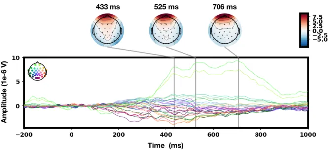
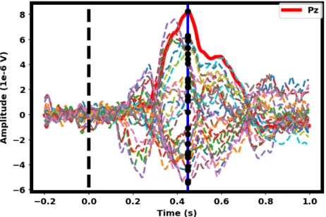
Figure 2.3: Example of ERP difference butterfly plot (target ERPs minus standard ERPs) across all trials after rejecting the eye-related artifacts. Plots were generated using common average reference (CAR). Both an early ERP (e.g., the N200) and a later ERP (e.g., the P300) can be seen in the plot. The N200 can be seen at occipital electrode sites peaking at 240 ms while the P300 can be seen at parietal electrode sites peaking at 450 ms. Channels are color added using the scalp mapping legends in the top left corner.
by the ERPs difference between target ERPs and standard ERPs using all trials (excluding eye-related artifacts). It can be seen that both an early ERP (e.g., the N200) and a later ERP (e.g., the P300) are clearly elicited. The N200 appears at the occipital electrode sites peaking at 240 ms while the P300 is detected at the parietal electrode sites peaking at 450 ms. These two types
![Figure 1.5: Schematic of action potentials and postsynaptic potential. Figure was constructed via [70]](https://thumb-us.123doks.com/thumbv2/123dok_us/1288195.2672843/29.892.140.804.534.1025/figure-schematic-action-potentials-postsynaptic-potential-figure-constructed.webp)

![Table 3.1: CNNs architectures in the literature. The architecture of DeepConvNet and ShallowConvNet in work [101] has been explained detailly in [103]](https://thumb-us.123doks.com/thumbv2/123dok_us/1288195.2672843/72.892.129.810.126.707/table-architectures-literature-architecture-deepconvnet-shallowconvnet-explained-detailly.webp)
![Table 4.1: Hyperparameter summary for each pipeline discussed in this chapter. For each hyperparame- hyperparame-ter, N f ∈ [1, 16] is searched from 1 to 16 spatial filters, α ∈ [1e − 6, 1e + 2] is sampled from uniform distribution, β ∈ [1e − 6, 1e + 2] is](https://thumb-us.123doks.com/thumbv2/123dok_us/1288195.2672843/85.892.230.707.399.697/hyperparameter-summary-pipeline-discussed-hyperparame-hyperparame-searched-distribution.webp)