eScholarship@UMMS
eScholarship@UMMS
GSBS Dissertations and Theses Graduate School of Biomedical Sciences
2017-12-12
Analysis, Visualization, and Machine Learning of Epigenomic Data
Analysis, Visualization, and Machine Learning of Epigenomic Data
Michael J. PurcaroUniversity of Massachusetts Medical School
Let us know how access to this document benefits you.
Follow this and additional works at: https://escholarship.umassmed.edu/gsbs_diss
Part of the Computational Biology Commons, Genomics Commons, and the Integrative Biology Commons
Repository Citation Repository Citation
Purcaro MJ. (2017). Analysis, Visualization, and Machine Learning of Epigenomic Data. GSBS Dissertations and Theses. https://doi.org/10.13028/M23T1Q. Retrieved from
https://escholarship.umassmed.edu/gsbs_diss/938
Creative Commons License
This work is licensed under a Creative Commons Attribution-Noncommercial 4.0 License
This material is brought to you by eScholarship@UMMS. It has been accepted for inclusion in GSBS Dissertations and Theses by an authorized administrator of eScholarship@UMMS. For more information, please contact
ANALYSIS, VISUALIZATION, AND MACHINE LEARNING OF EPIGENOMIC DATA
A Dissertation Presented By
MICHAEL JOSEPH PURCARO
Submitted to the Faculty of the
University of Massachusetts Graduate School of Biomedical Sciences, Worcester in partial fulfillment of the requirements for the degree of
DOCTOR OF PHILOSOPHY
DECEMBER 12, 2017
BIOINFORMATICS AND COMPUTATIONAL BIOLOGY M.D., PH.D. PROGRAM
ANALYSIS, VISUALIZATION, AND MACHINE LEARNING OF EPIGENOMIC DATA
A Dissertation Presented By
MICHAEL JOSEPH PURCARO
This work was undertaken in the Graduate School of Biomedical Sciences Bioinformatics and Computational Biology
Under the mentorship of
________________________________________________ Zhiping Weng, PhD, Thesis Advisor
________________________________________________ Elinor Karlsson, PhD, Member of Committee
________________________________________________ Manuel Garber, PhD, Member of Committee
________________________________________________ Robert Brewster, PhD, Member of Committee ________________________________________________
Ross Hardison, PhD, External Member of Committee ________________________________________________
Jeffrey Bailey, MD, PhD, Chair of Committee ________________________________________________
Anthony Carruthers, Ph.D.,
Dean of the Graduate School of Biomedical Sciences December 12, 2017
This work is dedicated to the most loving, understanding, and special woman in my world: my wife and best friend, Zemei.
It is also dedicated to my parents, who have always encouraged me
to pursue my dreams,
Acknowledgements
Being involved for 4.5 years in an undertaking as complex as the ENCODE Consortium complicates writing acknowledgments. With so many moving pieces—data, metadata, code, papers, conference calls, and Google Docs—to deal with, ENCODE, and especially the Weng Lab, is a swirl of activity and ideas; keeping track of the intellectual and
personal impact so many people have made on me would be a job in itself. Certain
people, though, were particularly important in my journey: Arjan van der Velde, Nicholas Hathaway, Henry Pratt, Nathaniel Erskine, and Eugenio Mattei (in no particular order) were critical in my survival, allowing me to test ideas (about code, science, medicine, and life in general) and get honest, always entertaining feedback. I learned much from the intellectual prowess of our lab mates: Jill Moore, Tyler Borrman, Jack Huey, Sweta Vangaveti, Sowmya Iyer, Xiao-Ou Zhang, Shikui Tu, Junko Tsuhi, Jie Wang, Hao Chen, Thom Vreven, Yu Fu, Adam Wespiser, and Wei Wang.
From the long suffering ENCODE DCC group, I would like to particularly thank "data heroes" Cricket Sloan, Kathrina Onate, and Jason Hilton for all their work in supporting us. From the IT department, I would like to thank David Plamondon, Lewis Robbins, and Charles Davidson for help and advice. I am very appreciative of mentoring and advice from Thomas Smith, who has helped put my work in a wider clinical
perspective. I am also very appreciative of my TRAC committee—Jeffrey Bailey, Elinor Karlsson, Konstantin Zeldovich, and Robert Brewster—for helping me steer through this process, as well as the MD/PhD program for giving me the opportunity to undertake training here for a career as a physician scientist.
Life in the lab would come to a crashing halt without the endless support, advice, and motherly watchfulness from Barbara Bucciaglia, Heidi Beberman, Christine
Tonevski, and Maureen Schulz. And, of course, none of this would have been possible without Zhiping Weng, whose endless generosity, curiosity, and intellectual insight have been deeply inspiring, and made the lab into an intellectual—and computational— playground.
Abstract
The goal of the Encyclopedia of DNA Elements (ENCODE) project has been to characterize all the functional elements of the human genome. These elements include expressed transcripts and genomic regions bound by transcription factors (TFs), occupied by nucleosomes, occupied by nucleosomes with modified histones, or hypersensitive to DNase I cleavage, etc. Chromatin Immunoprecipitation (ChIP-seq) is an experimental technique for detecting TF binding in living cells, and the genomic regions bound by TFs are called ChIP-seq peaks. ENCODE has performed and compiled results from tens of thousands of experiments, including ChIP-seq, DNase, RNA-seq and Hi-C.
These efforts have culminated in two web-based resources from our lab— Factorbook and SCREEN—for the exploration of epigenomic data for both human and mouse. Factorbook is a peak-centric resource presenting data such as motif enrichment and histone modification profiles for transcription factor binding sites computed from ENCODE ChIP-seq data. SCREEN provides an encyclopedia of ~2 million regulatory elements, including promoters and enhancers, identified using ENCODE ChIP-seq and DNase data, with an extensive UI for searching and visualization.
While we have successfully utilized the thousands of available ENCODE ChIP-seq experiments to build the Encyclopedia and visualizers, we have also struggled with the practical and theoretical inability to assay every possible experiment on every possible biosample under every conceivable biological scenario. We have used machine learning techniques to predict TF binding sites and enhancers location, and demonstrate machine learning is critical to help decipher functional regions of the genome.
Table of Contents
I.
Chapter I: Introduction ... 2
II.
Chapter II: Building and Visualizing an Encyclopedia of
ENCODE candidate Regulatory Elements ... 9
II.1 Preface ... 9
II.2 Summary ... 11
II.3 Introduction ... 11
II.4 Results ... 14
II.4.1 Summary of Encode Phase 3 Data Production ... 14
II.4.2 The Encode Portal and Uniformly Processed Data ... 18
II.4.3 The Encode Encyclopedia ... 19
II.4.4 Encyclopedia Ground Level ... 20
II.4.5 Encyclopedia Integrative Level ... 21
II.4.6 The Registry of candidate Regulatory Elements ... 23
II.4.7 Selection of cREs for the Registry ... 27
II.4.8 Comprehensiveness of the current Registry of cREs ... 28
II.4.9 Classifying cREs in the Registry ... 30
II.4.10 Relative abundance of cREs-PLS vs. cREs-ELS ... 34
II.4.11 Comparison between cREs and the corresponding ChromHMM states... 35
II.4.12 Cell and tissue type clustering ... 35
II.5 SCREEN: A Web Engine for Searching and Visualizing cREs ... 36
II.5.1 SCREEN Methods ... 40
II.5.2 SCREEN Usage ... 46
II.5.3 Testing SCREEN User Interface ... 49
II.6 Use Cases of the Encode Encyclopedia and SCREEN ... 52
II.6.1 Comparing cREs across mouse developmental timepoints ... 53
II.6.2 Using the Registry of cREs to annotate GWAS SNPs ... 54
II.6.3 Combining orthologous cREs to fine-map GWAS SNPs ... 57
II.7 Methods ... 59
II.7.1 Identifying rDHSs ... 59
II.7.2 Normalizing epigenomic signals ... 59
II.7.3 Saturation analysis of rDHSs ... 60
II.7.4 Overlap of cREs in cell types without DNase-seq data ... 61
II.7.5 Classifying cREs ... 61
II.7.6 Saturation of cREs group with increasing numbers of cell types ... 63
II.7.7 Overlap of cREs with ChromHMM states ... 63
II.7.8 Clustering cell types on the basis of their cRE activities ... 64
II.7.9 Enrichment of GWAS variants in cREs ... 64
II.7.10 Best single features for predicting tissue-specific enhancers ... 65
II.7.11 Combining signals accurately predicts active promoters ... 67
II.7.12 Evaluating the group classification of cREs in GM12878 cells ... 68
II.7.13 Uniform Data Processing and Data Quality Control ... 69
II.7.14 Testing single features for predicting tissue-specific enhancers ... 73
II.7.15 Prediction of expression levels ... 74
II.8 Discussion ... 75
II.9 Tables ... 78
III.
Chapter III: Factorbook V5: Peak-centric ENCODE
Visualizer ... 136
III.1 Preface ... 136 III.2 Introduction ... 137 III.3 Methods ... 138 III.4 Uses ... 142 III.5 Discussion ... 144 III.6 Figures ... 145IV.
Chapter IV: Machine Learning Epigenomic Data ... 152
IV.1 Preface ... 152
IV.2 Imputation of ChIP-seq TF Data ... 153
IV.2.1 Introduction ... 153
IV.2.2 Methods ... 155
IV.2.3 Results ... 164
IV.3 DREAM in vivo Transcription Factor Binding Site Prediction Challenge ... 166
IV.3.1 Introduction ... 166
IV.3.2 Methods ... 167
IV.3.3 Results ... 169
IV.4 Enhancer Prediction ... 170
IV.4.1 Introduction ... 170
IV.4.2 Methods ... 173
IV.4.3 Results ... 175
IV.5 Overall Machine Learning Discussion ... 176
IV.6 Tables ... 178
IV.7 Figures ... 179
V.
Chapter V: SnoPlowPy: Advanced ENCODE Data
Manipulation Tools ... 205
V.1 Preface ... 205
V.2 Introduction ... 205
V.3 Tools ... 206
V.3.1 Experiment and experiment file metadata objects ... 206
V.3.2 ENCODE submission system... 207
V.3.3 api.wenglab.org ... 208
V.3.4 JobRunner and JobMonitor ... 208
V.3.5 Helpers ... 209
V.4 Discussion ... 209
V.5 Figures ... 210
VI.
Chapter VI: Discussion ... 217
VI.1 Preface ... 217
VI.2 Introduction ... 217
VI.3 ENCODE Encyclopedia and SCREEN ... 217
VI.4 Factorbook ... 222
VI.5 Machine Learning Epigenomic Data ... 224
VI.6 SnoPlowPy ... 225
VI.7 Conclusion ... 226
VII.1 Preface ... 228 VII.2 Introduction ... 228 VII.3 Methods ... 228 VII.4 Results ... 229 VII.5 Tables ... 232 VII.6 Figures ... 233
VIII.
Appendix B: Encyclopedia V3 ... 242
VIII.1 Preface ... 242
VIII.2 Introduction ... 242
VIII.3 Visualizer ... 243
VIII.4 Figures ... 245
IX.
Chapter VII: Bibliography ... 250
List of Tables
Table II-1 | ENCODE Project data production as of June 20, 2017 ... 78
Table II-2 | Consolidated ChromHMM States ... 81
Table II-3 | GWAS Studies ... 82
Table II-4 | Minor Allele Frequency ... 84
Table II-5 | PR Curve Results ... 85
Table II-6 | Promoter Prediction ... 86
Table IV-1 | VISTA Datasets ... 178
List of Figures
Figure II-1 | ENCODE Phase III data production as of February 1, 2017 ... 87
Figure II-2 | New assays used in ENCODE Phase III... 88
Figure II-3 | RAMPAGE data signal at EP300 ... 89
Figure II-4 | DNA replication timing (RT) programs ... 90
Figure II-5 | DNA replication timing (RT) programs are cell type-specific ... 91
Figure II-6 | New assays used in ENCODE Phase III... 92
Figure II-7 | Overview of the ENCODE Encyclopedia ... 93
Figure II-8 | SCREEN display of gene and TSS expression levels. ... 94
Figure II-9 | | Enhancer prediction using the average ranks ... 95
Figure II-10 | In vivo validation of ENCODE-predicted enhancers ... 96
Figure II-11 | Validation rates ... 97
Figure II-12 | Prediction of mouse embryonic enhancers ... 98
Figure II-13 | Selection of cREs ... 99
Figure II-14 | Assignment of cREs to cell type-specific 9 state and 5 group ... 100
Figure II-15 | GM12878 cRE states ... 101
Figure II-16 | Overlap of cREs with chromHMM states ... 102
Figure II-17 | General architecture of SCREEN ... 103
Figure II-18 | Core of SCREEN’s PostgreSQL schema ... 104
Figure II-19 | Overview of SCREEN cRE-centric search view ... 105
Figure II-20 | Overview of SCREEN gene-centric view ... 106
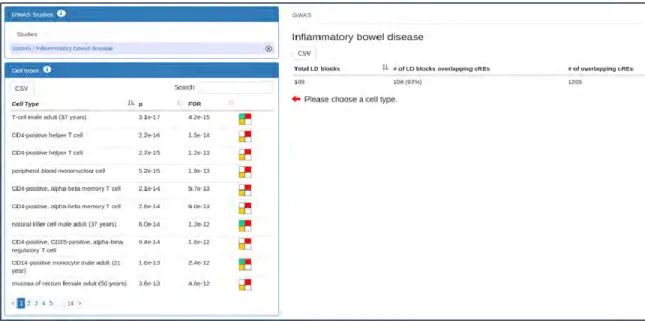
Figure II-22 | Results are shown here for a 2012 IBD GWAS study... 108
Figure II-23 | SCREEN Search Results ... 109
Figure II-24 | SCREEN and UCSC Genome Browser ... 110
Figure II-25 | SCREEN cRE Details View ... 111
Figure II-26 | Gene expression view for LSP1 ... 112
Figure II-27 | SCREEN Differential Gene Expression ... 113
Figure II-28 | Signals around Ogn locus ... 114
Figure II-29 | Overall cell type enrichments for variants reported by GWAS .... 115
Figure II-30 | Top cell type enrichments for variants reported by GWAS ... 116
Figure II-31 | Annotating GWAS variants using SCREEN ... 117
Figure II-32 | Annotating GWAS variants using SCREEN ... 118
Figure II-33 | SCREEN display of the ZMIZ1 gene and TSS levels ... 119
Figure II-34 | SCREEN display of PPIF gene and its TSS expression levels ... 120
Figure II-35 | SCREEN display of AGAP1 expression levels ... 121
Figure II-36 | Fine mapping GWAS variants using SCREEN ... 122
Figure II-37 | Fine mapping GWAS variants using SCREEN ... 123
Figure II-38 | EM10E0042440 H3K27ac signal across mouse tissues ... 124
Figure II-39 | Method for normalizing epigenomics signals ... 125
Figure II-40 | Precision-Recall (PR) curves for VISTA Enhancer prediction .... 126
Figure II-41 | PR curves for VISTA Enhancer prediction anchored on DHSs ... 127
Figure II-42 | Correlation of gene expression with epigenomic signals ... 128
Figure II-44 | EP300 signals for GM12878 cREs ... 130
Figure II-45 | cRE states cluster into groups ... 131
Figure II-46 | POL2 signals at cREs ... 132
Figure II-47 | UCSC Genome Browser views of cREs around the HNF4a TSS 133 Figure II-48 | UCSC Genome Browser views of cREs around the SPI1 TSS .... 134
Figure II-49 | UCSC Genome Browser views of cREs around NPAS4 TSS ... 135
Figure III-1 | Factorbook TF UML ... 145
Figure III-2 | Factorbook: ChIP-seq TF Index ... 146
Figure III-3 | Factorbook: Histone Profiles ... 147
Figure III-4 | Factorbook: Motifs ... 148
Figure III-5 | Factorbook: Histone Heatmaps ... 149
Figure III-6 | Factorbook: Transcription Factor Heatmaps ... 150
Figure III-7 | Factorbook: Nucleosome Profiles ... 151
Figure IV-1 | Validate predictions for known ChIP-seq experiments ... 179
Figure IV-2 | Imputation Flowchart ... 180
Figure IV-3 | Predict ATF3 binding sites ... 181
Figure IV-4 | CTCF PR Curve ... 182
Figure IV-5 | Actual vs Imputed TFBS in UCSC Genome Browser ... 183
Figure IV-6 Predicting TFBSs in 246 ChIP-seq Datasets ... 184
Figure IV-7 | Predicting TFBSs in 246 ChIP-seq Datasets ... 185
Figure IV-8 | Predicting TFBSs in HepG2 ... 186
Figure IV-10 | aucPR Curves for ChomImpute vs LR ... 188
Figure IV-11 | aucPR curves ChromImpute vs xGBoost ... 189
Figure IV-12 | DREAM Genomic Binning ... 190
Figure IV-13 | Machine Learning Approach... 191
Figure IV-14 | Kernel ... 192
Figure IV-15 | Gradient Boosting Tree Example ... 193
Figure IV-16 | Feature Importance... 194
Figure IV-17 | Performance Comparison ... 195
Figure IV-18 | Leaderboard Training Performance ... 196
Figure IV-19 | Leaderboard Training Performance ... 197
Figure IV-20 | DREAM Leaderboard Testing Performance ... 198
Figure IV-21 | Method Comparison for Predicting Forebrain Enhancers ... 199
Figure IV-22 | Method Comparison for Predicting Forebrain Enhancers ... 200
Figure IV-23 | Method Comparison for Predicting Heart Enhancers ... 201
Figure IV-24 | Method Comparison for Predicting Heart Enhancers ... 202
Figure IV-25 | Round 2 Midbrain Prediction Results ... 203
Figure IV-26 | Round 2 Midbrain Prediction Results ... 204
Figure V-1 | Exp and ExpFile Class Diagram... 210
Figure V-2 | File and Paths Class Diagram ... 211
Figure V-3 | Downloader Class Diagram ... 212
Figure V-4 | QueryDCC Class Diagram ... 213
Figure V-6 | Peaks Class Diagram ... 215
Figure V-7 | Utils Class Diagram ... 216
Figure VII-1 | Fraction of H3K27ac peaks that overlap DNase peaks (hg19) .... 233
Figure VII-2 | Fraction of DNase peaks overlapping H3K27ac peaks (hg19) ... 234
Figure VII-3 | Encyclopedia Master Peak Distances (mm10) ... 235
Figure VII-4 | Fraction of DNase peaks overlapping H3K27ac peaks (mm10) . 236 Figure VII-5 | Fraction of H3K27ac peaks that overlap DNase peaks (mm10) . 237 Figure VII-6 | | Cross-biosample master peak overlap... 238
Figure VII-7 | UCSC Visualization ... 239
Figure VII-8 | WashU Genome Browser ... 240
Figure VII-9 | Problems with early versions ... 241
Figure VIII-1 | Overview Of ENCODE Encyclopedia V3 ... 245
Figure VIII-2 | Visualizer Main Page ... 246
Figure VIII-3 | Dynamic trackhubs ... 247
Figure VIII-4 | UCSC Genome Browser... 248
List of copyrighted Materials Produced by the Author
List of Third Party Copyrighted Material
Imparo ancora
I.
Chapter I: Introduction
Our experience hitherto justifies us in trusting that nature is the realization of the simplest that is mathematically conceivable.
–Albert Einstein, Herbert Spencer Lecture, 1933
We, as humans, have an innate, natural curiosity about ourselves, how we work, and the myriad ways in which we malfunction. In many ways, this curiosity has been codified and matured by the scientific method into modern molecular biology. What we have found so far is that our human genome—our code—is beautifully innate and immensely complex. That there is structure in this code, though, is becoming clearer. The central dogma of molecular biology—that sequence information from DNA is transcribed into mRNA, and mRNA is ultimately translated into protein (Crick 1958)—belies an enormous amount of machinery controlling this biological flow of information.
A genome is composed of coding and non-coding regions of DNA. Coding regions get processed into protein products, while non-coding regions have a myriad of functions. Large eukaryotic genomes must be packaged and folded multiple times to fit into a cell nucleus. The first level of DNA packaging into chromatin occurs by winding DNA around histone proteins, forming structures called nucleosomes. Eight histone proteins form the core of the nucleosome, with each of 4 histone proteins (H2A, H2B, H3, and H4) found twice. Histone protein tails can undergo a large number of post-translational chemical modifications. For example, the 27th lysine residue of H3 can be acetylated (H3K27ac, for short), or the 4th lysine residue on H3 can be trimethylated (H3K4me3). These modifications have wide-ranging effects on cellular processes, regulating everything from gene expression and the cell cycle to DNA replication and
apoptosis (Wang et al. 2001; Koprinarova, Schnekenburger, and Diederich 2016; Eberharter and Becker 2002). Nucleosomes can then be further packaged into
increasingly compact and complex chromatin structure. This 3D structure enables gnomic elements separated by large linear genomic distance to suddenly be able to interact; almost any genomic location has a non-zero probability of interacting with any other portion of the genome (Dekker, Marti-Renom, and Mirny 2013). The winding and unwinding of chromatin all the way down to modifications of histone tails changes the accessibility of the gnome; local DNA accessibility changes influence where transcription factors can bind promoter and enhancer regions, affecting gene expression.
Experimentally, chromatin accessibility is indicated by DNase I (a nuclease) digestion (Neph et al., 2012). DNase digestion followed by next generation DNA sequencing (DNase-seq) (Boyle et al., 2008) is now a widely-used and reliable technique, with experimental data available for hundreds of biosamples in ENCODE.
Certain patterns or signatures of chromatin accessibility and histone modifications have been found to have association with certain events. For example, the H3K27ac histone mark in a DNA-accessible region typically indicates that one or more activator proteins (called transcription factors) can bind and increase protein translation (Rada-Iglesias et al. 2011). These regions that increase gene expression are typically within a ~1 MB window of the gene (upstream or downstream), and are called enhancer regions (Gillies et al. 1983). Similarly, the H3K4me3 histone mark in a DNA-accessible region, within +/- 1,000 bases upstream of where transcription is initiated (the Transcription Start
Site (TSS)), and on the same strand as the gene generally indicates a promoter region (Heintzman et al. 2007).
Transcription factors (TFs) are DNA-binding proteins that regulate transcription of genetic information from DNA to RNA. TFs have activating or repressing activity via many potential mechanisms: they may complex with other TFs (Maston, Evans, and Green 2006), coactivator proteins, RNA polymerase II, chromatin remodeling complexes, and/or noncoding RNA molecules (Phillips 2008). DNA-binding TFs bind short (6-15 base pair) fragments of genomic DNA; a particular location is called a motif site or TF binding site. These sites demonstrate high evolutionary conservation (Chen & Rajewsky, 2007). Motif sites show distinct cleavage patterns (called footprints) after digestion by DNase I. While DNase-seq provides footprint data reflecting the presence of any DNA-binding proteins, additional evidence of a particular TF-DNA bound complex can be experimentally determined through chromatin immunoprecipitation (ChIP) with massively parallel DNA sequencing (ChIP-seq) (Johnson, Mortazavi, Myers, & Wold, 2007). Thousands of ChIP-seq datasets are available for hundreds of DNA-binding TFs (Wang et al., 2012). There are, however, thousands of different DNA-bound TFs in the human genome (Wilson et al. 2008), and TF binding depends upon many factors, including cell type specificity, phase of development, and/or experimental design. It is increasingly clear that many diseases are a product of genetic variations in regulatory regions of the genome, frequently in regions impact regulatory TF binding (Lee and Young).
The role of genetics in understanding disease pathology has become a central aspect of medicine, with an explosion of research occurring in the past few years. It is increasingly apparent, however, that understanding how changes “above” the genome— in the epigenome—is central to both advancement of basic science and to the translation of these findings to clinical therapy. Around 90% of disease-associated Single Nucleotide Polymorphisms (SNPs) have been found to be in intronic or intergenic regions across multiple Genome Wide Association (GWAS) studies (Hindorff et al. 2009). These genetic variants outside of protein-coding regions indicate that disease pathology may be altered by changes in regulatory regions of the genome, in functional regions such as enhancers and promoters (Hrdlickova et al. 2014). Better understanding of the
epigenome, including building an encyclopedia of all functional elements that details how and why these elements work, is central to this advancement.
Deciphering this complex orchestra of histone modifications, chromatin
remodelers, transcription factors, etc. is central to better understanding the epigenome. Since 2003, the Encyclopedia of DNA Elements (ENCODE) project has collecting and analyzing data in a large-scale to characterize all the functional elements of the human genome. Thus far, ENCODE has successfully collected thousands of chromatin
accessibility, transcription factor, and histone modification experiments, finding hundreds of millions of regions of putative regulatory function across hundreds of different
biosamples. ENCODE has also been highly influential in developing and publishing standards guidelines for DNA-seq, ChIP-seq, and RNA-seq experiments, as well as cloud-scale, open-source pipelines for processing these experiments. ENCODE has also
collected hundreds of gene expression experiments, as well as developing (through GENCODE) a curated set of gene annotations.
Making an actual Encyclopedia of functional genomic elements, though, has proven difficult. The diversity of chromatin accessible regions, histone modification patterns, and TF binding sites across all the different cell and tissue types of the human body has made clear there is a combinatorial number of activation signatures in the genome, as well as millions of potential functional elements. In this thesis, we develop systematic methods of selecting cell-type specific candidate Regulatory Elements (cREs), and demonstrate the biological usefulness of these regions. We have also assigned stable IDs (called accessions) to these regions, with the intent to construct a stable, curated annotation of functional genomic elements, just as Ensembl does for genes (Birney et al. 2004; Aken et al. 2016).
As the visual analytics field has shown, just being able to display raw data is not useful: the extracted analysis products are where the real value is (Keim 2010). Just making an Encyclopedia of cREs is insufficient unless there are ways to visualize and understand the genomic and epigenetic context the putative functional elements exist in. The great importance of being able to visualize highly-multidimensional omic data is clear; there are a multitude of examples available, with even entire frameworks being developed for pathways and gene expression visualization (Streit et al. 2009). Some of the most exciting developments include a Google-maps view of 3D chromatin structure analysis products (Perkel 2017). In this thesis, we develop two visualizers—SCREEN
and Factorbook—to assist users in searching and viewing the millions of elements and data points available from the ENCODE data.
While we have successfully utilized the thousands of available ENCODE ChIP-seq experiments to build the Encyclopedia and visualizers, we have also struggled with one of the core limitations of ChIP-seq TF experiments. The binding of a transcription factor at a particular motif site in the genome depends upon a diverse number of factors. While experimental methods seq have begun to shed light on these binding patterns, fully understanding regulation via transcription factor binding, though, will require an
enormous number of ChIP-seq experiments. Given the wide variety of conditions affecting binding, millions of different experiments are required to comprehensively understand when and where transcription factors bind (Lee and Young). Accurately predicting transcription factor binding sites through statistical and machine learning methods could drastically reduce the number of experiments needed. Further, improved understanding of transcription factor binding would shed light on gene regulatory networks present during embryogenesis, development, and disease states.
Several different labs have developed predictive models for motif site binding over the last decade. Previous predictive models (such as CENTIPEDE and PIQ)
demonstrate the initial feasibility of probabilistically predicting the bound state of a motif site in the genome. For experimental input data, these models primarily utilize DNase-seq data for elucidation of chromatin state and, ultimately, motif site binding probability. In this thesis, we develop several predictive models based upon supervised learning methods. These models leverage ChIP-seq data already acquired by ENCODE
participants, as well as other ENCODE and Roadmap Epigenomics datasets. We also develop a large number of features for model training, using supervised approaches, and have achieved some success in predicting TF binding sites. We have also utilized some of these techniques while competing in the ENCODE-DREAM in vivo Transcription Factor Binding Site Prediction Challenge.
This thesis describes efforts toward better understanding and visualizing the epigenome. Chapter II will introduce our current version of the ENCODE Encyclopedia. In it, we demonstrate how we locate regions of the genome with open chromatin and enhancer-like or promoter like signatures based on histone modification marks and other genomic distance information. These regions—candidate Regulatory Regions (cREs)— are our first approximation of a systematic, accessioned, genomic-wide catalog detailing regions that are involved with functional control of the genome. Chapter II will introduce the latest version of an aggregated, peak-centric visualizer for transcription factor binding sites (TFBS). Chapter IV discusses our work on imputing entire epigenetic experiments, first focusing on predicting locations of TFBS. Lastly, Chapter V introduces SnoPlowPy, our tool driving metadata and job management functionality for large-scale analysis projects.
II.
Chapter II: Building and Visualizing an Encyclopedia of
ENCODE candidate Regulatory Elements
II.1
Preface
This research chapter encompasses work performed by Jill Moore, myself, Henry Pratt, Zhiping Weng, and >500 other collaborators in the ENCODE Consortium. The chapter combines one manuscript currently in review (as of December 2017) with another manuscript on SCREEN (with Henry Pratt) that is currently in draft.
With more than 14,000 experimental datasets, consuming more than 0.5 petabytes of storage space across hundreds of thousands of files, the ENCODE project has built a vast catalog of gene expression, chromatin accessibility, histone medication, and
transcription factor binding data. While investigating these data, I found the data difficult to utilize to answer fundamental biological questions, such as where putative enhancers and promoters are located, or how gene expression levels vary across disease conditions or developmental time points. These questions were impossible to answer without
manually curating, downloading, and processing the data. Just determining which files to use for such analyses was also non-intuitive and essentially undocumented, and the wide-variety of data processing techniques added many subtle problems when integrating data across labs, let alone different experiments. I decided to ameliorate these problems and allow straightforward analysis of the data and generation of actionable biological insights and hypotheses.
While individual tools to interrogate particular regions of the genome have existed for more than two decades (Kent et al. 2002), and epigenetic annotations have been available for years (Frankish et al. 2015), no tool has integrated as much genetic and epigenetic data in one location as SCREEN. SCREEN solves many of the problems (both biological and practical) described above. Inside of SCREEN, I have integrated and condensed thousands of ENCODE experiments into an easy-to-use product that allows researchers to intuitively explore the available data. I am the overall architect of
SCREEN, having designed and implemented the database system, data import pipeline, and much of the software architecture. SCREEN excels at allowing the user to develop hypotheses for potential functional regulation across millions of region in the human and mouse genomes.
SCREEN offers new solutions to help navigate the vast sea of data. I am also one of the first to build an online database of hundreds of millions of DNase and ChIP-seq peaks that could then be intersected with candidate regulatory regions at the click of a button. The accessioning system for peaks I implemented is the start of a critical new stage of epigenetics, where individual regions can be tracked not just through
publications, but across hundreds to thousands of experiments. Current projects like ENCODEproject.org are designed to ascension a few hundred thousand objects; the systems are not capable of supporting millions of objects. Comparing how these regions change across developmental or disease states becomes not just far more straightforward, but, in fact, doable in seconds, not hours or days it would take before. This approach of
systematically cataloging regions of the genome will become as integral to the field as plant taxonomy became for the field of botany.
II.2
Summary
Many human genomes have been sequenced, yet we still lack comprehensive maps of genomic functional elements and do not fully understand how they specify cell and tissue types. Such information is critical to assess how genomic variants affect development, ageing, and susceptibility to diseases. The goal of the Encyclopedia of DNA Elements (ENCODE) project is to discover and characterize the full repertoire of elements (www.encodeproject.org). Here, we summarize the data generated in Phase III of the project and introduce the ENCODE Encyclopedia, an evolving collection of annotations derived from assay-specific and integrative analyses. At the heart of the Encyclopedia is a new Registry of candidate Regulatory Elements (cREs), defined by a biochemical
signature that uses chromatin accessibility, histone modification and transcription factor occupancy data. The Registry currently contains 1.31 M human and 0.43 M mouse cREs, covering hundreds of biosample types. The cRE landscape recapitulates the current understanding of cellular identity, tissue composition, developmental progression, and disease-associated genetic variants. Aided by a dedicated visualization engine called SCREEN (screen.encodeproject.org), the Registry is a resource for exploring noncoding DNA elements and their variants.
II.3
Introduction
The genome contains the blueprint for organismal development and function.
that motivates many individual research labs and organized consortium efforts. Among these efforts is the Encyclopedia of DNA Elements (ENCODE) Project, launched by the National Human Genome Research Institute (NHGRI) in 2003. The overarching goal of ENCODE is to provide an integrated resource to aid the scientific community in studying mammalian biology and human diseases.
In pursuit of this goal, ENCODE develops and applies high-throughput experimental technologies and computational approaches to catalogue candidate functional elements in the human and mouse genomes, including transcripts and their regulatory elements. The pilot phase of ENCODE focused on 44 carefully selected regions covering 1% of the human genome using array-based techniques (Birney et al. 2007). Phase II used deep-sequencing-based biochemical assays to interrogate the entire human genome, producing 1,640 datasets, and integrative analyses of these datasets identified an extensive set of candidate functional elements (Consortium 2012). The related Mouse ENCODE (Yue et al. 2014) and modENCODE projects (Gerstein et al. 2010; Roy et al. 2010) performed thousands of genome-wide experiments on the mouse, fly, and worm. Complementary projects, including the NIH Epigenomics Project
(Kundaje et al. 2015) and the International Human Epigenome Consortium have also produced thousands of epigenomic maps for human cells and tissues (Stunnenberg, International Human Epigenome, and Hirst).
Despite this progress, the human and mouse genomes remain only partially annotated, limited by the depth of biochemical element types mapped for any one cell type and the breadth of cell types mapped for any single biochemical feature.
Accordingly, our understanding of the diversity of transcripts and their regulatory elements in each cellular context is far from complete. To begin to address these limitations, ENCODE Phase III expanded data collection in both depth and breadth— studying additional chromatin features, regulatory factors and RNA types with an emphasis on primary cells and tissue samples.
All data are submitted to the ENCODE Data Coordination Center, reviewed for quality and released to the scientific community via the freely accessible ENCODE web portal (www.encodeproject.org). ENCODE members have reported new findings
throughout the past five years based on data generated and released during Phase III. Additionally, we have now assembled the ENCODE Encyclopedia of predicted and confirmed functional elements, based on all ENCODE data collected during Phases II and III, supplemented by data from the NIH Epigenomics Project. This chapter describes the ENCODE Encyclopedia and presents illustrative examples of its use.
A new focus in ENCODE Phase III has been to build a Registry of candidate Regulatory Elements (abbreviated as cREs). This effort is guided by the current
understanding that robust biochemical signatures, including chromatin accessibility and particular histone modifications, are preferentially associated with major classes of noncoding regulatory DNA elements—transcriptional promoters, enhancers, insulators, and silencers. While the biochemical signatures are neither causal nor perfect predictors of element activity, they enable the selection of an enriched set of cREs that are
experimental data, for exploration by users via a specifically designed visualization tool called SCREEN (screen.encodeproject.org).
II.4
Results
II.4.1 Summary of Encode Phase 3 Data Production
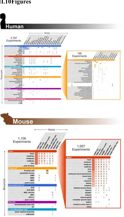
The ENCODE Consortium has produced data on three main aspects of genome activity— transcriptomes, DNA-based regulatory elements for transcription and replication, and RNA-based elements for post-transcriptional regulation. Phase III greatly expanded the number of experiments in each category and released 4,903 experiments (3,797 on human and 1,106 on mouse; see Figure II-1 on page 87). Table II-1 on page 78 summarizes these experiments by category. We define an experiment as the application of a genomic assay (such as ChIP-seq, RNA-seq, DNase-seq, or ATAC-seq) to a particular biosample type (such as a tissue, a cell line, primary cells, or stem cells). In this section, we summarize the new assays and highlight the results of Phase III data production.
New polyA and short RNA transcriptome data production has focused on primary cells from different body locations and various embryological origins. Single-cell long-RNA-seq was further developed for laser-capture microdissection of human and mouse brain tissues. To better define full-length transcripts, we analyzed captured RNAs using long-read sequencing. This effort, in collaboration with the GENCODE project,
improved the annotations of gene and transcript structures for 14,667 human and 8,708 mouse long noncoding RNAs (Lagarde et al., in review).
A new 5´-complete cDNA sequencing assay called RAMPAGE quantifies gene expression, identifies promoter locations, and assigns 5´ capped termini to their
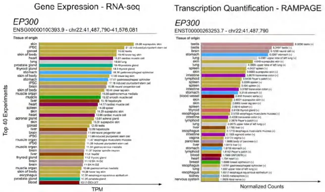
corresponding RNA isoforms (Batut et al. 2013). RAMPAGE yields data at single-nucleotide resolution and is more accurate than RNA-seq for quantifying expression (Batut et al. 2013)—advances which enable it to improve transcription start site (TSS) annotation and transcript quantification. For example, the gene ARHGAP23, which encodes Rho GTPase-activating protein 23, has 12 GENCODE-annotated transcripts and 11 different TSSs. RAMPAGE data revealed a novel TSS in the testis (Figure II-2a on page 88), located 9.2 kb upstream of the nearest annotated TSS, and another novel TSS in exon 7 specific to the spleen (Figure II-2b on page 88). As another example, two different TSSs, 824 bp apart, are annotated by GENCODE V26 and UCSC for EP300, which encodes a widely studied histone acetyltransferase important for enhancer activity. RAMPAGE data across six cell and tissue types showed that although both TSSs are active, one TSS is used far more frequently than the other (Figure II-3 on page 89).
The coverage of noncoding, biochemically marked DNA elements, many of which have potential regulatory functions, has been greatly expanded during ENCODE Phase III. We completed 163 new DNase accessibility maps, including deep sequencing DNase-seq datasets on hundreds of cell and tissue samples, thus facilitating the prediction of regulatory protein occupancy by footprinting (Hesselberth et al. 2009). The ATAC-seq assay (Buenrostro et al. 2013), which assesses chromatin accessibility via insertion by the Tn5 transposome, was conducted on tens of human and mouse tissues and primary cells. We expanded the application of ChIP-seq to map the locations of modified histones, histone variants, and 33 chromatin regulators and modifiers in a carefully selected collection of five human cell lines—K562, H1, GM12878, HepG2, and A549. Over 600
ChIP-seq experiments were completed in Phase III for 493 different transcription factors (TFs) in at least one cell type (1,622 experiments on 549 different TFs in Phases II and III combined). For these ChIP-seq experiments, we used either TF-specific antibodies or epitope-tagged TFs created by BAC transfections or CRISPR/Cas9 genome editing. ChIA-PET of Rad21 and CTCF, which are involved in the nuclear organization, along with Hi-C experiments, provide 3D linkage data that include many regulatory regions and cognate target genes. Through the ENCODE Portal (encodeproject.org/antibodies/), we provide quality metrics for all datasets as well as detailed information about the
antibodies used in our experiments to help users evaluate and use the data most effectively.
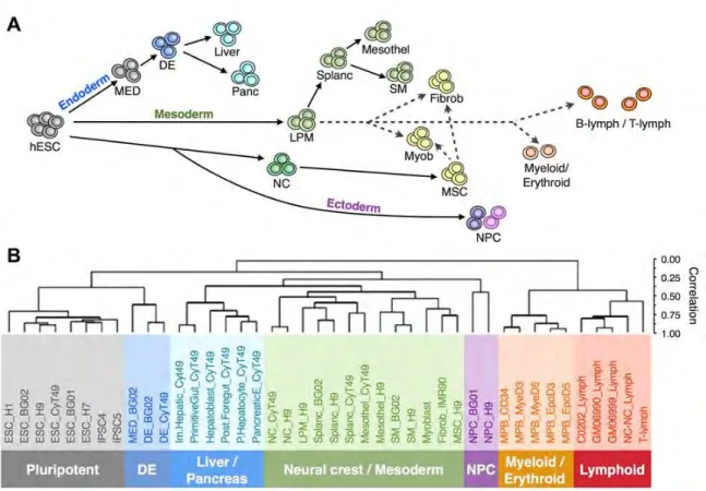
DNA replication timing provides insights into gene regulation and spatiotemporal genome compartmentalization (Gilbert 2002). We measured replication timing during fate commitment of human embryonic stem cells, thus yielding 84 datasets for 26 cell types representing the embryonic layers endoderm, mesoderm, ectoderm, and neural crest (Rivera-Mulia et al. 2015) (see Figure II-4 on page 90). Because replication timing differs across cell types, we expected that clustering of these datasets would recapitulate their developmental lineages, and that was indeed observed (see Figure II-5 on page 91).
The mouse component of ENCODE Phase III focused on embryo development at daily intervals between embryonic day 10.5 (e10.5) and postnatal day 0 (p0), with 6-12 tissues sampled per day. RNA-seq of polyA RNAs and miRNAs, ChIP-seq for eight histone modifications, ATAC-seq, and whole-genome bisulfite sequencing were
performed on all the samples of the mouse embryonic developmental series, augmented by DNase-seq and ChIP-seq of three TFs in selected samples.
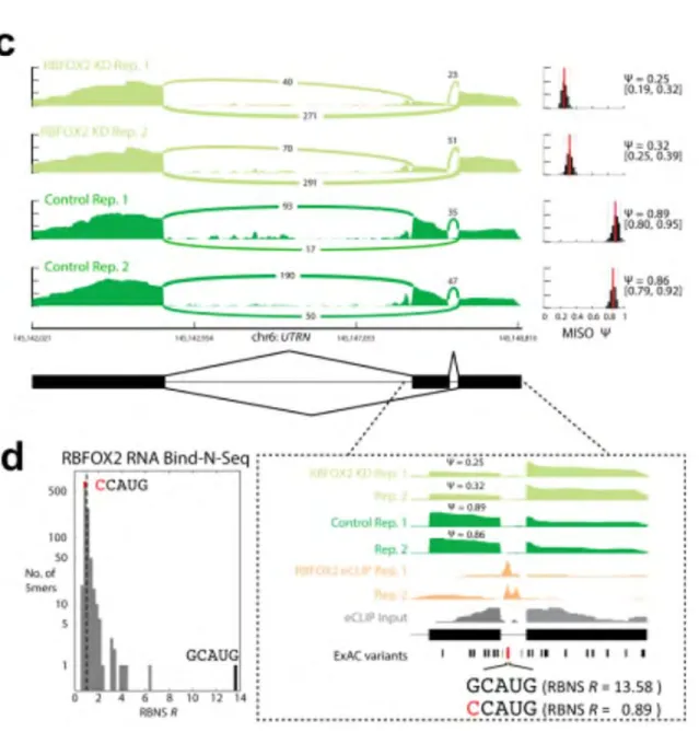
A new project in ENCODE Phase III was to identify and characterize functional RNA elements bound by RNA-binding proteins (RBPs) (van Nostrand et al., in
preparation). Four types of related data were generated: RIP-seq and enhanced UV crosslinking and immunoprecipitation of RBPs followed by sequencing (eCLIP-seq) (Van Nostrand et al. 2016) to identify bound RNAs in vivo and pinpoint the portions of these RNAs involved in binding interactions; RNA-seq on cells depleted of specific RBPs by shRNA or CRISPR; RNA Bind-N-Seq (RBNS)(Lambert et al. 2014) to determine the relative binding affinity of RBPs in vitro for all possible RNA sequences; and subcellular localization of RBPs by immunostaining.
The breadth of our RBP data enables integrative analyses to relate genetic variation to RBP regulation. For the 18 RBPs with eCLIP, RBNS and, RBP-knockdown RNA-seq data, we identified 26 variants from the Exome Aggregation Consortium (ExAC)(Lek et al. 2016) that overlapped an eCLIP peak, disrupted an RBNS motif, and produced a splicing change upon knockdown of the corresponding RBP (van Nostrand et al., in preparation). For example, intron 66 of UTRN (dystrophin-related protein 1) harbors an RBFOX2 eCLIP peak downstream of an alternatively spliced exon (Figure II-6c on page 92), which overlaps an ExAC variant (Lek et al. 2016). This G→C variant disrupts the RBFOX2 binding motif (GCAUG) at the first position. RBNS data reveal that this variant substantially changes the RBFOX2 binding site—the top 5-mer has an enrichment value of 13.58 for the major G allele but 0.89 for the C variant (Figure II-6d
on page 92), thus suggesting that the mutation disrupts RBFOX2 binding in vivo. To determine whether the disruption of RBFOX2 binding would alter splicing, we performed RNA-seq on HepG2 cells after knocking down RBFOX2. In wild-type cells, the upstream exon was included in 87% of messages, whereas the inclusion was decreased to 28% in the RBFOX2 knockdown cells (Figure II-6c on page 92). Taken together, these data argue that this G→C variant disrupts RBFOX2 binding, leading to decreased inclusion of the upstream exon in over half of UTRN messages, and resulting in an altered
composition of protein isoforms. Overall, the actual number of variants that influence RNA metabolism is larger than the 26 ExAC variants identified in this way, because they may affect aspects of RNA biology other than splicing.
II.4.2 The Encode Portal and Uniformly Processed Data
The ENCODE portal (www.encodeproject.org) is the primary interface for retrieving all ENCODE data, metadata, data standards, and experimental protocols (Sloan et al. 2016). It also provides entry to the ground and integrative levels of the ENCODE Encyclopedia (Figure II-7 on page 93), which is described in the next section. The Portal is designed to provide users with extensive metadata that describe how ENCODE experiments were performed, processed, and connected in common biological themes (Hong et al. 2016). All experiments followed data production guidelines
(www.encodeproject.org/about/experiment-guidelines/#guideline). An experiment typically comprises two biological replicates, with some exceptions; in the case of single-cell assays or assays utilizing human donor tissues of limited availability, for example, no cell is a conventional replicate of another. A released experiment includes the “raw”
sequencing data (typically FASTQ files) and all analysis output files (such as alignment files, signal files, or peak files) from the uniform processing pipelines. These pipelines are central to ENCODE data, and the major pipelines are available for users to apply to their own data, either by downloading the code and running it locally or by accessing the pipelines at the DNAnexus cloud provider.
The Portal was completely redesigned during Phase III for better data access and metadata clarity. The homepage presents summaries of the numbers and types of
experiments, with intuitive links for data access. Experiments are annotated by key features (called facets) so that users can easily find experiments via a faceted search. A matrix view displays the search results (encodeproject.org/matrix/?type=Experiment; see Figure II-1 on page 87), which can be switched to list or table views. Entries in the matrix are hyperlinked to underlying datasets, along with metadata and quality metrics.
II.4.3 The Encode Encyclopedia
The raw data described above and their signal maps across the human and mouse
genomes are valuable for interrogating genome function in myriad ways, from browsing individual loci to large-scale data integration. To aid users in data mining and hypothesis building, we have derived summaries of key aspects of the raw data and organized them into the ENCODE Encyclopedia. The Encyclopedia presently has two levels of
annotations (Figure II-7 on page 93). The ground level includes peaks and quantifications produced by the uniform data-processing pipelines for individual data types, and the integrative level contains annotations derived from combined analyses across multiple data types and ground-level annotations.
II.4.4 Encyclopedia Ground Level
The ground level currently has nine components (Figure II-7 on page 93). The chromatin accessibility component contains DNase hypersensitive sites (DHSs)—genomic regions significantly enriched in DNase-seq reads—and their constituent DNase peaks, as well as ATAC-seq peaks. Locations of histone marks and histone variants are provided in the histone modification component as histone peaks, which are regions of the genome significantly enriched in histone ChIP-seq reads. The transcription factor binding
component provides TF peaks, or genomic regions significantly enriched in TF ChIP-seq reads; these peaks are further characterized by enriched sequence motifs (identified using the MEME-ChIP tool (Machanick and Bailey 2011)) and the average histone mark ChIP signals and nucleosome occupancy signal surrounding them in each cell type. The TF peaks and associated information can be viewed in the wiki-style web resource
Factorbook (see page 136). The gene and TSS expression components give quantitative estimates of the abundance of the various types of RNA molecules in each of the assayed cell types based on ENCODE RNA-seq and RAMPAGE data. These estimates are provided at the gene and TSS levels for GENCODE-annotated genes, plus activity levels for novel TSSs identified by RAMPAGE. Gene or TSS expression profiles across cell types can be visualized using the SCREEN tool described below (see Figure II-8 on page 94).
The RNA binding protein (RBP) component provides RBP peaks, which are regions of the transcriptome enriched for binding by an RBP, as determined by the
account variations in transcript abundance and processing(Van Nostrand et al. 2016). The DNA methylation component analyzes whole-genome bisulfite sequencing data and provides the methylation state for each cytosine in the genome. The 3D chromatin interaction component provides interaction frequency estimates between genomic loci, such as between promoters and distal enhancers, as computed from ChIA-PET data. Finally, the component for chromatin domains and compartments provides topologically associated domains (TADs) and A/B compartments called using Hi-C data.
New data are processed and added to the ground level of the Encyclopedia as soon as they are available. Thus the ground level is continually updated ("live"), and these updates do not constitute new versions. More components will be added as additional analysis pipelines are developed and existing pipelines are improved. Components of the integrative level of the Encyclopedia are versioned as described below.
II.4.5 Encyclopedia Integrative Level
A longstanding goal of functional genomics is to discover and map the full regulatory element repertoire of the genome and then to delineate which elements are active or repressed in individual cell types. In pursuit of this goal, ENCODE and Roadmap Epigenomics Consortia have now produced basic epigenetic signals broadly in hundreds of human and mouse cell types and tissues. ENCODE has also examined a few cell types much more extensively for diverse transcription factor occupancy, genome-wide DNA methylation, RNA-binding protein occupancy and other “deep” assays. These differences in assay breadth versus depth have motivated two complementary computational
approaches to build catalogues of candidate transcriptional regulatory elements, and our Encyclopedia offers both.
The first approach started in ENCODE II. It uses machine learning methods such as ChromHMM(Ernst and Kellis 2010; Ernst and Kellis 2012) and Segway (Hoffman et al. 2012) to integrate many different types of epigenetic signals. ChromHMM and Segway are unsupervised probabilistic models that integrate a specified number of epigenetic signals to define a large repertoire of chromatin states, many of which
correlate with known functional element types and activity levels, e.g., active promoters, enhancers, or heterochromatin domains. ChromHMM have been augmented to
accommodate cell types with some missing assays and then applied to the contemporary Roadmap (Ernst and Kellis 2015a) and ENCODE III cell types and tissues that achieved sufficient assay coverage. A strategy was developed in ENCODE III to train separate Segway models on each cell type—allowing for different assay coverages in different cell types—and then automatically interpret these results across all cell types using a Random Forests classifier. The chromatin states of 164 human cell types have been annotated using this strategy by integrating 1,615 genomics datasets (Libbrecht et al. 2016). We similarly applied ChromHMM to the mouse embryo development series—66 complete epigenomes each assayed by ChIP-seq of eight histone marks— and defined 15
chromatin states that showed coordinated changes with gene expression measured by RNA-seq for each of the 66 samples (Gorkin et al., in preparation, Tsuji et al., in
preparation). The resulting chromatin state maps from this section are all included in the integrative level of the Encyclopedia.
The second approach is motivated by the substantially increased number of experiments on primary cells and tissues during ENCODE Phase III. The limited
quantities of primary cells and tissues have led to incomplete assay coverage for many of these samples. Thus we have developed an approach that uses a highly parsimonious combination of just four types of assays to maximize the coverage of cell and tissue types, though at the expense of subtler inferences about each element’s possible activity. The rest of the chapter focuses on the second approach that has led to the new Registry of candidate Regulatory Elements.
II.4.6 The Registry of candidate Regulatory Elements
Given the breadth of biosamples in the union of ENCODE and Roadmap data, we aspired to build an initial Registry covering a majority of cREs in the genome. The most direct approach to identifying cREs would be to include all relevant epigenetic signals in a comprehensive statistical model and then train the model with experimentally validated regulatory elements. Indeed, such methods have been developed (Rajagopal et al. 2013; Erwin et al. 2014). However, at this time, relatively few enhancers and insulators have been systematically tested across many cell environments with functional assays: without such a “gold standard,” it is not possible to train a general statistical model that remains predictive in new cell types.
Therefore, we pursued a different approach that is based on just four epigenetic signals that we found to be most predictive of regulatory elements: chromatin
accessibility (measured by DNase-seq), the histone modifications H3K4me3 and
work in the field. DNase hypersensitive sites delineate all the main classes of cis-regulatory elements in a cell-type-specific manner, including promoters, enhancers, insulators, and locus control regions (Thurman et al. 2012). H3K4me3 and H3K27ac are the two histone marks most enriched at promoters and enhancers respectively (Heintzman et al. 2007; Visel et al. 2009). CTCF is the established insulator binding protein in
mammals (Kim et al. 2007) and its binding sites are enriched at interacting chromatin loci (Rao et al. 2014).
To further test our selection, we compared the effectiveness of ten different types of epigenetic signals in predicting enhancers in the corresponding tissue: DNase
hypersensitivity, eight histone marks (H3K4me1, H3K4me2, H3K4me3, H3K27ac, H3K9ac, H3K9me3, H3K36me3, and H3K27me3), and DNA methylation. These epigenetic signals were all assayed with specific mouse e11.5 tissues during ENCODE III, and the tissue-specific e11.5 enhancers tested using in vivo transgenic assays were from obtained the VISTA database (Visel et al. 2007). We found that DNase and
H3K27ac were the best single features for predicting tissue-specific enhancers. We then used RNA-seq to evaluate the effectiveness of these same epigenetic signals in predicting gene expression levels and found H3K4me3 to the best single feature. We found that DNase offers high spatial precision in defining cREs: DHSs are ~350 bp long and typically correspond to the core of regulatory elements. In contrast, the H3K27ac and H3K4me3 signals are more diffuse: they tend to be low at the center of a regulatory element—presumably because of the lack of a nucleosome there—but are elevated at flanking nucleosome positions. DNase, therefore, presents the best localization of a cRE,
while H3K27ac and H3K4me3 suggest the recent activity state, and the coincidence of significant signals from at least two assay types increases the overall confidence in the cRE.
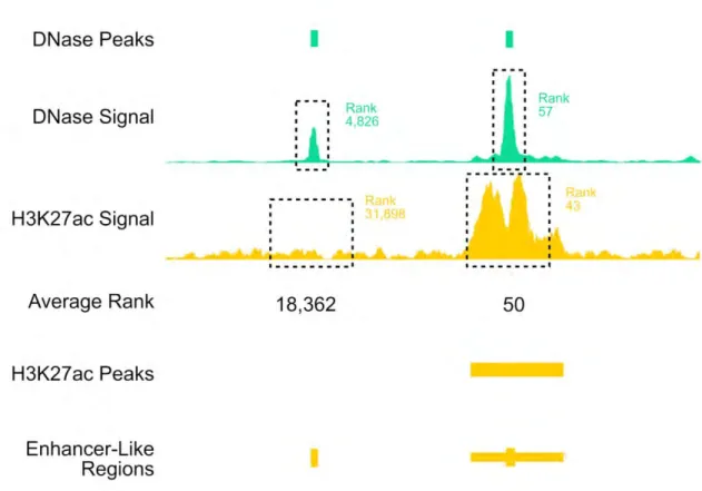
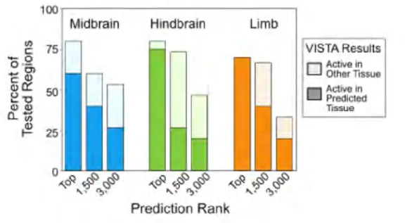
To experimentally test the enhancer branch of our predictor, we used the average rank of the DNase and H3K27ac signals to identify previously untested TSS-distal (> 2 kb from the nearest TSS) candidate enhancers in the mouse e11.5 hindbrain, midbrain, and limb. The boundaries for the predicted regions were defined using the H3K27ac ChIP-seq peaks called by the MACS2 algorithm (Zhang et al. 2008) (see Figure II-9 on page 95). For each tissue, we tested 20, 15 and 15 new regions around the ranks 1-20, 1500-1520, and 3000-3020, respectively. In total, we tested 151 regions (for results, see online Supplementary Table 4). Representative e11.5 transgenic embryos for the
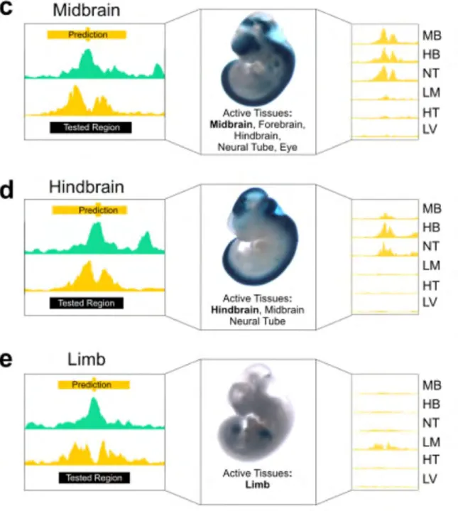
enhancers that validated in the expected tissues are shown in Figure II-10 on page 96. Consistently, higher ranking regions were more likely than lower ranking regions to show enhancer activity in their predicted tissue (Figure II-11 on page 97; e.g., 75%, 26.6%, and 20% for the hindbrain). When enhancers were active in multiple tissues, these tissues also had high H3K27ac signals across the predicted enhancer regions (Figure II-12c-e on page 98). For example, a predicted enhancer in the hindbrain was also active in the midbrain and neural tube; accordingly, high H3K27ac signals were observed in all three tissues (Figure II-12d on page 98). In contrast, an enhancer active almost exclusively in the limb (Figure II-12e on page 98) did not show high H3K27ac signals in other tissues assayed. These results suggest that combining DNase and H3K27ac can identify active enhancers in a particular tissue and quantify their tissue selectivity patterns.
In aggregate, our evaluations showed that combining DNase with two histone marks, H3K4me3 and H3K27ac, is an effective way to build a first version of the
Registry of candidate promoters and enhancers active in specific cell types. We extended this predictor by adding CTCF, a highly conserved architectural protein that binds to insulators and contributes to the establishment and maintenance of three-dimensional chromatin structure (Ong and Corces 2014). Our final algorithm anchors cREs on a representative set of all DHSs, and then evaluates cRE types and activities based on H3K4me3, H3K27ac, and CTCF signals. To maximize coverage, we applied the
algorithm to all cell types interrogated by at least one of these assays, making it possible to include data from 301 human cell types (620 when primary cells or tissues from different donors are counted separately) and 58 mouse cell types (138 with
developmental time-points counted separately) with all ENCODE and Roadmap data considered. It is thus important to note that we distinguish two classes of cREs displaying no activity in a given cell type: cREs for which necessary assays are missing in the cell type, and cREs for which the necessary assays are present but the associated signals did not score as significantly positive.
The first release of the Registry presented here includes 1.31 million human cREs and 0.43 million mouse cREs; future versions will be released periodically, and are already under development. Based on the levels of the four core epigenetic signals and the distance to the nearest annotated TSS, we also classify cREs as those that have promoter-like signatures (PLS) or enhancer-like signatures (ELS) or as those that lack these signatures but are bound by the insulator-binding protein CTCF.
II.4.7 Selection of cREs for the Registry
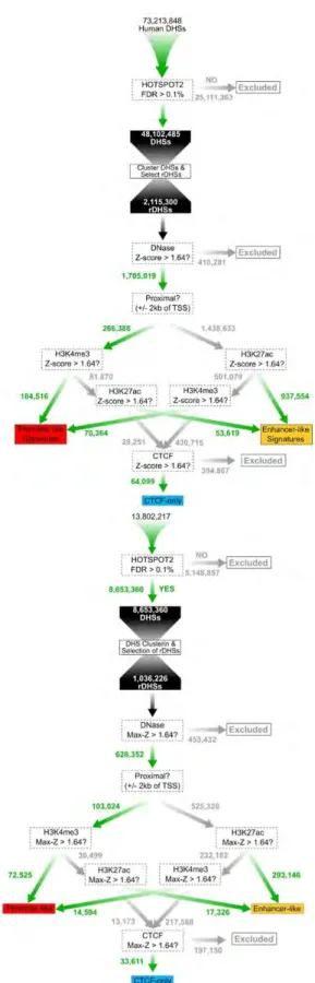
We define cREs as DHSs supported by at least one additional type of epigenetic signal among H3K4me3, H3K27ac, and CTCF in at least one cell type. We first condensed all DHSs from individual samples into a set of non-overlapping representative DHSs
(rDHSs) as described in Methods. We then filtered out the rDHSs with Z-scores less than 1.64—a threshold corresponding to the 95th percentile of a one-tailed test.
Approximately 1.6 M human and 0.63 M mouse rDHSs remained. The rDHSs that have high H3K4me3, H3K27ac, or CTCF signals (a high signal is defined as a Z-score > 1.64 throughout) in at least one cell type are designated cREs. In total, there are 1,310,152 human cREs (Figure II-13 on page 99) and 431,202 mouse cREs. Among them, 724,590 human cREs and 228,027 mouse cREs have high DNase and high H3K4me3, H3K27ac, or CTCF in the same cell type, and these cREs are recognized for having "concordant" support, labelled with an asterisk by their accessions in SCREEN. The remaining 585,562 human and 203,175 mouse "non-concordant" cRE result from high DNase signal in one cell type and high H3K4me3, H3K27ac, or CTCF signals in a different cell type. As more data become available, we anticipate that many of the non-concordant cREs will move into the concordant class, and will be updated to reflect that.
cREs are further designated as TSS proximal if they lie within ±2 kb of a
GENCODE-annotated TSS. There are 242,739 TSS-proximal cREs in human and 92,405 in mouse. The cREs that overlap a TSS are called TSS-containing cREs; there are 46,749 and 24,549 TSS-containing cREs in human and mouse respectively. TSS-overlapping cREs are significantly longer than the rest of the TSS-proximal cREs and TSS-distal
cREs (median length = 548, 317, 342 for human and 589, 320, 339 for mouse; Wilcoxon test p-values < 2.2E-16 for all tests).
II.4.8 Comprehensiveness of the current Registry of cREs
In defining the Registry of cREs based on rDHSs, our working hypothesis is that a collection of rDHSs derived from hundreds of DNase-seq experiments will represent a large fraction of all cREs in the genome and that a new cell type is likely to use as its cRE repertoire a subset of the cREs already in the Registry. To test this hypothesis, we set out to analyse how comprehensive the Registry is in three ways.
First, we examined how many of the GENCODE-annotated TSSs (V19 for human and M4 for mouse) were covered by the current version of the Registry of cREs. To the extent that GENCODE is a mature repository of expressed RNAs across all cell types and states in the human and mouse life cycle, this test provides an informative estimate for the completeness of promoters and promoter-proximal regulatory elements in our Registry. For human, 67% (121,692/181,177) of all annotated TSSs and 72%
(105,196/145,671) of the TSSs of protein-coding genes overlap a cRE in the Registry. For mouse, 61% (57,459/93,719) of all annotated TSSs and 66% (52,066/78,782) of the TSSs of protein-coding genes overlap a cRE in the Registry.
Second, we analyzed how rapidly the total number of unique rDHSs saturated as more and more cell types were added. In ENCODE Phase II, we modelled DHS
saturation using a Weibull distribution and estimated that we had discovered around half of the total DHSs. We performed this analysis again using all human DNase-seq data generated by ENCODE and Roadmap projects. The saturation curves of rDHSs continue
to follow Weibull distributions, revealing at the plateau 1.66 M rDHSs with FDR < 0.1% and Z-score > 1.64. Because only a subset of such rDHSs can be cREs—those with a high H3K4me3, H3K27ac, or CTCF Z-score in at least one cell type—we have identified at least 78.9% cREs in human. We performed the same saturation analysis for mouse but could not reach a reliable estimate due to the smaller number of input tissue types.
Third, we computed the Registry's coverage of H3K27ac, H3K4me3, and CTCF peaks (FDR<0.01) in those cell types with the corresponding ChIP-seq data but without DNase-seq data. The Registry covered 90 ± 8% of H3K4me3 peaks (74 cell types), 87 ± 5% of H3K27ac peaks (54 cell types), and 99 ± 1% of CTCF peaks (31 cell types). The coverage was equally high for mouse, despite a smaller number of DNase-seq
experiments for building the mouse Registry: 88 ± 5% of H3K27ac peaks (69 tissue– time-points) and 96 ± 8% of H3K4me3 peaks (74 tissue–time-points) were accounted for. (There were no cell types with CTCF but without DNase data for mouse.) The coverages for H3K4me3 peaks were low for several human and mouse cell types. The average -log(FDR) of the H3K4me3 peaks in these datasets were low. We visually inspected the two datasets with the lowest coverage (CD-1 megakaryocyte and GR1-ER4 in mouse) and confirmed that the peaks that were not covered by the Registry had low signals and were likely false positives by the peak calling algorithm.
In conclusion, the human Registry appears to be comprehensive: by the above criteria, it covers two-thirds of all cREs and 85% of elements marked by H3K4me3 or H3K27ac or bound by CTCF in any cell type. A cautionary note is that we do not yet know the extent of coverage on highly cell-type-specific cREs active in rare cell types
(numerically minor in their tissues of origin) that have not yet been sensitively assayed. The mouse Registry is less comprehensive than the human Registry, but we expect that it will continue to grow with experiments performed on additional cell types.
II.4.9 Classifying cREs in the Registry
Gene catalogues such as GENCODE define gene models irrespective of their varying expression levels and alternative transcripts across different cell types. By analogy, we provide a general, "cell type agnostic" classification of cREs based on the maximal Z-score of each feature across all cell types with ENCODE and Roadmap data, abbreviated henceforth as max-Z. The goal is to provide a useful overview of the entire cRE
landscape by integrating all input cell types for the four epigenetic features. We then classify cREs according to these four features at two levels of detail—the state classification and group classification—described below in turn.
As described above, all cREs must have a high DNase max-Z and furthermore must have a high max-Z for least one of three epigenetic signals—H3K4me3, H3K27ac, or CTCF. The state classification is simply a delineation of all possible combinations of high (max-Z ≥ 1.64) or low (max-Z < 1.64) H3K4me3, H3K27ac, and CTCF signals, with each combination called a state. This classification captures the fact that while some cREs are marked by just one high signal (41% of human and 59% of mouse cRE), many cREs have two or three high signals (Figure II-14 on page 100). Because the all-low state is not allowed, a cRE can adopt one of seven states. Furthermore, each cRE is classified as being proximal or distal (within or outside the ±2 kb window) to the nearest
states a cRE is in and is displayed in SCREEN with a color code alongside the
information on TSS proximity and whether the cRE is supported by concordant signals from the same cell type.
The group classification is an abbreviated abstraction that assigns each cRE to a group according to its biochemically dominant signature. As reported above for
transgenic mouse enhancer assays, the intensity of biochemical signals is positively but modestly predictive of functional enhancer activity. We define broad, mutually exclusive groups of elements in the expectation that they will be enriched in the respective
promoter-like, enhancer-like, or CTCF-mediated functions. We currently use three groups assigned in the following order (Figure II-13 on page 99):
1. cREs with promoter-like signatures (cRE-PLS) must have high H3K4me3 max-Zs. If they are TSS-distal, they must also have low H3K27ac max-max-Zs.
2. cREs with enhancer-like signatures (cRE-ELS) must have high H3K27ac max-Zs. If they are TSS-proximal, they must also have low H3K4me3 max-max-Zs.
3. CTCF-only cREs are the remaining cREs. They do not fall into either of the first two groups and thus by definition must have high CTCF max-Zs to qualify as cREs.
Classifications are assigned in the above order; thus, a cRE possessing high histone mark signals and a high CTCF signal will be classified as either PLS or ELS. This simplified classification scheme is designed to give users a first-cut idea of the most likely function for each cRE, although we are acutely aware that regulatory elements are known to play multiple roles. For example, the IFITM3 promoter is bound by CTCF and a SNP that interrupts the binding is associated with severe influenza risk in humans
(Allen et al. 2017). Massively parallel reporter assays indicate that some promoters also have enhancer activities while some enhancers also have promoter activities (Nguyen et al. 2016). A tiling-deletion-based CRISPR screen of the 2-Mb POU5F1 locus identified 45 cis-regulatory elements, among which 17 are promoters of functionally unrelated genes (Diao et al. 2017). CapStarr-seq data revealed that 2-3% of the coding-gene promoters display enhancer activity in a given mammalian cell line (Diao et al. 2017). Thus, the group classification is intended to ease analysis and simplify discussion, and we emphasize that many cREs belong to multiple groups.
As currently formulated, the Registry does not explicitly define negative elements, but we aim to include them in the next version of the Registry. We note that some of the cREs in the Registry may be repressive in the appropriate cellular contexts. Repression can be achieved through diverse mechanisms: binding a sequence-specific repressor, replacing the binding of a strongly activating transcription factor by a weakly activating one, competing for transcription factors with low abundance, attracting repressive epigenetic regulator such as Polycomb group proteins, or establishing DNA methylation. Indeed, depending on the cellular context, 25% of Drosophila
developmental enhancers can also function as Polycomb response elements, silencing transcription in a Polycomb-dependent manner (Erceg et al. 2017). Such findings underscore the notion that many cREs belong to multiple groups.
We analyzed the fraction of the genome covered by each group of cREs,
considering only regions of the genome which are mappable by 36-nt long sequences in DNase-seq experiments (~2.65 billion bases for human and 2.29 billion bases for mouse).