IDENTIFICATION OF T-CELL ANTIGENIC DETERMINANTS IN
THE PROTEINS OF HUMAN PAPILLOMAVIRUS
Dharmendra Kumar Chaudhary
1,2, Indra Mani
1,3, Vijai Singh
1,4 *1. National Bureau of Fish Genetic Resources, Canal Ring Road, PO- Dilkusha, Lucknow, 226002, India 2. Department of Biotechnology, H.N.B. Garhwal University, Srinagar Garhwal, India
3. Department of Biochemistry, Faculty of Science, Banaras Hindu University, Varanasi, 221005, India 4. Molecular Diagnostics & Biotechnology Laboratory, Division of Crop Protection
Central Institute for Subtropical Horticulture, Lucknow -227107, India ABSTRACT
BaCkgroUnd: Human papillomavirus (HPV) infection is one of the most common sexually transmitted viral infections which cause the cervical cancer in women globally. Identification of HPV T-cell epitopes would be an important not only for our understanding of the protective immune response but also in the development of vaccines for immunotherapy. Thus, there is an urgent need for HPV T-cell specific antigen for appropriate diagnostic and vaccination.
METhodology: We have used several bioinformatics tools for analysis of HPVs genome. Propred and Propred1 have been used for identification of T-cell epitopes in all protein sequences of HPVs.
rEsUlTs: Total 48 promiscuous T-cell epitopes in HPV 16 and 60 promiscuous T-cell epitopes in HPV 18 were identified against major histocompatibility complex (MHC) Class I molecule. Whereas, total 41 T-cell epitopes in HPV 16 and 45 epitopes in HPV 18 were also identified for MHC class II molecule. These epitopes showed highest binding score at optimal threshold and also verified using the existing data.
ConClUsion: The characterization of novel T-cell epitopes and long lasting epitope specific memory T-cells may be useful for development of a potential epitope based vaccine against HPV. These finding provide new insight for accelerating immunology and immunotechnology for development of vaccines and monoclonal antibody for immunotherapy. keywords: Human Papillomavirus; T-cell; epitope; immunotherapy; major histocompatibility complex; vaccine 1. inTrodUCTion
Human papillomaviruses (HPVs) are causative agents of tumor (papilloma) and also causes cervical cancer in women. It is major concern of public health globally. Cancer of the uterine cervix is second most common malignant tumor in world (CANCER Mondial [http:// www-dep.iarc.fr/], 2005). Approximately, 100,000-1,40,000 women are suffering from cervical cancer every year constituting about 16% of the world’s annual incidence.[1] However, cervical carcinomas are unfortunate *Corresponding author:
Molecular Diagnostics & Biotechnology Laboratory Division of Crop Protection
Central Institute for Subtropical Horticulture Rehmankhera, Lucknow -227107 (U.P.), India
Tel: +91 522 2841022 Fax: +91 522 2841025
E-mail: [email protected]
complications of long standing infections with high risk in women.[2] In a meta-analysis of cervical squamous cell carcinomas compared to high grade squamous intraepithelial lesions HPV16, HPV18 and HPV45 appeared to display an elevated prevalence in cervical cancer.[3] A second meta-analysis revealed that HPV16 and HPV18 are more prevalent in Squamous Cell
Carcinoma (SCC) than in low-grade Squamous Intraepithelial Leison (SIL).[3] There is an increasing the demand for diagnostic for the detection of HPV and also requires the suitable and potent vaccines for control of infection. There are two vaccines such as Gardasil and Cervarix against both highly pathogenic HPV types 16 and 18 which give protection for 70% of cervical cancers, 80% of anal cancers, 60% of vaginal cancers, and 40% of vulvar cancers.[4] Therefore, we require more protection against HPV and now bioinformatics tools have been developed that can help for designing of more vaccines.
Recently, development of several immunoinformatics and computational biology tools are useful for identification of antigenic regions (epitopes) in the protein sequences which can accelerate the wet laboratory practices. These tools have been developed on the basis of existing and validated data with specific algorithms.[5] An epitopes are also known as antigenic determinant in the protein sequences which is recognized by the major histocompatibility complex (MHC) molecules. These epitopes are short peptides between 8 and 11 amino acids in lengths. The advances immunoinformatics tools enable the systematic identification of potential antigen from pathogenic microorganisms that is possible to develop a safe and effective vaccine against any infectious disease.[6,7] These tools assist the designing of subunit vaccines that can start from prediction of antigenic epitope in protein sequences. [8] Identification of T-cell epitopes in proteins of human papilomavirus using Propred and Propred1 are the newer strategies for the identification of antigenic peptide that can use in diagnostic and further vaccines development.[9,10] Therefore, one of major risk for working with pathogenic microorganism can not be easily culture thus; it requires high level biosafety containment facility. The culture of HPV also requires the cell line for growing and all these steps are time consuming, laborious and cost effective. [11] Therefore, antigen preparation of HPV is not an easy process thus; immunoinformatics tools were helpful for identification of antigenic site from complete protein sequences which are available in databank. The aim of present study was to identify and map the T-cell epitopes in the complete genome encoding putative proteins sequences of Human papilomavirus type 16 and 18. 2. METhodology
There are an increasing the availability of genes and genomes sequencing data of microbes that can help to accelerate our wet laboratory experiments. We have used Bioinformatics and Immunoinformatics tools for analysis of whole of genome of Human papillomavirus. The complete genome sequence of HPV type 16 and 18 were retrieved from NCBI-GenBank (http://www.ncbi.nlm.
nih.gov/genbank/). The open reading frames (ORF) were
identified and verified from the whole genome sequences using Gene Runner, ORF finder, APE and DNAstar softwares. ApE, plasmid editor software was used to design the genome of HPV. The expected molecular weight and isoelectric point (pI) value of proteins were also verified using Gene Runner and ExPasy (http://www.expasy. org/). We have used Propred (http://www.imtech.res.in/
raghava/propred/) and Propred1 (http://www.imtech.res.
in/raghava/propred1/) immunoinformatics tools which
are available for identification of epitopes in the protein sequences. There are significant efforts have been made in last decade by several groups devoted their research toward the development of procedures and algorithms that allow more effective and accurate prediction of MHC binding affinity. These tools cover maximum number of human leukocyte antigen (HLA) comparison to other immunoinformatics tools. Thus, we have considered the parameters during epitopes prediction such as 4% threshold with maximum binding score to HLA molecules.[9,10] The predicted T-cell epitopes were also used for verification and homology matching with other amino acids by NCBI protein blast.
3. rEsUlTs
3.1 Physicochemical analysis of HPVs
Fig. 1: Position of putative genes sequences represented in genome of Human Papillomavirus. (a): HPV type 16 and (b): HPV type 18. The complete genome size of HPV 16 was 7904 bp but the proteins encoding region was started from 83 for E6 to 7154 for L1 protein. While in HPV 18 was 7857 bp and proteins encoding region was started from 105 for E6 to 7132 bp for L1 protein. The map of genome for HPV type 16 and 18 were shown (figure 1a and 1b) which indicates the some part of protein coding region is fused with other part of genes. No significant similarity was observed.
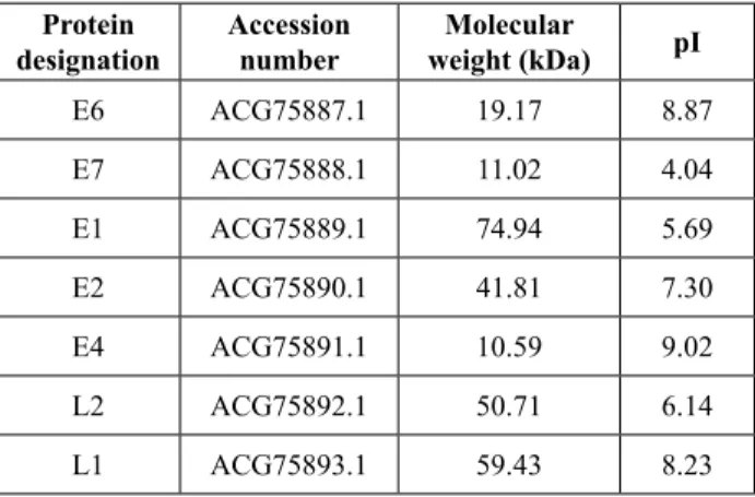
Table 1: Physicochemical properties of different putative proteins of hPv type 16.
Protein
designation accession number weight (kda)Molecular pi
E6 ACG75887.1 19.17 8.87 E7 ACG75888.1 11.02 4.04 E1 ACG75889.1 74.94 5.69 E2 ACG75890.1 41.81 7.30 E4 ACG75891.1 10.59 9.02 L2 ACG75892.1 50.71 6.14 L1 ACG75893.1 59.43 8.23
In the present study, seven proteins of HPV type16 and eight proteins of HPV type 18 were used for physicochemical analysis such as molecular weight, isoelectric point (pI value) and antigenic nature. E1 protein showed the highest molecular weight 74.94 kDa while the lowest molecular weight was 10.59 kDa of E4 protein. Isoelectric point of these proteins was ranged between 4.04 to 9.02. The physicochemical properties of all 7 proteins of HPV type 16 were given (Table 1).
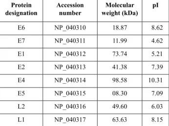
Table 2: Physicochemical properties of different putative proteins of hPv type 18.
Protein
designation accession number weight (kda)Molecular pi
E6 NP_040310 18.87 8.62 E7 NP_040311 11.99 4.62 E1 NP_040312 73.74 5.21 E2 NP_040313 41.38 7.39 E4 NP_040314 98.58 10.31 E5 NP_040315 08.30 7.09 L2 NP_040316 49.60 6.03 L1 NP_040317 63.63 8.15
The E4 protein of HPV type 18 having the highest molecular weight 98.58 kDa and the lowest molecular weight was 8.30 kDa of E5 protein. Isoelectric point of these proteins was ranged between 4.62 to 10.31. The physicochemical properties of all 8 proteins of HPV-18 were given (Table 2).
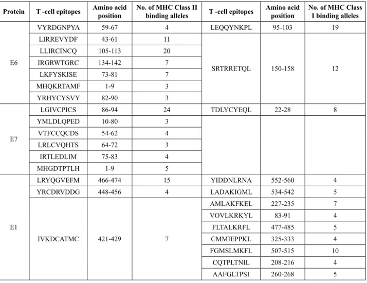
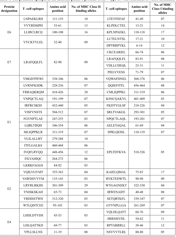
3.2 Identification and analysis of T-cells epitopes In the present study, we have used all these protein which is encoded by genome of HPV for identification and mapping of T-cell epitopes. The identification of epitopes in E6, E7, E1, E2, E4, L2, and L1 proteins of HPV 16 were investigated by PROPRED and PROPRED1 tools. Total 48 epitopes were predicted for class I MHC and 41 epitopes for class II MHC molecules in these proteins. The predicted epitopes having the MHC alleles for putative proteins from HPV-16 were performed (Table 3). The other most important type 18 of HPV was used for prediction of epitopes in E6, E7, E1, E2, E4, E5, L2, and L1 proteins. Total 60 epitopes were predicted for class I MHC and 45 epitopes for class II MHC molecules in these proteins. The predicted epitopes having the MHC molecules for proteins from HPV-18 were investigated (Table 4). Here, we have preferred these tools due to it cover the maximum human
leukocyte antigen (HLA) alleles as compare to other existing tools.
4. disCUssion
We have used the complete genome for identification of T-cell epitopes of all proteins in HPV type 16 and 18. Here, we have shown the minimal/optimal amino acid sequences and the HLA restricting molecules of these dominant CD4 and CD8 T-cell epitopes in both HPVs. Major dominant CD4 T-cell epitopes in HPV-16 were recognized in E7 86-94(LGIVCPICS; restricted by the HLA-DRB1_1104 molecule), E6 105-113 (LLIRCINCQ; restricted by HLA-DRB1_0813), E1 466-474 (LRYQGVEFM; restricted by HLA-DRB1_0301 molecule), E2 210-218 (IRQHLANHS; restricted by HLA-DRB1_1327 molecule), E4 1-9 (YVLHLCLAA; restricted by HLA-DRB1_1502 molecule), L2 451-459 (YYMLRKRRK; restricted by HLA-DRB1_0101 molecule) and in L1 4-12 (FIYILVITC; restricted by HLA-DRB1_1128 molecule) were identified with highest affinity. In HPV-18 dominant CD4 T-cell epitopes were recognized in E7 82-90 (LRAFQQLFL; restricted by the HLA-DRB1_1501 molecule), E6 100-108 (LLIRCLRCQ; restricted by HLA-DRB1_0806), E1 418-426 (YRRAQKRQM; restricted by HLA-DRB1_1120 molecule), E2 301-309 (LRYRLRKHS; restricted by HLA-DRB1_0804 molecule), E4 58-66 (IVDLSTHFS; restricted by HLA-DRB1_0410 molecule), E5 36-44 (FVYIVVITS; restricted by HLA-DRB1_1305 molecule), L2 371-389 (FKYSPTISS; restricted by HLA-DRB1_0421 molecule) and in L1 49-57 (LRNVNVFPI; restricted by HLA-DRB1_0703 molecule) were also identified with highest affinity
The homologous peptides IHSMNSTIL and IHSMNSSIL derived from L1 HPV type 16 and 18 proteins respectively, and with high specificity for particular allele, according with an algorithm prediction program that induced T-cell stimulation in patients with advanced cervical cancer positive for HPV-16 or 18 infections. T-cell derived from a patient with HPV type 18 infection which is stimulated with peptide IHSMNSTIL have capable to kill a cervical cancer cell line.[12] In another recent report, the identification of T-cell epitopes from secretory and cell surface proteins of M. tuberculosis H37Rv strain. Total 69 promiscuous nanomer T-cell epitopes have been identified for MHC Class II and 47 promiscuous epitope has been identified for MHC class I molecules.[13]
The prediction of T-cell epitopes in highly virulence surface proteins such as hemagglutinin and neuraminidase from Influenza A virus H5N1 has been earlier reported. [10] Recently, the T-cell epitopes have been identified
in thermostable hemolysin protein of Aeromonas hydrophila that can use in immunodiagnostic reagent.[14] In our study, we have identified the major dominant CD8 T-cell epitopes of HPV-16 in E7 20-28 (TDLYCYEQL; restricted by the HLA-A2 molecule), E6 95-103 (LEQQYNKPL; restricted by HLA-B*2705), E1 507-515 (FGMSLMKFL; restricted by HLA-B*5102molecule), E2 207-215 (SPEIIRQHL; restricted by HLA-CW*0401 molecule), E4 11-19 (TKYPLLKLL; restricted by HLA-DRB1_1502 molecule), L2 78-86 (RPPTATDTL; restricted by HLA-B*2705 molecule) and in L1 488-496 (FPLGRKFLL; restricted by HLA-MHC-kd molecule). In HPV-18 major dominant CD8 T-cell epitopes were identified in E7 23-31 (VDLLCHEQL; restricted by the HLA-MHC-kd molecule), E6 110-118 (KPLNPAEKL; restricted by HLA-B*2705), E1 456-464 (QQIEFITFL; restricted by HLA- MHC-kd molecule), E2 75-83 (KAIELQMAL; restricted by HLA-B*2705 molecule),
E4 38-46 (RPTARRRLL; restricted by HLA- B*2705 molecule), E5 56-64 (CFLLPMLL; restricted by HLA- MHC-kd molecule), L2 327-335 (APSPEYIEL; restricted by HLA-B*2705 molecule) and in L1 66-74 (RPSDNTVYL; restricted by HLA-A20 molecule). The evaluation of synthetic peptides as vaccine candidate for flavivirus has been investigated. The prediction of epitopes using computational tools and synthetic peptides from E glycoprotein of Murray Valley encephalitis (MVE) and DEN 2 viruses have been earlier reported and their immunogenicity has been evaluated in mice.[15] The T-cell epitopes of E6 protein have been identified from HPV type 16 infected woman.[16] Recently, T-cell epitopes of hepatitis B virus of seven proteins such as polymerase, large-S- protein, middle –S- protein, S- protein, X- protein, precore/core protein, core and E- antigen proteins have been earlier reported.[17]
Table 3: The predicted epitopes in the different putative proteins of hPv type 16 with reference to MhC molecules. Protein T -cell epitopes amino acid position no. of MhC Class ii binding alleles T -cell epitopes amino acid position no. of MhC Class i binding alleles
E6 VYRDGNPYA 59-67 4 LEQQYNKPL 95-103 19 LIRREVYDF 43-61 11 SRTRRETQL 150-158 12 LLIRCINCQ 105-113 20 IRGRWTGRC 134-142 7 LKFYSKISE 73-81 7 MHQKRTAMF 1-9 3 YRHYCYSVY 82-90 3 E7 LGIVCPICS 86-94 24 TDLYCYEQL 22-28 8 YMLDLQPED 10-80 3 VTFCCQCDS 54-62 4 LRLCVQHTS 64-72 3 IRTLEDLIM 75-83 4 MHGDTPTLH 1-9 5 E1 LRYQGVEFM 466-474 15 YIDDNLRNA 552-560 4 YRCDRVDDG 448-456 4 LADAKIGML 534-542 5 IVKDCATMC 421-429 7 AMLAKFKEL 227-235 7 VOVLKRKYL 83-91 4 FLTALKRFL 477-485 5 CMMIEPPKL 325-333 4 FGMSLMKFL 507-515 10 CQTPLTNIL 208-216 4 AAFGLTPSI 260-268 5
E2 LHRDSVDSA 256-264 8 SPEIIRQHL 207-215 9 VILCPTSVF 191-199 5 ATHTKAVAL 221-229 5 IRQHLANHS 210-218 22 QDVSLEVYL 95-103 6 VKIPKTTTV 349-357 6 QYSNEKWTL 86-94 3 VCQDKILTH 9-17 5 DSVDSAPIL 260-268 5 LAVSKNKAL 62-70 6 VHAGGQVIL 186-194 5 SNTTPIVHL 283-291 4 KHKSAIVTL 327-335 3 RQHLANHSA 212-220 4 E4 YVLHLCLAA 1-9 30 TKYPLLKLL 11-19 10 LKLLGSTWP 15-23 3 GLTVIVTLH 86-94 5 WTVLQSSLH 69-77 3 L2 LTVDPVGPS 92-100 8 IPKVEGKTI 33-41 11 IRYSRIGNK 300-308 6 SMGVFFGGL 50-58 8 IVALHRPAL 285-293 7 RPPTATDTL 78-86 15 FFSDVSLAA 464-472 5 PTFTDPSVL 161-169 3 FVTTPTKLI 241-249 6 NITDQAPSL 419-427 7 YYMLRKRRK 451-459 23 NIAPDPDFL 276-284 3 FTLSSSTIS 180-188 3 SRIGNKQTL 304-312 6 VEGKTLADQ 35-43 3 IDPAEEIEL 333-341 6 YGSMGVFFG 47-55 3 VPSVPSTSL 382-390 11 LGLYSRTTQ 224-232 4 VHYYYDFST 323-331 3 L1 YRFVTSQAI 443-451 10 VEVGRGQPL 131-139 8 ISMDYKQTQ 172-180 8 FPLGRKFLL 488-496 17 FIYILVITC 4-12 15 LPSEATVYL 31-39 13 MTYIHSMNS 413-421 10 QYRVFRIHL 95-103 5 LFFYLRREQ 271-279 14 FGHPLLNKL 144-152 6 FVRHLFNRA 281-289 5 YHAGTHRLL 61-69 5 LELINTVIQ 213-221 7 YIKGSGSTA 302-310 4 YHIFFQMSL 20-28 8 LVPKVSGLQ 86-94 5 VNLKEKFSA 474-482 15 LKAKPKFTL 499-507 4 LQFIFQLCK 397-404 5 YRVFRIHLP 95-103 5
Table 4: The predicted epitopes in the different putative proteins of hPv type 18 with references to MhC molecules. Protein
designation T -cell epitopes amino acid position no. of MhC Class ii binding alleles T -cell epitopes amino acid position
no. of MhC Class i binding alleles E6 LNPAEKLRH 111-119 10 LTEVFEFAF 41-49 07 VVYRDSIPH 53-61 13 KLPDLCTEL 13-21 14 LLIRCLRCQ 100-108 16 KPLNPAEKL 110-118 17 VYCKTVLEL 32-40 09 LCTELNTSL 17-21 10 DPTRRPYKL 6-14 12 E7 LRAFQQLFL 82-90 09 CKCEARIEL 66-74 06 LRAFQQLFL 83-91 08 VDLLCHEQL 23-31 11 PIELVVESS 71-79 07 E1 VMGDTPEWI 338-346 06 VQWAFDNEL 368-376 06 LVRNFKSDK 228-236 07 QQIEFITFL 456-464 08 YRRAQKRQM 418-426 20 CMLIQPPKL 311-319 06 VNPQCTLAQ 191-199 07 KSNCQAKYL 401-409 03 IRFRCSKID 432-440 05 FKDTYGLSF 218-226 04 VISFVNSTS 504-512 07 SRLTVAKGL 293-301 05 FGVNPTLAE 247-255 03 NPQCTLAQL 193-201 07 LQRLTIIQH 346-354 06 AELETAQAL 61-69 04 MLIQPPKLR 311-319 07 SPRLQEISL 110-119 07 VLILALLRY 279-288 10 EPLTDTKVA 518-526 05 ITFLGALKS 460-468 06 IVQFLRYQQ 448-456 12 FILYAHIQC 264-272 06 LKRKFAGGS 84-92 03 E2 VQILVGYMT 355-363 04 KAIELQMAL 75-83 17 VAWDSVYYM 135-143 03 RYKTEDWTL 90-98 09 LRYRLRKHS 301-309 29 WTGAGNEKT 322-330 04 YNISKSKAH 65-73 04 IRWENAIFF 40-48 08 YRDISSTWH 312-320 03 SETQRTKFL 339-347 07 WTLQDTCEE 95-103 03 GTVNPLLGA 261-269 07 E4 LHDLDTVDS 45-53 03 VQLHLQATT 68-76 09 SRRSSIVDL 54-62 11 LHLQATTKD 69-77 03 RPTARRRLL 38-46 12 YPLLSLLNS 11-19 08 NSVVVTLRL 80-88 05
E5 FVYIVVITS 36-44 38 CFLLPMLLL 56-64 14 YVFCFLLPM 52-60 21 HIHAILSLQ 65-73 09 LHIHAILS 63-71 15 L2 VRRVAGPRL 217-225 06 SLGIFLGGL 49-57 08 VRFSRLGQR 293-301 05 VSVANPEFL 234-242 16 LTFDPRSDV 262-270 07 APSPEYIEL 327-335 17 FKYSPTISS 371-389 20 KRKRVPYFF 447-455 04 FYHDISPIA 318-326 03 AGTTTPAVL 133-141 08 FVGTPTSGT 180-188 04 VPKVEGTTL 32-40 11 IGIHGTHYY 427-435 13 IIQWSSLGI 43-51 06 IRLHRPALT 279-287 12 LQTFASSGT 196-204 03 YIELQPLVS 331-339 05 YYFIPKKRK 440-448 05 MVSHRAARR 1-9 03 L1 YRFVQSVAI 479-487 10 SGHPFYNKL 179-187 09 LRNVNVFPI 49-57 23 VEIGRGQPL 166-174 09 WNVDLKEKF 508-516 07 RPSDNTVYL 66-74 20 LVYMVHIII 32-40 15 CGHYIILFL 42-50 06 IVTSDSQLF 359-367 04 MGDTVDQSL 328-336 05 VSVDYKQTQ 207-215 06 LPDPNKFGL 138-146 14 WRPSDNTVY 64-72 04 VDTTPSTNL 395-403 06 LRRKPTIGP 535-543 08 YHAGSSRLL 96-104 03 LTICASTQS 402-410 03 MSYIHSMNS 449-457 04 VTVVDTTPS 391-399 03 WNRAGTMGD 321-329 03 YRVFRVQLP 130-138 05 MFFCLRREQ 306-314 06 ILHYHLLPL 8-16 03 5. ConClUsion
Disease diagnosis is one of the major challenges for HPV by conventional techniques that can not be able to detect the viruses at early stages. Immunoassays are good choice for earlier diagnostic but it requires purified antigens. Thus;
these finding provide a better choice for synthesis of epitopes that can help for diagnosis without in vitro grow and purify HPV to minimize the time, labor and cost of diagnosis. The further study will be needed to test our identified T-cell epitopes for immunodiagnostic and development of peptide based vaccines against cervical cancer.
aUThors' ConTriBUTions: DKC, IM and VS designed the research work and performed experiments. All authors analyzed the data, wrote the paper and approved the final manuscript.
ConfliCT of inTErEsTs: There is no conflict of interest.
aCknowlEdgMEnTs
The authors are thankful to A.K. Singh, Pritee Singh and Reena for providing the suggestions, fruitful discussion and encouragement during experiment and preparation of the manuscript.
rEfErEnCEs
1. Luthra UK, Prabhakar AK, Seth P, Aggarwal SS, Murthy NS, Bhatnagar P, Sharma BK. Natural history of precancerous and early cancerous lesions of the uterine cervix. Acta Cytologica, 31: 226-6, 1987.
2. Walboomers JM, Jacobs MV, Manos MM, Bosch FX et al. Human papillomavirus is a necessary cause of invasive cervical cancer worldwide. Journal of Pathology, 189: 12-9, 1999.
3. Clifford GM, Rana RK, Franceschi S, Smith JS, Gough G, Pimenta JM. Human papillomavirus genotype distribution in low-grade cervical lesions: comparison by geographic region and with cervical cancer. Cancer Epidemiology, Biomarkers & Prevention, 14: 1157-4, 2005.
4. De Vuyst H, Clifford GM, Nascimento MC, Madeleine MM, Franceschi S. Prevalence and type distribution of human papillomavirus in carcinoma and intraepithelial neoplasia of the vulva, vagina and anus: A meta-analysis. International Journal of Cancer,124: 1626–1636, 2009.
5. Petrovsky N, Silva D, Brusic V. The future for computational modelling and prediction systems in clinical immunology. NovartisFoundation symposium,254: 23-2, 2003.
6. Pizza M, Scarlato V, Masignani V, Giuliani MM et al.
Identification of vaccine candidates against serogroup B
Meningococcus whole genome sequencing. Science,287: 1816-20, 2000.
7. Rappuoli R. Reverse vaccinology, a genome-based approach to vaccine development. Vaccine, 19:2688-91, 2001. 8. de Groot AS, Rappuoli R. Genome-derived vaccines. Expert
Review of Vaccines, 3: 59-6, 2004.
9. Singh H, Raghava GPS. ProPred: prediction of HLA-DR binding sites. Bioinformatics,17: 1236-7, 2001.
10. Singh H, Raghava GPS. ProPred1: prediction of promiscuous MHC class-I binding sites. Bioinformatics,
19: 1009-4, 2003.
11. Somvanshi P, Singh V, Seth PK. Prediction of epitopes in
hemagglutinin and neuraminidase proteins of Influenza
A virus H5N1 strain: A clue for diagnostic and vaccine development. OMICS: Journal of Integrative Biology,12: 61-69, 2008.
12. Monroy-García A, Weiss-Steider B, Hernández-Montes J, Ortiz-Navarrete VF et al. Identification of two homologous
antigenic peptides derived from L1 HPV-16 and 18 proteins
specific for the HLA-B*3901allele. Archives of Virology,
147: 1933-42, 2002.
13. Somvanshi P, Singh V, Seth PK. In silico prediction of epitopes in virulence proteins of Mycobacterium tuberculosis H37Rv for diagnostic and subunit vaccine design. Journal of Proteomics and Bioinformatics,1: 143-53, 2008.
14. Singh V, Mani I, Chaudhary DK, Somvanshi P. Molecular detection and cloning of thermostable hemolysin gene from
Aeromonas hydrophila. Molecular Biology, 45: 551-60, 2011.
15. Gao XM, Liew XY, Tite JP. A dominant the epitope in
influenza nucleoprotein, analysis of the fine specificity and
functional repertoire of T-cells. Journal of Immunology, 144: 2730-7, 1990.
16. Nakagawa M, Kim KH, Moscicki AB. Patterns of CD8 T-cell epitopes within the human papillomavirus type 16 (HPV 16) E6 protein among young women whose HPV 16 Infection has become undetectable. Clinical and Diagnostic Laboratory Immunology,12: 1003-5, 2005.
17. Singh V, Mani I, Chaudhary DK, Somvanshi P. HLA Class I and II binding promiscuity of the T-cell epitopes in putative proteins of Hepatitis B virus. JournalofComputer Science & Systems Biology,2: 69-3, 2009.