Structure and function of complement
component C3 and its fragments
Krisztián Papp
2008
Supervisor:
Prof. Anna Erdei D.Sc.
Immunology Programme
Department of Immunology
Institute of Biology
Contents
Chapter 1 Introduction
1.1. The complement system 1.2. The C3 molecule
1.3. Diagnostic methods for the detection of complement activation 3 5 13 20 Chapter 2 Objectives 29
Chapter 3 Summary of new scientific results 32
Chapter 4 Novel monoclonal antibodies against mouse C3 interfering with complement activation: description of fine specificity and application to various immunoassays
Molecular Immunology 2004. 40:1213-1221
35
Chapter 5 Dynamic structural changes during complement C3 activation analysed by hydrogen/deuterium exchange mass spectrometry
Molecular Immunology 2008
45
Chapter 6 B lymphocytes and macrophages release cell membrane deposited C3-fragments on exosomes with T cell response-enhancing capacity
Molecular Immunology 2008. 45: 2343–2351
60
Chapter 7 On-chip complement activation adds an extra dimension to antigen microarrays
Molecular & Cellular Proteomics 2007. 6:133-140
70
Chapter 8 Two-dimensional immune profiles improve antigen microarray-based characterization of humoral immunity
Submitted
79
Chapter 9 General discussion 93
Summary 101
Hungarian summary – Összefoglalás 102
Abbreviations 103 Acknowledgements 104
Chapter 1
Introduction
Introduction
During their life, organisms are continually endangered by foreign pathogens and altered self structures that threaten the existence of the host. An effective apparatus had to evolve for preserving the consistency of the genetic code and this way making the survival of the species possible. The machinery which distinguishes self and non-self or altered self and fights against the causative agent is the immune system. During evolution, primitive pathogens, obeying the selection pressure, kept developing novel apparatus for the infection. The immune system also had to suit to these newer demands in protection of the host and this fight resulted the very complex and effective defence system of the mammals. The ancient arm of the immune system, innate immunity, did not disappear during evolution; on the contrary, it still tightly cooperates with the later formed adaptive immunity in protection of the vertebrates.
Pathogens, by breaking the physical barrier that separates the host form the environment, make contact with the components of the innate immune system. This ancient part of the immune system remained the first, instantly activated line of protection against invading pathogens. Cellular (macrophages, dendritic cells, granulocytes) and humoral (antibacterial peptides, cytokines, complement system) components of innate immunity destroy most of the pathogens in a short time. A certain proportion of invading agents survives the encounter with the innate immunity and challenge adaptive immunity, which provides an effective response in about two weeks following activation. It has to be emphasised that the cellular (B cells, T cells) and the humoral (antibodies, lymphokines) components of adaptive immunity are unable to work separately from the innate system. Components of the latter modulate adaptive responses and also take a crucial part in the effector phase. Some researches concentrate only on adaptive immunity, however without knowing the exact mechanisms of innate immunity some basic phenomena of the immune system can not be understood.
In this study, we focused on the complement system, the major humoral component of innate immunity. We have analyzed in great detail the third component (C3) of this system and its activation fragments which interact with several other molecules yielding a diverse set of biological responses.
1.1.1. Activation of the complement system
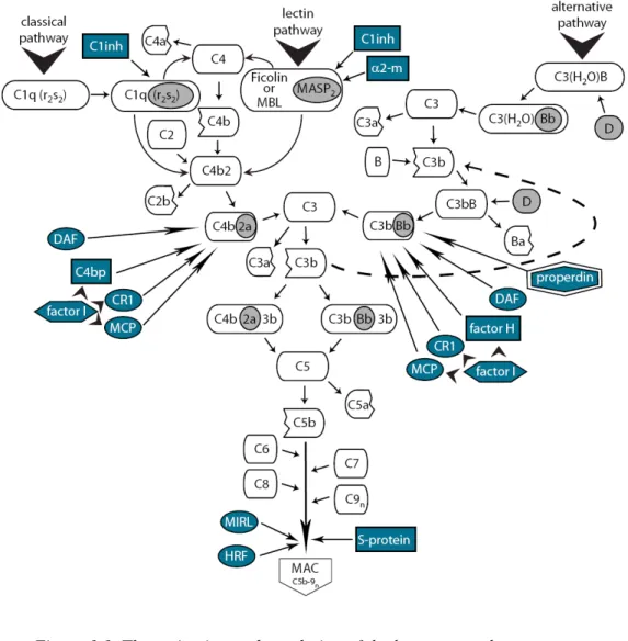
The complement system is an ancient component of the innate immune system; some of its major proteins appeared in echinoderms and exist in each deuterostome (Nonaka, 2001;Smith et al., 1999). The complement system contains more than 30 proteins in various tissue fluids and on the surface of cells, including proteins of the enzymatic cascade, regulators and receptors. The cascade can be initiated by any of the three activation routes: the classical, lectin or alternative pathway (Figure 1.1). These pathways are analogous to the coagulation, fibrinolysis and kinin serine protease systems. The three routes converge at the point of the cleavage of the third component, C3, which is followed by the common terminal pathway. Proteins of the classical pathway are designated C1 through C9, while the components of the alternative pathway are called factors, followed by a letter (e.g. factor B, factor H). Several complement components are cleaved during activation of the system and normally the larger fragments are designated with ‘b’ while the smaller with ‘a’ suffixes (in the case of C2, the large fragment is designated C2a and the small C2b for historical reasons) (reviewed by (Walport, 2001)).
The classical pathway is initiated by the interaction of C1q, the subunit of the C1-complex with antigen-bound antibodies. The C1 complex consists of C1q and the two serine proteases, C1r and C1s, held together by Ca2+. Conformational changes in the complex activate C1r, then C1s which first cleaves C4 and the larger C4b fragment binds covalently to the activating surface (bacteria, cell, etc.). C2 attaches to bound C4b, then it is cleaved by C1s leading to the formation of the C4b2a enzyme complex which is the C3 convertase of the classical pathway.
The lectin pathway is initiated by binding of the complex of mannose-binding lectin (MBL) or ficolins (Matsushita et al., 2000) to arrays of sugar groups on the surface of bacterial cells. The mannose-binding lectin-associated serine protease 2 (MASP2) acts in a fashion similar to C1s, leading the formation of the C3 convertase enzyme, C4b2a (Matsushita and Fujita, 1992).
The alternative pathway is initiated by surface bound C3b fragments or by spontaneously hydrolysed C3 (C3H2O) (Pangburn et al., 1981), which in the presence of Mg2+ binds factor B, a protein homologous to C2. Factor D cleaves C3b-bound factor B and this way C3bBb, the C3 convertase of the alternative pathway is formed (Lesavre and Muller-Eberhard, 1978). The labile convertase complex has short half-life and is stabilised over 10-fold by the binding of properdin (Fearon and Austen,
1975;Hourcade, 2006). The C3 convertase enzymes cleave many C3 molecules and some of the generated C3b fragments bind covalently to the activating surface. These bound fragments are able to activate again the alternative pathway, making a positive feedback loop in the system, which increases dramatically the amount of activated C3b fragments. C3b is not able to distinguish between self and potentially harmful non-self structures, but other factors make this discrimination. The carbohydrate environment of the surface on which C3b fragments are deposited, determines the relative affinity of C3b for factor B or for a complement control protein, factor H. On the host cell surface that is abundant in polyanions such as sialic acid, factor H binds to C3b with higher affinity than does factor B and halts the formation of C3 convertases. On microbial surfaces that lack a polyanionic coating, factor B binds to C3b with a higher affinity than does factor H, and this leads amplified C3 cleavage (Meri and Pangburn, 1990).
In the terminal pathway, the C3b fragment binds to the C4b or C3b containing C3 convertases and forms the C5 convertase. This newly bound C3b acts as an acceptor site for C5 which will be cleaved to anaphylatoxin C5a and a metastabile C5b fragment (Takata et al., 1987). C6 binds to C5b and forms a bimolecular complex, which remains loosely bound to C3b on the target cell surface until it reacts with C7. The C5b-7 complex then undergoes hydrophilic-amphiphilic transition and anchors itself firmly in the lipid bilayer (Preissner et al., 1985). Membrane inserted C5b-7 functions as a receptor for C8. The binding of C8 changes its conformation and allows the α chain to penetrate into the lipid bilayer and finally the whole C5-8 complex drops deeper into the hydrophobic phase (Esser et al., 1979), and forms a small functional channel of ~ 30 Å in diameter. The C5b-8 complex can mediate the binding of multiple C9 molecules (1-18), which incorporate into the membrane and form a channel of ~100 Å diameter (Tranum-Jensen et al., 1978). This terminal complex (C5b-9), called membrane attack complex (MAC), causes cell lysis by disrupting the cell membrane.
Figure 1.1. The activation and regulation of the human complement system. The initiation of the complement cascade can take place in three different ways (classical, lectin or alternative pathways), but following the convergence of these pathways at the level of the C3 molecule, a single route proceeds until the formation
of the membrane attack complex. The positive feedback loop of the alternative pathway is illustrated by dashed line. The enzymatically active fragments are depicted
1.1.2. Regulation of the complement cascade
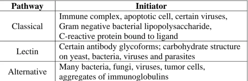
The complement system is a very effective apparatus against invading pathogens, but because of its effectiveness, it has to be well orchestrated or otherwise it can also destroy the host. The regulation starts at the level of the activators of the three pathways. The molecules or cells which initiate the cascade are listed in table 1.1.
Table 1.1. Initiators of the three complement pathways
The complement activating properties of the antibodies are influenced by their isotype (Table 1.1) (Erdei, 2008;Janeway, 2001;Roitt, 1997), avidity and glycosylation (Arnold et al., 2006).
The action of these activators should not result inevitable completion of the cascade, because this would cause unnecessary harm in the host, as well. There are many soluble and membrane bound complement control proteins (CCP) which are responsible for the proper functioning of this system. Three major targets in the complement cascade exist, where CCPs can exert their inhibitory effect; these are the serine proteases of the recognition complex, the C3 convertases and finally the MAC.
Pathway Initiator Classical
Immune complex, apoptotic cell, certain viruses, Gram negative bacterial lipopolysaccharide, C-reactive protein bound to ligand
Lectin Certain antibody glycoforms; carbohydrate structure on yeast, bacteria, viruses and parasites
Alternative Many bacteria, fungi, viruses, tumor cells, aggregates of immunoglobulins
Isotype (human) Isotype (mouse) Complement activation
IgM IgM +++++ IgG1 IgG2a,c +++ IgG2 IgG3 ++ IgG2b +++ IgG3 +++ IgG4 IgG1 +
IgA1, IgA2 IgA + ?
The first control point is at the level of the initiator serine proteases (C1r, C1s, MASP1, MASP2). The C1 inhibitor (C1inh) irreversibly inactivates these enzymes (Munkvad et al., 1990), so it blocks the classical and lectin pathway, as well. The other inhibitor at this point is α2-macroglobulin that can inactivate only the MASP enzymes (Terai et al., 1995).
The major amplification of the cascade is carried out by the C3 convertases, where two mechanisms are involved in the regulation. The first mechanism prevents formation or accelerates dissociation of the C3 convertase by a process known as decay-accelerating activity. The second mechanism consists of the cleavage of the C3b or C4b molecule into iC3b or iC4b that can no longer participate in the formation of the C3 convertase enzymes (Molina, 2002). A serine protease, factor I, in the presence of cofactors, is responsible for the cleavage of the haemolytically active C3b and C4b fragments. The further listed CCPs, except for the membrane cofactor protein (MCP) (only cofactor) and accelerating factor (DAF) (only decay-accelerating activity) are able to control the complement system by both mechanisms. Among the soluble cofactors of factor I, the C4 binding protein (C4bp) takes part in C4b, while factor H in C3b inactivation, while the membrane bound complement receptor 1 (CR1) and MCP cofactors inhibit the formation of both C4b2a and C3bBb. CR1 facilitates the complete degradation of C3, while the MCP helps only in the first cleavage of factor I (Muller-Eberhard, 1988). CR2 exhibits cofactor activity only on iC3b and helps its cleavage into C3dg and C3c fragments (Mitomo et al., 1987). The decay-accelerating factor (DAF, CD55) prevents the assembly of the C3 convertase and dissociates the formed enzyme (Medof et al., 1987). Interestingly, MCP has limited expression in mice, compared with the widespread expression in human tissues; here an additional CCP, called complement receptor 1 related protein (Crry), blocks the unnecessary formation of C3 convertases on self cells (Molina, 2002).
The formation of the MAC is the final event of the cascade and, in the same time, the last point where it can be stopped. The S-protein competes with the membrane binding site of MAC and this is the primary soluble MAC inhibitor of the serum (Podack and Muller-Eberhard, 1979); it allows binding of C8 and C9, but prevents C9 polymerization (Muller-Eberhard, 1988). The homologous restriction factor (HRF) is highly effective in inhibiting complement mediated channel formation, including the C5b-8 and MAC (Zalman et al., 1986). The membrane
inhibitor of reactive lysis protein (MIRL, CD59) inhibits the incorporation of C9 into C5b-9 complexes formed in the membrane and reduces C9 polymerization (Rollins and Sims, 1990).
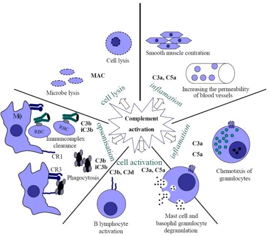
1.1.3. Biological effects of the complement system
Activation of the complement system initiates various physiological activities that help the host to protect its integrity against invading pathogens or altered self structures (Figure 1.2). The importance of the complement system is not restricted to innate immunity, but extends to adaptive responses as well, by forming a bridge between these two systems (Erdei et al., 1991).
A remarkable feature of the complement system – which was actually the basis of its discovery – is its capability of lysing microbes. During the first contact with a pathogen, the alternative or lectin dependent pathway (or sometimes even the classical route – e.g. HIV) can be activated without the presence of antibodies, so the complement system can destroy the invading pathogens promptly as they appear in the body. If the host had already encountered a specific pathogen, the previously produced pathogen specific antibodies may also initiate the classical pathway, which is strongly augmented by the feed-back loop. The generated MAC disrupts the integrity of the pathogen cell membrane by pore forming, and thus eliminates the invading agent. Of course some pathogens try to avoid this protecting line, so during evolution several escape mechanisms have been developed by the pathogens, which hinder their clearance.
Every complement activating route results in C3 activation and a certain portion of the newly generated C3b fragments bind covalently to the activating surface. Following opsonisation, the covalently bound C3b and iC3b fragments serve as ‘tags’ on the pathogen. CR1 and CR3 receptors on the surface of macrophages bind to pathogen bound C3b and iC3b fragments increasing their phagocytosis and in this way facilitating presentation of antigens to T cells. On Kuppfer cells another receptor, the newly discovered complement receptor of the immunoglobulin superfamily (CRIg), is required for the efficient binding of C3-opsonized particles (Helmy et al., 2006).
Complement activation also generates small peptides, called anaphylatoxins (C3a, C4a, C5a), that induce inflammation, promoting the effectivity of the immune
(Gergely and Erdei, 2000;Johnson and Chenoweth, 1985). Anaphylatoxins induce the degranulation of the serosal type mast cells and basophils and the released histamine increases the permeability of blood vessels. Both C3a and C5a anaphylatoxins have chemotactic effect on eosinophils, while C5a attracts directly neutrophils to the site of inflammation, where these cells effectively eliminate microbes (Daffern et al., 1995).
Formation of immune-complexes is a physiological phenomenon leading to the clearance of the antigen. In various pathological conditions however, excessive amounts of immune-complexes may be formed and can deposit in blood vessels, causing harmful inflammation. In the clearance of the immune complexes the complement system plays crucial role in two ways. On one hand C3 fragments can force apart the complexes by binding to them, and this way the large immune-complexes will be solubilised. On the other hand, the C3b fragments bound to the antigen-antibody complexes bind to human red blood cells via CR1. These cells then transfer the complexes to liver Kuppfer cells that phagocytose them, so they disappear from the bloodstream (Helmy et al., 2006). In mice this clearance is not functioning, since mouse erythrocytes do not carry CR1 receptor on their surface (Kinoshita et al., 1988).
Activation of the complement system also has an effect on acquired immunity. Antigen bound C3d fragments function as molecular adjuvant: the CD19/CR2/TAPA-1 complex augments signalling through the B cell antigen receptor. The blockade of CR2 receptor suppresses the primary antibody response (Dempsey et al., 1996;Fearon and Carter, 1995).
CR1 and CR2 on the surface of follicular dendritic cells (FDCs) are critical for the generation of normal humoral immunity, especially during the later stages of the primary immune response and the generation of B cell memory. The FDC plays a crucial role in germinal center reactions, like somatic hypermutation of Ig genes, Ig isotype switching and the generation of memory cells, by trapping immune complexes via CR1 and CR2 receptors (Fang et al., 1998).
C3 fragments fixed to the antigen presenting cells enhance the proliferation of antigen-specific T cells by interacting with the C3 receptors expressed on activated T lymphocytes. C3 fragments form a bridge between the antigen presenting cell (APC) and T cells, enhancing cell-to-cell contact and intracellular signalling (Kerekes et al., 1998).
Figure 1.2. The multiple effects of the complement system (Gergely and Erdei, 2000)
The role of the complement system is not restricted only to the protection against invading pathogen, but it also modulates several other biological processes (Mastellos and Lambris, 2002). It is suggested that C3 and C3-binding proteins facilitate gamete membrane fusion and thereby promote sperm penetration and oocyte fertilization (Anderson et al., 1993). The involvement of C3 molecules was suggested also in the dedifferentiation process and the muscle differentiation in the limb regeneration of urodele species (Del Rio-Tsonis et al., 1998). The role of C5 was proved in liver regeneration following toxic injury (Mastellos et al., 2001). There is evidence, which suggests that C3a also has a role in the maturation and lineage commitment of hematopoetic progenitors (Mastellos and Lambris, 2002).
Because of the multiple roles of the complement system, it is reasonable to make any effort to understand every detail of this ancient part of humoral immunity.
1.2.1 Structure of C3
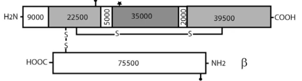
Component C3 is the most abundant complement protein in the serum (chuman C3 ~1.2 mg/ml, cmouse C3 ~ 0.5 mg/ml). Its main source is the hepatocyte (Alper et al., 1969), but macrophages (Zimmer et al., 1982) and, with less efficiency, endothelial cells (Warren et al., 1987) can also secrete this crucial complement component. The C3 molecule belongs to the α2-macroglobulin family, whose members, like C4 and the proteinase inhibitor α2-macroglobulin contain an internal thioesher group and have important roles in the immune response. C5 is a homologue of C3 and C4, but lacks the thioesther group. Human and mouse C3 have 77% amino acid identity (de Bruijn and Fey, 1985;Fey et al., 1984) and their genes are located on chromosome 19 (Whitehead et al., 1982) and 17, respectively. The molecule consists of α and β chains which are linked by a disulphide bond and non-covalent forces (Figure 1.3).
Figure 1.3. Schematic representation of the mature human C3.
The molecular massses of the fragments (in Da) have been calculated from amino acid sequences and do not include carbohydrate content. The location of the thioester group (asterix) and N-linked carbohydrate (closed balloon) sites are also indicated in
the figure.
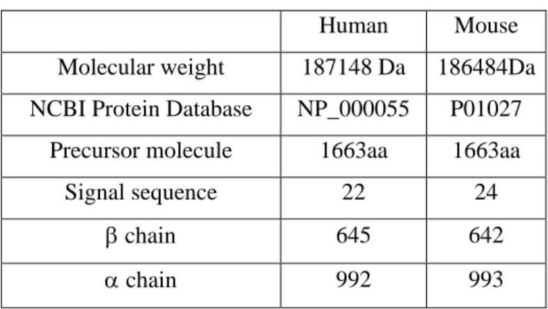
The human C3 molecule is translated into a precursor molecule, the prepro-C3 of 1663 amino acid residues. At the NH2 terminal end there is a 22-residue signal sequence which is followed by the β and the α chain sequence separated by tetra-arginine residues (Table 1.3). During maturation a furin-like enzyme cleaves out the four arginine residues between the β and α chain (Misumi et al., 1991), so the molecule can attain its final structure. In the Golgi, N-linked carbohydrate moieties also attach at asparagine residue 917 of the α chain [Man8(GlcNAc)2 +
Man9(GlcNAc)2] and 63 of the β chain [Man5(GlcNAc)2 + Man6(GlcNAc)2] and together account for 1.5% of the molecular weight of human C3 (Hirani et al., 1986;Sahu and Lambris, 2001).
Human Mouse
Molecular weight 187148 Da 186484Da NCBI Protein Database NP_000055 P01027
Precursor molecule 1663aa 1663aa
Signal sequence 22 24
β chain 645 642
α chain 992 993
Table 1.3. Major properties of human and mouse C3
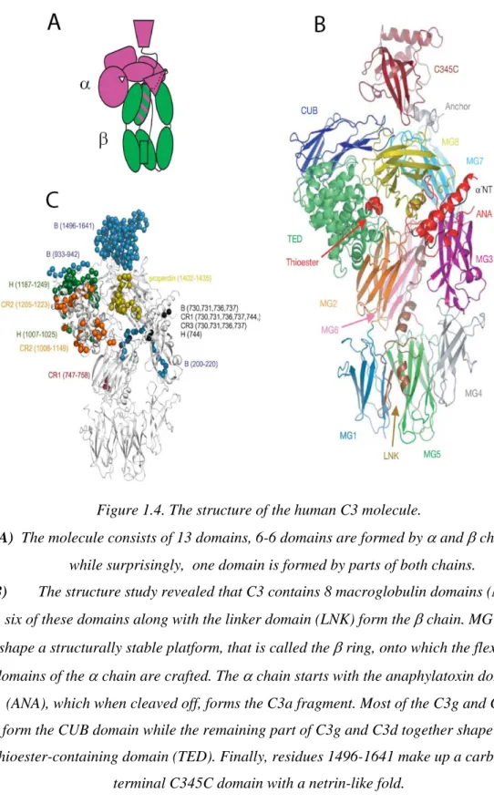
The crystal structure of the C3 is already solved (Janssen et al., 2005), the structure of its 13 domains are shown in figure 1.4.
Figure 1.4. The structure of the human C3 molecule.
A) The molecule consists of 13 domains, 6-6 domains are formed by α and β chains,
while surprisingly, one domain is formed by parts of both chains.
B) The structure study revealed that C3 contains 8 macroglobulin domains (MG); six of these domains along with the linker domain (LNK) form the β chain. MG1-6 shape a structurally stable platform, that is called the β ring, onto which the flexible domains of the α chain are crafted. The α chain starts with the anaphylatoxin domain
(ANA), which when cleaved off, forms the C3a fragment. Most of the C3g and C3f form the CUB domain while the remaining part of C3g and C3d together shape the thioester-containing domain (TED). Finally, residues 1496-1641 make up a
carboxyl-terminal C345C domain with a netrin-like fold.
1.2.2. The function of the internal thioester in the C3 molecule
One of the most arresting feature of the C3 molecule is that following activation the C3b fragment can bind covalently to the activating surface (Law and Levine, 1977). For this attribution, a thioester bound is responsible which is formed during post-translational modification, as a result of intramolecular transacylation between the thiol group of cysteine and the γ-amide group of the glutamine within the sequence Cys1009-Gly-Glu-Gln1012 (Sahu and Lambris, 2001;Tack et al., 1980).
Figure 1.5. Composition of the C3 thiolactone ring (Law and Dodds, 1997)
The process of covalent binding goes through short half-life intermediers. First a histidine at position 1126 attacks the thioester to form an acyl-imidasole intermedier that is possibly stabilized by Glu1128. The released thiol then acts as a base to catalyse the transfer of the acyl group to hydroxyl-nucleophiles, including water (Figure 1.6) (Dodds et al., 1996).
The circulating native C3 is an inert molecule, the highly reactive thioester is shielded in a hydrophobic/aromatic pocket, between the MG8 and TED domains, 85Å away from the solvent, and the His1126 and Glu1128 that are necessary for the formation of the reactive intermedier are also far from the thioester group. The ANA domain has critical role in protecting the thioester, it may serve to keep MG8 in a correct position for interaction with TED and possibly, to induce a conformation of MG8 that enhances interaction with TED. The removal of ANA, yielding C3b, weakens the interactions between MG8 and TED, thereby allowing TED to swing out of its nested position (Figure 1.7) (Janssen et al., 2005;Janssen et al., 2006). The thioester completely exposes to the surface of the C3b molecule and it can bind preferentially to hydroxyl and at much more less extent to amide groups, forming an ester or amide linkage to the targeted surface on which complement activation is occurring. The half-life of C3b is only ~60μs (Sim et al., 1981), the binding reaction is not efficient, typically only about 10% of generated C3b binds to target while the remaining 90% remains in fluid phase, where it is inactivated rapidly (Law and Dodds, 1997). The metastable C3b does not react randomly or non-specifically, rather it prefers specific OH groups and specific residues on proteins. Yet it needs to be emphasized that the nascent C3b fragment does not have the ability to discriminate between self and non-self structures.
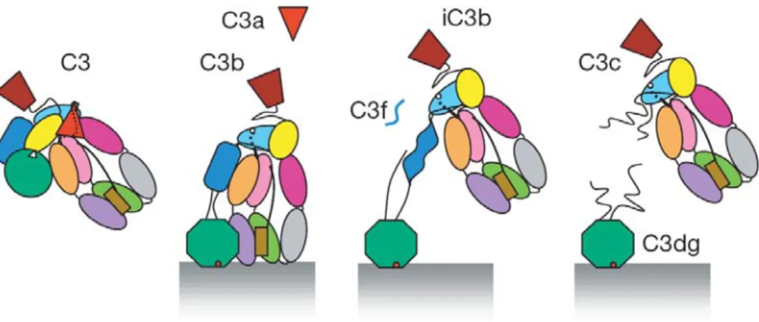
Figure 1.7. Proposed model for the conformational pathway of C3.
The native C3 is an inactive molecule, but following activation, the formerly buried thioester bond appears on the exterior of the molecule and binds covalently to hydroxyl groups of the activator surface. In the next steps factor I and cofactors
cleave off the small C3f then the large C3c fragment, so finally only C3dg stays attached to the surface (Janssen et al., 2006).
1.2.3. Limited proteolysis of the C3 molecule
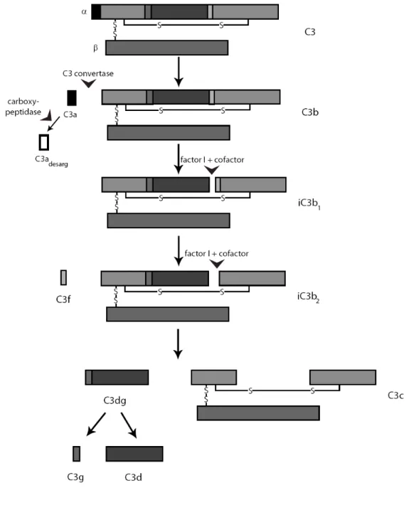
The three pathways of complement activation converge at the C3 glycoprotein, when fragments are cleaved off from the molecule as a result of limited proteolysis. The formation of classical (C4b2a) or alternative (C3bBb) C3 convertases leads to the cleavage of native C3 between residues 726 and 727 (Arg-Ser), thus the anaphylatoxin C3a and the major fragment C3b are generated (Figure 1.8). A carboxypeptidase is responsible for the inactivation of the C3a anaphylatoxin by the removal of the C terminal arginine and formation of the inactive C3adesArg molecule (Bokisch and Muller-Eberhard, 1970;Zwirner et al., 1998).
The formation of C3b results conformational changes (Janssen et al., 2006;Wiesmann et al., 2006) and binding sites appear on the molecule for the C5, properdin (P), factors H, B and I, complement receptor1 (CR1) and the membrane co-factor protein (MCP). Inactivation of C3b by co-factor I proceeds in three steps with the help of co-factor molecules (MCP, CR1, or factor H). The cleavage between residues 1281 and 1282 (Arg-Ser) results in the formation of iC3b1 and the cleavage between residues 1298 and 1299 (Arg-Ser) liberates the small C3f fragment and yields iC3b2. The third factor I cleavage site, cut with the help of CR1 or factor H, is at residues 932-933 (Arg-Glu) of the α chain. This cleavage generates the C3c and C3dg fragments.
Figure 1.8. The limited proteolysis of the C3 molecule Only disulphide bridges relevant for interchain bonds are shown.
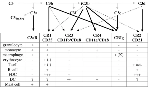
1.2.4. Receptors for various C3 fragments
Native C3 does not bind to any receptors, thus does not exhibit biological activity, while the C3 degradation products interact with various proteins, yielding a diverse set of biological responses. The following six cell surface receptors responsible for the various effects of the C3 fragments have been identified: C3aR, CR1, CR2, CR3, CR4 (Gergely and Erdei, 2000) and the newly discovered CRIg (Gros et al., 2008;Helmy et al., 2006;Wiesmann et al., 2006). The major properties of these receptors are listed in table 1.4.
C3 C3b iC3b C3d C3a C3f C3c
C3desArg
Table 1.4. Tissue distribution of the C3 fragment specific receptors K: Kupffer cells; brackets refer to murine expression
(Helmy et al., 2006)
1.3. Diagnostic methods for the detection of complement activation
In the last several decades routine complement analysis in the clinic was usually associated only with the quantification of C3 and C4, measurement of C1-inhibitor and screening for complement activity. In recent years, the field of complement analysis has expanded considerably and new methods were developed makeing the survey of this enzyme cascade easier, faster and more precise (Mollnes et al., 2007). C3aR CR1 CD35 CR3 CD11b/CD18 CR4 CD11c/CD18 CRIg CR2 CD21 granulocyte + + + + - - monocyte + + + + - - macrophage + + + + + (K) - erythrocyte - + (-) - - - T cell - + (-) - - + act. B cell - + - - + FDC - +++ + - +++ DC ? ? +/- + - ? Mast cell + +
The functional assays of the complement system assess the integrity of the individual activation pathways. These assays give global view about this system and with a subsequent analysis of various components the cause of the complement deficiency can be assigned. The haemolytic assay is a commonly used technique for the measurement of the classical pathway. The result of the CH50 test is expressed as a reciprocal dilution of the sample needed to achieve 50% lysis of fixed amount of antibody-coated sheep erythrocytes. In a similar setup, where rabbit or guinea pig erythrocytes are used in the presence of Mg2+ but not Ca2+, the activity of the alternative pathway and the value of AH50 can be determined. Several variants of these assays are known, which are easier to perform and are able to handle large numbers of various samples (Nilsson and Nilsson, 1984;Truedsson et al., 1981). The haemolytic assays are frequently used, but one has to keep in mind some of its disadvantages, e.g. it does not give information about the lectin pathway and properdin deficiency; furthermore, the use of erythrocytes can be problematic. A new commercially available ELISA based method (Wielisa®, Wieslab, Lund, Sweden) for the functional assay of the complement system can provide a solution for the limitations of the haemolytic assays. This method is based on the observation that following initiation of the complement pathway, complement components (even the terminal complex) bind to the plastic surface of the ELISA plate. For the activation of the classical, lectin and alternative pathways, IgM, mannan and LPS coat is used, respectively. During the separate detection of a pathway, the other two have to be blocked. Using the suitable ions (Ca2+ or Mg2+) or selective blocking of a pathway (anti-C1q antibody for the blockade of classical beside lectin route measurement) guarantees the selective measurement of a pathway. The readout of these measurements is the detection of the incorporated C9 by a suitable antibody (Roos et al., 2003;Seelen et al., 2005). These assays are suitable for measuring only the overall condition of the complement system. In the future it will be necessary to develop a suitable technique for the detection of complement activation when it reacts with a specific pathogen derived antigen or with an autoimmune disease related autoantigen (Atkinson and Frank, 2006).
When a pathway is defective, measurement of the concentration of complement components can assign the missing molecule. Various tests like Western blot, ELISA, nephelometry, turbidometry, radial immundiffusion, are available for the quantitative or semi-quantitative measurement of the complement proteins. When
complement deficiency is present but the levels of complement proteins are in the physiological range, the presence of a non-functional or dysfunctional variant of the complement component is suspected. The best way to detect the functional activity of a single component is to test the capacity of the sample to reconstitute the total complement activity of a serum that is deficient for a known component. This can be done either by haemolytic or ELISA assays (Seelen et al., 2005). The measurement of the regulatory proteins has clinical relevance, as well. For the diagnosis of haemolytic uraemic syndrome, the functional detection of C1-inhibitor is suggested by an assay which is based on the action of C1inh as an enzyme inhibitor and the use of chromogenic substrate (Munkvad et al., 1990). Another important complement regulator protein is factor H. For the detection of factor H deficiency, unsensitised sheep erythrocytes are incubated with the serum in question. In normal serum, factor H binds to the erythrocytes and protects them from lysis, whereas sera lacking functional factor H cause erythrocyte lysis (Sanchez-Corral et al., 2004).
The haemolytic activity and individual component measurements are useful as a first level screening technique but are not suitable for the measurement of the pathologically increased complement activation in vivo. For assessing in vivo complement activation the measurement of the various complement activation products is the better solution. One always has to consider the half-life of the measured active component in these assays. The half-life of C5a, SC5b-9 and C3 activation products are 1min, 50-60 min and a few hours, respectively. (Mollnes et al., 2007). ELISA and flow cytometry microbeads are suitable techniques for the quantification. Both techniques need monoclonal antibodies which recognize only those neoepitopes which are exclusively exposed on active split products, thus inactive proteins do not disturb the quantification.
Genetic analysis of complement components is also available, but these techniques are more expensive and harder to use for screening purpose. The determination of point mutations and polymorphic variants in genes of complement component is carried out usually for research and not diagnostic purpose.
Reference List
1. Alper,C.A., Johnson,A.M., Birtch,A.G., Moore,F.D., 1969. Human C'3: evidence for the liver as the primary site of synthesis. Science 163, 286-288. 2. Anderson,D.J., Abbott,A.F., Jack,R.M., 1993. The role of complement
component C3b and its receptors in sperm-oocyte interaction. Proc. Natl. Acad. Sci. U. S. A 90, 10051-10055.
3. Arnold,J.N., Dwek,R.A., Rudd,P.M., Sim,R.B., 2006. Mannan binding lectin and its interaction with immunoglobulins in health and in disease. Immunol. Lett. 106, 103-110.
4. Atkinson,J.P., Frank,M.M., 2006. Bypassing complement: evolutionary lessons and future implications. J. Clin. Invest 116, 1215-1218.
5. Bokisch,V.A., Muller-Eberhard,H.J., 1970. Anaphylatoxin inactivator of human plasma: its isolation and characterization as a carboxypeptidase. J. Clin. Invest 49, 2427-2436.
6. Daffern,P.J., Pfeifer,P.H., Ember,J.A., Hugli,T.E., 1995. C3a is a chemotaxin for human eosinophils but not for neutrophils. I. C3a stimulation of neutrophils is secondary to eosinophil activation. J. Exp. Med. 181, 2119-2127.
7. de Bruijn,M.H., Fey,G.H., 1985. Human complement component C3: cDNA coding sequence and derived primary structure. Proc. Natl. Acad. Sci. U. S. A 82, 708-712.
8. Del Rio-Tsonis,K., Tsonis,P.A., Zarkadis,I.K., Tsagas,A.G., Lambris,J.D., 1998. Expression of the third component of complement, C3, in regenerating limb blastema cells of urodeles. J. Immunol. 161, 6819-6824.
9. Dempsey,P.W., Allison,M.E., Akkaraju,S., Goodnow,C.C., Fearon,D.T., 1996. C3d of complement as a molecular adjuvant: bridging innate and acquired immunity. Science 271, 348-350.
10. Dodds,A.W., Ren,X.D., Willis,A.C., Law,S.K., 1996. The reaction mechanism of the internal thioester in the human complement component C4. Nature 379, 177-179.
11. Erdei,A., 2008 Immunológiai módszerek. Medicina Könyvkiadó Zrt., Budapest.
12. Erdei,A., Fust,G., Gergely,J., 1991. The role of C3 in the immune response. Immunol. Today 12, 332-337.
13. Esser,A.F., Kolb,W.P., Podack,E.R., Muller-Eberhard,H.J., 1979. Molecular reorganization of lipid bilayers by complement: a possible mechanism for membranolysis. Proc. Natl. Acad. Sci. U. S. A 76, 1410-1414.
14. Fang,Y., Xu,C., Fu,Y.X., Holers,V.M., Molina,H., 1998. Expression of complement receptors 1 and 2 on follicular dendritic cells is necessary for the generation of a strong antigen-specific IgG response. J. Immunol. 160, 5273-5279.
15. Fearon,D.T., Austen,K.F., 1975. Properdin: binding to C3b and stabilization of the C3b-dependent C3 convertase. J. Exp. Med. 142, 856-863.
16. Fearon,D.T., Carter,R.H., 1995. The CD19/CR2/TAPA-1 complex of B lymphocytes: linking natural to acquired immunity. Annu. Rev. Immunol. 13, 127-149.
17. Fey,G.H., Lundwall,A., Wetsel,R.A., Tack,B.F., de Bruijn,M.H., Domdey,H., 1984. Nucleotide sequence of complementary DNA and derived amino acid sequence of murine complement protein C3. Philos. Trans. R. Soc. Lond B Biol. Sci. 306, 333-344.
18. Gergely,J., Erdei,A., 2000 Immunológia. Medicina Könyvkiadó Rt., Budapest.
19. Gros,P., Milder,F.J., Janssen,B.J., 2008. Complement driven by conformational changes. Nat. Rev. Immunol. 8, 48-58.
20. Helmy,K.Y., Katschke,K.J., Jr., Gorgani,N.N., Kljavin,N.M., Elliott,J.M., Diehl,L., Scales,S.J., Ghilardi,N., van Lookeren,C.M., 2006. CRIg: a macrophage complement receptor required for phagocytosis of circulating pathogens. Cell 124, 915-927.
21. Hirani,S., Lambris,J.D., Muller-Eberhard,H.J., 1986. Structural analysis of the asparagine-linked oligosaccharides of human complement component C3. Biochem. J. 233, 613-616.
22. Hourcade,D.E., 2006. The role of properdin in the assembly of the alternative pathway C3 convertases of complement. J. Biol. Chem. 281, 2128-2132. 23. Janeway,C., 2001 Immunobiology. Garland Publishing, New York.
24. Janssen,B.J., Christodoulidou,A., McCarthy,A., Lambris,J.D., Gros,P., 2006. Structure of C3b reveals conformational changes that underlie complement activity. Nature 444, 213-216.
25. Janssen,B.J., Gros,P., 2007. Structural insights into the central complement component C3. Mol. Immunol. 44, 3-10.
26. Janssen,B.J., Huizinga,E.G., Raaijmakers,H.C., Roos,A., Daha,M.R., Nilsson-Ekdahl,K., Nilsson,B., Gros,P., 2005. Structures of complement component C3 provide insights into the function and evolution of immunity. Nature 437, 505-511.
27. Johnson,R.J., Chenoweth,D.E., 1985. Structure and function of human C5a anaphylatoxin. Selective modification of tyrosine 23 alters biological activity
28. Kerekes,K., Prechl,J., Bajtay,Z., Jozsi,M., Erdei,A., 1998. A further link between innate and adaptive immunity: C3 deposition on antigen-presenting cells enhances the proliferation of antigen-specific T cells. Int. Immunol. 10, 1923-1930.
29. Kinoshita,T., Takeda,J., Hong,K., Kozono,H., Sakai,H., Inoue,K., 1988. Monoclonal antibodies to mouse complement receptor type 1 (CR1). Their use in a distribution study showing that mouse erythrocytes and platelets are CR1-negative. J. Immunol. 140, 3066-3072.
30. Law,S.K., Dodds,A.W., 1997. The internal thioester and the covalent binding properties of the complement proteins C3 and C4. Protein Sci. 6, 263-274. 31. Law,S.K., Levine,R.P., 1977. Interaction between the third complement protein
and cell surface macromolecules. Proc. Natl. Acad. Sci. U. S. A 74, 2701-2705. 32. Lesavre,P.H., Muller-Eberhard,H.J., 1978. Mechanism of action of factor D of
the alternative complement pathway. J. Exp. Med. 148, 1498-1509.
33. Mastellos,D., Lambris,J.D., 2002. Complement: more than a 'guard' against invading pathogens? Trends Immunol. 23, 485-491.
34. Mastellos,D., Papadimitriou,J.C., Franchini,S., Tsonis,P.A., Lambris,J.D., 2001. A novel role of complement: mice deficient in the fifth component of
complement (C5) exhibit impaired liver regeneration. J. Immunol. 166, 2479-2486.
35. Matsushita,M., Endo,Y., Fujita,T., 2000. Cutting edge: complement-activating complex of ficolin and mannose-binding lectin-associated serine protease. J. Immunol. 164, 2281-2284.
36. Matsushita,M., Fujita,T., 1992. Activation of the classical complement pathway by mannose-binding protein in association with a novel C1s-like serine protease. J. Exp. Med. 176, 1497-1502.
37. Medof,M.E., Walter,E.I., Rutgers,J.L., Knowles,D.M., Nussenzweig,V., 1987. Identification of the complement decay-accelerating factor (DAF) on epithelium and glandular cells and in body fluids. J. Exp. Med. 165, 848-864.
38. Meri,S., Pangburn,M.K., 1990. Discrimination between activators and
nonactivators of the alternative pathway of complement: regulation via a sialic acid/polyanion binding site on factor H. Proc. Natl. Acad. Sci. U. S. A 87, 3982-3986.
39. Misumi,Y., Oda,K., Fujiwara,T., Takami,N., Tashiro,K., Ikehara,Y., 1991. Functional expression of furin demonstrating its intracellular localization and endoprotease activity for processing of proalbumin and complement pro-C3. J. Biol. Chem. 266, 16954-16959.
40. Mitomo,K., Fujita,T., Iida,K., 1987. Functional and antigenic properties of complement receptor type 2, CR2. J. Exp. Med. 165, 1424-1429.
41. Molina,H., 2002. The murine complement regulator Crry: new insights into the immunobiology of complement regulation. Cell Mol. Life Sci. 59, 220-229. 42. Mollnes,T.E., Jokiranta,T.S., Truedsson,L., Nilsson,B., Rodriguez de,C.S.,
Kirschfink,M., 2007. Complement analysis in the 21st century. Mol. Immunol. 44, 3838-3849.
43. Muller-Eberhard,H.J., 1988. Molecular organization and function of the complement system. Annu. Rev. Biochem. 57, 321-347.
44. Munkvad,S., Jespersen,J., Gram,J., Overgaard,K., Ranby,M., 1990. Effects of methylamine and heparin on a rapid chromogenic assay of C1-esterase inhibitor in plasma. Clin. Chem. 36, 737-741.
45. Nilsson,U.R., Nilsson,B., 1984. Simplified assays of hemolytic activity of the classical and alternative complement pathways. J. Immunol. Methods 72, 49-59. 46. Nonaka,M., 2001. Evolution of the complement system. Curr. Opin. Immunol
13, 69-73.
47. Pangburn,M.K., Schreiber,R.D., Muller-Eberhard,H.J., 1981. Formation of the initial C3 convertase of the alternative complement pathway. Acquisition of C3b-like activities by spontaneous hydrolysis of the putative thioester in native C3. J. Exp. Med. 154, 856-867.
48. Podack,E.R., Muller-Eberhard,H.J., 1979. Isolation of human S-protein, an inhibitor of the membrane attack complex of complement. J. Biol. Chem. 254, 9808-9814.
49. Preissner,K.T., Podack,E.R., Muller-Eberhard,H.J., 1985. The membrane attack complex of complement: relation of C7 to the metastable membrane binding site of the intermediate complex C5b-7. J. Immunol. 135, 445-451.
50. Roitt,I., 1997 Roitt's Essential Immunology. Blackwell Science.
51. Rollins,S.A., Sims,P.J., 1990. The complement-inhibitory activity of CD59 resides in its capacity to block incorporation of C9 into membrane C5b-9. J. Immunol. 144, 3478-3483.
52. Roos,A., Bouwman,L.H., Munoz,J., Zuiverloon,T., Faber-Krol,M.C., Fallaux-van den Houten FC, Klar-Mohamad,N., Hack,C.E., Tilanus,M.G., Daha,M.R., 2003. Functional characterization of the lectin pathway of complement in human serum. Mol. Immunol. 39, 655-668.
53. Sahu,A., Lambris,J.D., 2001. Structure and biology of complement protein C3, a connecting link between innate and acquired immunity. Immunol. Rev. 180, 35-48.
54. Sanchez-Corral,P., Gonzalez-Rubio,C., Rodriguez de,C.S., Lopez-Trascasa,M., 2004. Functional analysis in serum from atypical Hemolytic Uremic Syndrome patients reveals impaired protection of host cells associated with mutations in
55. Seelen,M.A., Roos,A., Wieslander,J., Mollnes,T.E., Sjoholm,A.G., Wurzner,R., Loos,M., Tedesco,F., Sim,R.B., Garred,P., Alexopoulos,E., Turner,M.W., Daha,M.R., 2005. Functional analysis of the classical, alternative, and MBL pathways of the complement system: standardization and validation of a simple ELISA. J. Immunol. Methods 296, 187-198.
56. Sim,R.B., Twose,T.M., Paterson,D.S., Sim,E., 1981. The covalent-binding reaction of complement component C3. Biochem. J. 193, 115-127.
57. Smith,L.C., Azumi,K., Nonaka,M., 1999. Complement systems in invertebrates. The ancient alternative and lectin pathways. Immunopharmacology 42, 107-120. 58. Tack,B.F., Harrison,R.A., Janatova,J., Thomas,M.L., Prahl,J.W., 1980. Evidence
for presence of an internal thiolester bond in third component of human complement. Proc. Natl. Acad. Sci. U. S. A 77, 5764-5768.
59. Takata,Y., Kinoshita,T., Kozono,H., Takeda,J., Tanaka,E., Hong,K., Inoue,K., 1987. Covalent association of C3b with C4b within C5 convertase of the classical complement pathway. J. Exp. Med. 165, 1494-1507.
60. Terai,I., Kobayashi,K., Matsushita,M., Fujita,T., Matsuno,K., 1995. alpha 2-Macroglobulin binds to and inhibits mannose-binding protein-associated serine protease. Int. Immunol. 7, 1579-1584.
61. Tranum-Jensen,J., Bhakdi,S., Bhakdi-Lehnen,B., Bjerrum,O.J., Speth,V., 1978. Complement lysis: the ultrastructure and orientation of the C5b-9 complex on target sheep erythrocyte membranes. Scand. J. Immunol. 7, 45-46.
62. Truedsson,L., Sjoholm,A.G., Laurell,A.B., 1981. Screening for deficiencies in the classical and alternative pathways of complement by hemolysis in gel. Acta Pathol. Microbiol. Scand. [C. ] 89, 161-166.
63. Walport,M.J., 2001. Complement. First of two parts. N. Engl. J. Med. 344, 1058-1066.
64. Warren,H.B., Pantazis,P., Davies,P.F., 1987. The third component of complement is transcribed and secreted by cultured human endothelial cells. Am. J. Pathol. 129, 9-13.
65. Whitehead,A.S., Solomon,E., Chambers,S., Bodmer,W.F., Povey,S., Fey,G., 1982. Assignment of the structural gene for the third component of human complement to chromosome 19. Proc. Natl. Acad. Sci. U. S. A 79, 5021-5025. 66. Wiesmann,C., Katschke,K.J., Yin,J., Helmy,K.Y., Steffek,M., Fairbrother,W.J.,
McCallum,S.A., Embuscado,L., DeForge,L., Hass,P.E., van Lookeren,C.M., 2006. Structure of C3b in complex with CRIg gives insights into regulation of complement activation. Nature 444, 217-220.
67. Zalman,L.S., Wood,L.M., Muller-Eberhard,H.J., 1986. Isolation of a human erythrocyte membrane protein capable of inhibiting expression of homologous complement transmembrane channels. Proc. Natl. Acad. Sci. U. S. A 83, 6975-6979.
68. Zimmer,B., Hartung,H.P., Scharfenberger,G., Bitter-Suermann,D., Hadding,U., 1982. Quantitative studies of the secretion of complement component C3 by resident, elicited and activated macrophages. Comparison with C2, C4 and lysosomal enzyme release. Eur. J. Immunol. 12, 426-430.
69. Zwirner,J., Gotze,O., Sieber,A., Kapp,A., Begemann,G., Zuberbier,T., Werfel,T., 1998. The human mast cell line HMC-1 binds and responds to C3a but not C3a(desArg). Scand. J. Immunol. 47, 19-24.
Chapter 2
Objectives
The complement system is a fundamental element of humoral innate immunity but it has an important effect on adaptive response, as well. Complement proteins form a bridge between these two arms of the immune system. The most versatile molecule of this cascade is its third component, C3. This complement protein is situated in a key position of the complement cascade and it exerts several effects through different receptors specific to various activation fragments.
Studying the structural and functional properties of the C3 molecule we set the following four goals:
1. Generation and characterization of monoclonal antibodies recognizing various mouse C3-fragments.
For the better understanding of the human complement system, animal models are necessary. Our aim was to produce and characterize a series of monoclonal antibodies specific to various C3 fragments; these antibodies can help in the study of the mouse complement system.
2. Defining conformational changes of the C3-C3b conversion in fluid phase. As a consequence of complement activation the C3a fragment is deliberated from C3 by the convertase enzymes. Following this cleavage, approximately one-tenth of nascent C3b binds covalently to the activator surface and induces multiple effects through binding to its receptors. Our goal was to study the conformational changes of the C3-C3b conversion in fluid phase; these results could complete the already known static picture which was derived from x-ray crystallographic experiments.
3. Following the fate of cell surface bound C3-fragments.
C3 fragments can bind not only to the surface of the pathogens, but onto the membrane of various cells, as well. C3 fragments deposited on the cell membrane of antigen presenting cells facilitate their antigen presenting capability to T cells in the presence of suboptimal antigen dose. Our aim was to follow the fate of these cell membrane-bound C3 fragments.
4. Developing a new technique for the simultaneous detection of antibody binding and complement activation by various antigens.
The complement system is known to have a role in the pathogenesis of several diseases, so the measurement of its activation has an important diagnostic value. Our aim was to develop a new method which makes easier the parallel detection of the complement activating properties of antigens and antibodies.
Chapter 3
1. Generation and characterization of monoclonal antibodies recognizing various mouse C3-fragments.
We generated 8 rat monoclonal antibodies which are specific to various fragments of mouse C3. Clone 3/11 detects C3a, while the others recognize the larger fragments. Usability of these antibodies was determined in ELISA, Western-blot, cytofluorimetry and immunhistochemistry. Two of the eight antibodies are capable of modulating the complement system; they can inhibit the lysis of red blood cells in hemolytic assay. Antibodies produced by clone 3/26 were found to increase C3 deposition following incubation of B cells in autolougos serum (chapter 4.).
2. Defining conformational changes of the C3-C3b conversion in fluid phase. H/D exchange method coupled with mass spectrometry was carried out for the comparison of the conformational changes of C3-C3b conversion. By combining two forms of mass spectrometry (MALDI, ESI) we could reach 61% coverage on the C3b sequence. Based on our result most part of the molecule has the same conformation both in C3 and C3b. Eight out of the 82 detected peptides showed higher solvent accessibility in C3b, in good agreement with the crystallographic result. These peptides concentrated on TED, CUB, C345C and MG8 domains which are the binding sites for factor H, factor B and properdin, i.e. those components that bind only to C3b. In contrast, seven peptides were found that are more exposed in C3 than in C3b, what does not fit to the known 3d structure of these molecules. Surprisingly, these peptides cover the binding site for the CRIg receptor (chapter 5.).
3. Following the fate of cell surface bound C3-fragments.
Our results revealed that in mice the CR1/2 receptor is not the major acceptor-site for the nascent C3 fragments – in contrast to its human counterparts. Following the fate of cell surface deposited C3-fragments we found that they are concentrated in small patches and their amount decreases in time. We demonstrated that the cells remove these fragments by forming exosomes, which are able to increase the antigen presenting properties of these small vesicles (chapter 6.).
4. Developing a new technique for the simultaneous detection of antibody binding and complement activation by various antigens.
We developed a protein chip based method for the measurement of complement activation which allows the parallel measurement of the complement activating properties of many antigens and antibodies easily. We proved that following serum treatment on a protein array, bound antibodies and their complement activating capacity is simultaneously measurable. Drawing data of IgG and C3 measurements on the same 2D plot has immunological representation; in this way the Th1 or Th2 dominance following immunization is easily distinguishable. These results show the possible applicability of this technique in diagnostic (chapter 7-8.).
Chapter 4
Dimitrios Mastellos, József Prechl, Glória László, Krisztián Papp, Eszter Oláh, Emelia Argyropoulos, Silvia Franchini, Ruxandra Tudoran, Maciej Markiewski,
John D. Lambris, Anna Erdei
Novel monoclonal antibodies against mouse C3 interfering
with complement activation: description of fine specificity
and application to various immunoassays
Molecular Immunology 40 (2004) 1213–1221
Novel monoclonal antibodies against mouse C3 interfering with
complement activation: description of fine specificity and
applications to various immunoassays
Dimitrios Mastellos
a, József Prechl
b,c, Glória László
b, Krisztián Papp
b, Eszter Oláh
b,
Emelia Argyropoulos
a, Silvia Franchini
a, Ruxandra Tudoran
a,
Maciej Markiewski
a, John D. Lambris
a, Anna Erdei
b,c,∗aDepartment of Pathology and Laboratory Medicine, University of Pennsylvania, 401 Stellar Chance, Philadelphia, PA 1910, USA bDepartment of Immunology, University of Loránd Eötvös, Pázmány Péter s. 1/C, 1117 Budapest, Hungary
cImmunology Research Group of the Hungarian Academy of Sciences, University of Loránd Eötvös, Pázmány Péter s. 1/C, 1117 Budapest, Hungary
Received 30 July 2003; received in revised form 20 October 2003; accepted 21 October 2003
Abstract
The role of complement proteins in various pathophysiological settings has been studied primarily using mouse models of disease. However, the specific contribution of C3-derived fragments to these biologic processes has not been addressed in a rigorous manner because of a lack of antibodies that can selectively recognize mouse C3 or any of its degradation fragments. Here we report the generation and characterization of a panel of rat monoclonal antibodies reacting with mouse C3 and its degradation products. We describe their performance in various immunological assays such as ELISA, Western blotting, flow cytometry and immunohistochemistry. Of all the antibodies generated, one selectively recognized the C3a anaphylatoxin, and all other reacted with C3c. Furthermore, two monoclonal antibodies preferentially reacted with the cleaved C3 fragments C3b/iC3b/C3c but not native C3. Except for the one recognizing C3a, all antibodies were suitable for detecting C3 deposited on cells and tissues, two effectively inhibited the hemolytic activity of mouse complement and one enhanced C3-deposition to the cell membrane. These novel monoclonal antibodies may serve as useful reagents for elucidating functions mediated by C3-derived fragments in various pathophysiological conditions.
© 2004 Elsevier Ltd. All rights reserved.
Keywords: Mouse; C3; Monoclonal antibody; ELISA
1. Introduction
C3, the most abundant complement protein in serum, plays a central role in the complement activation cascade. Its cleavage product, C3b, forms an integral part of the C3 and C5 convertases (Rawal and Pangburn, 2001), promot-ing complement activation and the subsequent formation of the membrane attack complex. Covalent attachment of C3b to the activating surface results in opsonization of for-eign antigens and provides vital co-stimulatory signals to elements of the acquired immune response via specific in-teractions with complement receptor-bearing cells (Nielsen and Leslie, 2002; Barrington et al., 2001; Sahu and Lambris, 2001; Volanakis, 2002). C3a, a peptide released from the N-terminus of the␣-chain of C3, possesses
ana-∗Corresponding author. Tel.:+36-1-3812-175; fax:+36-1-3812-176.
E-mail address: [email protected] (A. Erdei).
phylatoxic as well as various immunoregulatory properties (Hugli, 1990).
Apart from serving as an essential link between in-nate and adaptive immunity and acting as an inflam-matory mediator, C3 has recently been implicated in developmental and non-inflammatory processes such as hematopoiesis, skeletal and vascular development, and reproduction (Mastellos and Lambris, 2002). Comple-ment activation also occurs in numerous pathological and clinical conditions (e.g., infection, autoimmunity, and transplantation) and is thereby associated with detrimen-tal effects such as sustained tissue injury and excessive inflammation.
C3 deficiency results in impaired immune responses against a variety of antigens and pathogens (Singer et al., 1994). A critical role for C3 and its receptors in regulat-ing acquired immunity is also underscored by the inability of mice deficient in CR1/2 (Haas et al., 2002) or CR3
1214 D. Mastellos et al. / Molecular Immunology 40 (2004) 1213–1221 (Rosenkranz et al., 1998) to mount a proper immune
re-sponse to pathogens.
The use of appropriate transgenic mouse models has facil-itated the study of C3-mediated functions in vivo. However, a lack of antibodies that can selectively recognize murine C3 or any of its degradation fragments has considerably im-peded the in-depth characterization of C3-mediated func-tions. Thus, developing tools for the detection of C3 and its split products in mice has become a necessity for comple-ment researchers, to better understanding the functions of the complement system both in physiological and pathologi-cal conditions. Moreover, antibodies inhibiting complement activation in mice may provide useful therapeutic tools once successfully tested in appropriate mouse models. Here we describe the fine specificity and possible applications of a set of novel monoclonal antibodies (mAbs) generated against mouse C3.
2. Materials and methods
2.1. Animals
Eight to 12-week-old female LOU/M/WSL rats and Balb/c mice obtained from the National Institute of On-cology, Budapest were used in all experiments performed according to EC regulations.
2.2. Generation of hybridomas
Rats were immunized by intraperitoneal injection of mouse C3-coated Sepharose beads (Tosic et al., 1989) and 100g purified mouse C3 emulsified in complete Freund’s adjuvant for priming or in incomplete Freund’s adjuvant for boosting. Mouse C3—purified according to Van Berg et al. (1989)—was dissolved in phosphate-buffered saline and administered intravenously three days before the fu-sion of rat spleen cells and Sp2/0-Ag14 mouse myeloma cells in the presence of PEG 6000 (Sigma–Aldrich, Hun-gary) using standard techniquesTosic et al. (1989). Clones producing specific anti-mouse C3 antibodies were se-lected based on antibody reactivity with the target anti-gen, assessed by solid-phase enzyme assay and Western blotting.
2.3. ELISA to measure the binding of rat mAbs to mouse C3 fragments
Ninety-six-well microtiter plates (Propilén Kft., Pécs, Hungary) coated with purified mouse C3 (5g/ml) were used for screening hybridoma supernatants for the presence of monoclonal antibodies. Serial dilutions of rat hybridoma supernatanats were added, and biotinylated mouse or goat anti-rat IgG and streptavidin-peroxidase (Sigma–Aldrich, Hungary) were used to detect bound mAb. For the selection of hybridomas reacting with mouse C3c, microtiter plates
were coated with purified mouse C3c (5g/ml), and bound mAbs were detected by the addition of HRP-conjugated goat anti-rat IgG. Similarly, for selecting mAbs reacting with synthetic mouse C3a (Spruce et al., unpublished ob-servations), plates were coated with mouse C3a (1g/ml), and bound mAbs were detected with HRP-conjugated goat anti-rat IgG. Plates coated with rat isotype-specific, puri-fied mouse mAbs (Serotec) were used to determine the IgG subclass of the selected mAbs.
2.4. ELISA to measure native and activated C3 fragments in mouse plasma
Microtiter wells coated with 50l of 2.3g/ml anti-rat IgG Fc (ICN Cappel) in PBS pH 7.4 for 2 h at 25◦C or overnight at 4◦C were then saturated with 200l of 10 mg/ml BSA (Sigma) in PBS (blocking buffer) The rat anti-mouse C3 mAb supernatants of clone 2/11 and 2/16, were diluted in blocking buffer and added at their optimal concentration based on previous titration experiments.
Mouse plasma was collected by intracardiac puncture of isoflurane anesthetized mice using 50g/ml Lepirudin (Refludan®, Aventis) (Mollnes et al., 2002). A portion of the plasma was activated with 2 mg/ml zymosan for 30 min at 37◦C. Samples of activated, non-activated, and 20 mM EDTA-treated plasma were serially diluted in blocking buffer (starting dilution 1:1000) and added to wells. Bound mouse C3 was detected with 3.2g/ml of HRP-conjugated goat anti-mouse C3 in blocking buffer (ICN Pharmaceu-ticals, Inc., OH). The reaction was developed by adding ABTS (Roche), and 0.033% H2O2 in 0.1 M CH2COONa pH 4.2. The optical density was measured in an ELISA reader at 405 nm. All incubations following the blocking step were performed at 25◦C for 1 h, and unbound proteins were removed by washing with PBS, pH 7.4, containing 0.05% Tween 20.
To assess which antibodies recognize specifically the C3b fragment of mouse C3, degradation of C3b to iC3b in zy-mosan treated serum was prevented using K76COOH as pre-viously described (Hong et al., 1981). Briefly, one volume of mouse serum was mixed with three volumes of 4 mg/ml K76COOH (kindly provided by Dr. T. Kinoshita, Osaka, Japan) dissolved in GVB++ or PBS as control and incu-bated with rotation for 30 min at 25◦C. The generated C3 fragments were detected as described above.
2.5. Western blotting
Electrophoresis of purified mouse C3 (1g; a kind gift of Barbara Uzonyi, ELTE, Budapest, Hungary) was performed under reducing or non-reducing conditions with 7.5% poly-acrylamide gels. Proteins were transferred onto a nitrocel-lulose membrane (Bio-Rad), which was then blocked with 5% milk and incubated with supernatants of anti C3 mAbs diluted 1:10 for 1 h at room temperature. Biotin-conjugated rabbit anti-rat IgG (Vector Laboratories, Burlingame, CA)
37
D. Mastellos et al. / Molecular Immunology 40 (2004) 1213–1221 1215 diluted 1:1000 was used as a secondary antibody and the
detection was carried out using chemiluminescence.
2.6. Flow cytometric detection of C3 fragments fixed to the cell membrane
Mouse splenocytes (3×10⁵) were incubated in 100l of 10×-diluted autologous, freshly drawn mouse serum at 37◦C for 1 h, and washed three times with PBS. Cells were then incubated with 80l of supernatants of anti-C3 mAbs for 20 min at 4◦C, then washed again three times. FITC-conjugated goat anti-rat IgG (Organon Teknika Cappel, Durham, NC) was used to detect bound pri-mary antibody. After washing, cells were incubated with Cy3-conjugated goat anti-mouse kappa light chain (South-ern Biotechnology, Birmingham, USA) for 20 min at 4◦C. FACS analysis was carried out on a FACS Calibur flow cytometer (Becton Dickinson, San Jose, CA) and data were analyzed by WinMDI software.
2.7. Immunohistochemistry
Frozen kidney sections (5m) from mice treated with anti-glomerular basement membrane antibodies (generous gift of Dr. Wen-Chao Song) (Sogabe et al., 2001) were fixed in acetone for 5 min at room temperature. Endogenous per-oxidases were quenched by incubation at room temperature in freshly prepared 3% hydrogen peroxide in methanol. Avidin and biotin blocking steps were performed using a Vectastain avidin–biotin blocking kit (Vector Laboratories, Burlingame, CA), followed by incubation with 4% rabbit serum in Tris Base Saline (TBS). Culture supernatants con-taining anti-C3 antibodies, diluted 1:5–1:25 in 1% bovine serum albumin in TBS, were applied to sections and in-cubated overnight at 4◦C. Biotinylated rabbit anti-rat IgG (Vector Laboratories), diluted 1:200 in 1% bovine serum albumin in TBS, was added to tissue sections. Standard per-oxidase ABC reagent and 3,3-diaminobenzidine substrate incubations were performed according to manufacturer’s instructions (Vector Laboratories). Slides were counter-stained with hematoxylin and assessed by light microscopy (Olympus BX 60).
2.8. Hemolytic assay to assess the ability of mAbs to inhibit mouse complement activation
The procedure described by Tanaka et al. (1986) was used with minor modifications. Antibody against rabbit erythrocytes (RE) was produced in guinea pigs. 300l of a 1% suspension of RE mixed with the same amount of complete Freund’s adjuvant was injected subcutaneously, followed by seven sequential injections with incomplete adjuvant at weekly intervals. Seven days after the last injection, animals were exsanguinated under ketamin anes-thesia by cardiac puncture. Serum was inactivated for 1 h at 56◦C and stored at −20◦C until use. RE were
sensi-tized by adding antiserum diluted four-fold to 3×10⁸ml−1 RE in 10 mM EDTA∗GVB solution at a 1:1 ratio. After incubation for 30 min at 37◦C, the RE were washed three times with PBS, and their concentration was adjusted to 1.5×10⁸cells/ml.
Hemolytic assays were carried out in 96-well U-bottom plates using 8.3l mouse serum diluted four-fold in GVB and 33l of 1.5×10⁸cells/ml sensitized RE. Hybridoma supernatants were added at the indicated dilutions, and samples were incubated for 1 h at 35◦C. Reactions were stopped by placing the plates on ice for 10 min. After centrifugation, 10l of the supernatants were mixed with 100l tetra-methyl-benzidine solution. The reaction was stopped with 100l 2 N H2SO4, and the OD of the samples was measured at 450 nm.
2.9. Modulation of C3 deposition on B cells by mAb 3/26
Mouse splenocytes (3×10⁵) were incubated in a mix-ture of 10l freshly drawn mouse serum and 80 or 20l mAb supernatant, made up to 100l with complete RPMI medium. After 1 h of incubation at 37◦C, cells were washed three times with PBS. FITC conjugated goat anti-mouse C3 F(ab)2fragment (Cappel) was used to detect cell-bound C3. After washing, cells were incubated with Cy3-conjugated goat anti-mouse kappa light chain (Southern Biotechnology, Birmingham, USA) for 20 min at 4◦C. FACS analysis was carried out on FACS Calibur flow cytometer (Becton Dick-inson, San Jose, CA) and data were analyzed by WinMDI software.
3. Results
3.1. Reactivity of mAbs with mouse C3 and its degradation fragments
The clones producing anti-mouse C3 antibodies were selected based on reactivity with ELISA plate-bound puri-fied C3. However, when we used puripuri-fied C3c fragments or zymosan-activated mouse serum instead of purified C3 in the detection assays, we saw noteworthy differences among the hybridomas. All mAbs except clone 3/11 recognized C3c (Fig. 1A and B). This finding prompted us to test the reactivity of clone 3/11 with the C3a anaphylatoxin, and, as demonstrated in Fig. 1C, clone 3/11 was indeed posi-tive for binding to C3a (Table 1). Adsorption of C3 to a plastic surface can result in conformational modifications that expose the mAb site(s) of the molecule (Andersson et al., 2002). As a result, we were unable to distinguish between reactivity with the native and fragments of C3. Two separate approaches were employed in order to over-come this problem. First, a sandwich ELISA was designed with the mAbs captured by anti-rat IgG-Fc antibodies. The mAbs bound various C3 forms present in non-activated or zymosan-activated serum. Subsequently, a polyclonal
1216 D. Mastellos et al. / Molecular Immunology 40 (2004) 1213–1221
Fig. 1. mAb 3/11 specifically recognizes mouse C3a. Plates were coated with C3, C3c, and BSA (A and B) or C3a and BSA (C), and plate-bound proteins were detected with mAbs 3/11 (A and C) or 3/26 (B). Note that 2/11 is representative of all other mAbs except 3/11.
antibody raised against mouse C3 was applied to detect all forms of C3 generated in the serum bound by the mAbs. Antibody 2/11 preferentially reacted with activated C3 frag-ments as shown inFig. 2A. In the second approach, serum
Table 1
Summary of mAb reactivities
Clone (isotype) Fragment specificitya K76 COOH testb N/A C3c SB-C3b/iC3bd Inhibition of hemolysise WB-NR WB-R
2/1 (IgG1) C3/iC3b/C3c − N+A + n.d. + +
2/11 (IgG1) C3b/iC3b/C3c + A + Yes + −
2/16 (IgG1) C3/iC3b/C3c – N+A + No + +
2/19 (IgG1) C3/iC3b/C3c − N+A + No + +
2/20 (IgG1) C3/iC3b/C3c − N+A + n.d. + +
2/26 (IgG1) C3/iC3b/C3c − N+A + No + +
3/11 (IgG2a) C3a − − − n.d. + −
3/26 (IgG2a) C3b/iC3b/C3c + − + Yes + −
aSummary of all tests by ELISA using purified C3c and synthetic C3a.
bBy ELISA after pretreatment of zymosan-activated serum with K76COOH inhibitor.
cAntibody against rat Fc was used to capture anti-C3 mAbs. After adding non-treated or zymosan-activated mouse serum, the C3-fragments were
detected with an HRP-conjugated goat anti-mouse C3. N, native; A, activated.
dTested by indirect cytofluorimetry using B cells incubated with fresh mouse serum (1:10 dilution). Binding of various mAbs to cell-bound C3 was
visualized using FITC-labeled anti-rat Ig.
eHemolytic assay using rabbit erythrocytes sensitized with antibodies generated in guinea pig.
Fig. 2. mAb 2/11 preferentially recognizes activated C3 fragments. mAb 2/16 (A) or 2/11 (B) was bound to the plate by anti-rat IgG Fc antibody, then zymosan-activated or non-activated plasma was added. Captured C3 and C3 fragments were detected with HRP-conjugated goat anti-mouse C3 IgG.
was pretreated with the monocarboxylic acid K76COOH to prevent cleavage of C3b to iC3b during zymosan-mediated activation of serum. Two of the antibodies, 2/11 and 3/26, showed significant reactivity with K76COOH-treated sera (Fig. 3).
D. Mastellos et al. / Molecular Immunology 40 (2004) 1213–1221 1217
Fig. 3. Reactivity of mAbs with C3 fragments in K76COOH treated and non-treated sera. The goat anti-mouse C3 polyclonal antibody was used as positive control. Two mAbs—2/11 and 3/26—showed stronger reactivity with K76COOH-treated sera than the others.
3.2. Mapping of mAb recognized epitopes to mouse C3 chain
All eight mAbs that were found to recognize C3 by ELISA were also able to detect the entire C3 molecule under non-reducing conditions (Fig. 4). Some of the mAbs also recognized the -chain of purified C3 under reduc-ing conditions in Western blottreduc-ing experiments (Fig. 4 and Table 1).
Fig. 5. Flow cytometric analysis of selected mAb binding to C3 fragments fixed on B cells. Splenocytes were incubated in autologous fresh mouse serum (shaded histogram) or EDTA-complemented serum (thick lines) to allow deposition of C3 on the cells, then the indicated monoclonal antibodies and FITC-conjugated goat anti-rat IgG were used to detect bound C3 fragments. Thin or dotted lines indicate fluorescence of unstained cells. Live B cells were gated on the basis of light scatter characteristics and Ig kappa chain positivity. Fluorescein conjugated polyclonal goat anti-mouse C3 antibody was used as a control.
Fig. 4. Reactivity of monoclonal mAbs in Western blotting with purified C3 run under non-reducing (NR) or reducing (R) conditions.
3.3. Reactivity of mAbs with C3 fragments deposited on cells
The deposition of C3 fragments on the surface of B cells is the consequence of incubation of those cells with au-tologous serum and subsequent activation of the comple-ment system via the alternative pathway (Kerekes et al.,
1218 D. Mastellos et al. / Molecular Immunology 40 (2004) 1213–1221 1998). This model was applied in our experimental
set-ting. Mouse splenocytes preincubated with serum from the same animal wer