Original Research
A tailored molecular profiling programme for children
with cancer to identify clinically actionable genetic
alterations
Sally L. George
a,b,*
,1, Elisa Izquierdo
c,d,1, James Campbell
e,
Eleni Koutroumanidou
c, Paula Proszek
c, Sabri Jamal
c,
Deborah Hughes
c, Lina Yuan
c, Lynley V. Marshall
a,b,
Fernando Carceller
a,b, Julia C. Chisholm
a,b, Sucheta Vaidya
a,b,
Henry Mandeville
b, Paola Angelini
b, Ajla Wasti
b, Tomas Bexelius
b,
Khin Thway
f, Susanne A. Gatz
b,g,h, Matthew Clarke
d,
Bissan Al-Lazikani
e, Giuseppe Barone
i, John Anderson
i,j,
Deborah A. Tweddle
k, David Gonzalez
c,l, Brian A. Walker
c,m,
Jack Barton
j, Sarita Depani
i, Jessica Eze
i,n, Saira W. Ahmed
i,n,
Lucas Moreno
b,o,p, Andrew Pearson
b, Janet Shipley
g, Chris Jones
d,
Darren Hargrave
h,i, Thomas S. Jacques
i,n, Michael Hubank
c,
Louis Chesler
a,ba
Paediatric Tumour Biology, Division of Clinical Studies, The Institute of Cancer Research, London, UK
bChildren and Young People’s Unit, Royal Marsden NHS Foundation Trust, London, UK
cMolecular Diagnostics Department, The Institute of Cancer Research and Clinical Genomics, The Royal Marsden NHS
Foundation, London, UK
dGlioma Team, Division of Molecular Pathology and Cancer Therapeutics, The Institute of Cancer Research, London, UK eBioinformatics Core Facility, The Institute of Cancer Research, London, UK
fPathology Department, Royal Marsden NHS Foundation Trust, London, UK
gSarcoma Molecular Pathology Team, Divisions of Molecular Pathology and Cancer Therapeutics, The Institute of Cancer
Research, London, UK
hCancer Research UK Clinical Trials Unit, Institute of Cancer and Genomic Sciences, University of Birmingham,
Birmingham, UK
i
Department of Haematology and Oncology, Great Ormond Street Hospital for Children NHS Foundation Trust, London, UK
j
Developmental Biology and Cancer Programme, UCL GOS Institute of Child Health, London, UK
k
Northern Institute for Cancer Research, Newcastle University, Newcastle, UK
l
Centre for Cancer Research and Cell Biology, Queens University Belfast, Belfast, UK
m
Myeloma Center, University of Arkansas for Medical Sciences, Little Rock, AR, USA
n
Department of Histology, Great Ormond Street Hospital for Children NHS Foundation Trust, London, UK
*Corresponding author. Paediatric Tumour Biology, Division of Clinical Studies, The Institute of Cancer Research, London, UK. E-mail address:[email protected](S.L. George).
1 These authors contributed equally.
https://doi.org/10.1016/j.ejca.2019.07.027
0959-8049/Crown Copyright ª 2019 Published by Elsevier Ltd. This is an open access article under the CC BY-NC-ND license (http:// creativecommons.org/licenses/by-nc-nd/4.0/).
Available online atwww.sciencedirect.com
ScienceDirect
o
HNJ-CNIO Clinical Research Unit, Hospital Universitario Nino Jesus, Madrid, Spain
p
Paediatric Oncology & Haematology, Vall d’Hebron University Hospital, Barcelona, Spain Received 18 April 2019; received in revised form 27 June 2019; accepted 23 July 2019 Available online 19 September 2019
KEYWORDS Paediatric oncology; Clinical targeted sequencing; Personalised medicine; Circulating tumour DNA
Abstract Background:For children with cancer, the clinical integration of precision medi-cine to enable predictive biomarkerebased therapeutic stratification is urgently needed. Methods:We have developed a hybrid-capture next-generation sequencing (NGS) panel, spe-cifically designed to detect genetic alterations in paediatric solid tumours, which gives reliable results from as little as 50 ng of DNA extracted from formalin-fixed paraffin-embedded (FFPE) tissue. In this study, we offered an NGS panel, with clinical reporting via a molecular tumour board for children with solid tumours. Furthermore, for a cohort of 12 patients, we used a circulating tumour DNA (ctDNA)especific panel to sequence ctDNA from matched plasma samples and compared plasma and tumour findings.
Results:A total of 255 samples were submitted from 223 patients for the NGS panel. Using FFPE tissue, 82% of all submitted samples passed quality control for clinical reporting. At least one genetic alteration was detected in 70% of sequenced samples. The overall detection rate of clinically actionable alterations, defined by modified OncoKB criteria, for all sequenced samples was 51%. A total of 8 patients were sequenced at different stages of treatment. In 6 of these, there were differences in the genetic alterations detected between time points. Sequencing of matched ctDNA in a cohort of extracranial paediatric solid tumours also iden-tified a high detection rate of somatic alterations in plasma.
Conclusion:We demonstrate that tailored clinical molecular profiling of both tumour DNA and plasma-derived ctDNA is feasible for children with solid tumours. Furthermore, we show that a targeted NGS panelebased approach can identify actionable genetic alterations in a high proportion of patients.
Crown Copyrightª2019 Published by Elsevier Ltd. This is an open access article under the CC BY-NC-ND license (http://creativecommons.org/licenses/by-nc-nd/4.0/).
1. Introduction
In adult malignancies, precision medicine initiatives
enabling standardised, high-throughput molecular
profiling and predictive biomarkerebased stratification have been implemented to maximise clinical efficacy of targeted therapeutics [1e7]. Similar initiatives are ur-gently needed for childhood cancer, which remains the primary cause of death in children after infancy[8].
In children, comprehensive molecular profiling pro-grammes have incorporated whole-exome sequencing (WES) and RNA sequencing (RNA-seq) and, in some cases, copy number analysis, whole-genome sequencing (WGS), microarray or methylation arrays. Such initia-tives have detected potentially actionable findings in 46e60.9% of patients [9e11]. However, logistical and financial practicalities limit large-scale implementation of this approach in most health-care settings. Targeted next-generation sequencing (NGS) panels are typically more
cost-effective and can be tailored to the study
population and standardised according to regulatory re-quirements. Therefore, this may present a more suitable alternative for implementation into health-care systems.
Generic adult cancer gene panels have been used in
children [12,13]; however, the spectrum of mutations
differs between adult and paediatric tumours. For example, recurrent H3 mutations are a hallmark of
paediatric high-grade glioma [14,15], and
rearrange-ments upstream to theTERTpromoter are frequent in
neuroblastoma [16]. These differences necessitate a
tailored approach to determine common and actionable events; hence, we have developed and clinically vali-dated a paediatric-specific solid tumour NGS panel for use in precision medicine[17].
In children with relapsed/refractory cancer, access to
adequate biopsy material remains challenging [18,19].
Therefore, our strategy has been to optimise the paedi-atric panel for use on formalin-fixed paraffin-embedded (FFPE) tissue if frozen tissue is unavailable and, in parallel, begin evaluating more-easily accessible sources of tumour DNA, such as plasma.
Plasma-derived circulating tumour DNA (ctDNA) has been shown to be an alternative to repeat biopsy in common adult malignancies[20e23]. ctDNA analysis is minimally invasive, amenable to serial sampling and
regarding tumour heterogeneity[24,25]. Limited studies in children with cancer have detected somatic mutations in small volumes of plasma[26e30].
Here, we report the development of version 2 of our paediatric solid tumourespecific NGS panel and the na-tional implementation of clinical NGS panel sequencing. We report on assay performance and the clinical relevance of the findings. In parallel, we evaluate the feasibility of performing targeted sequencing of ctDNA in a clinical laboratory setting using a ctDNA-specific NGS panel.
2. Materials and methods
2.1. Patients
A Royal Marsden Hospital (RM) pilot study for patients aged24 years with solid tumours treated at our Children and Young People’s Unit commenced in March 2016 and was subsequently expanded nationally for children aged
16 years. Ethical approval was obtained from the
Na-tional Research Ethics Service (reference: 15/LO/07) and the Biological Studies Steering Group of the Children’s Cancer and Leukaemia Group (reference: 2015 BS 09). Participants and/or guardians gave informed consent. Pa-tients were eligible to enrol at any time including diagnosis and relapse/progression. Blood was taken for germline DNA analysis, and archival tissue was retrieved from the most recent surgery, or if indicated, a repeat biopsy could be requested at the treating clinician’s discretion.
2.2. Sample preparation and sequencing
Sample preparation, DNA extraction, library prepara-tion and sequencing were performed according to established protocols[17,31]. Two different panels were used: version 1 (v1, 78 genes, 311 kb) and version 2 (v2, 91 genes, 473 kb) (Table S1). The custom hybridisation panel is capable of detecting single-nucleotide variants (SNVs), small insertions and deletions (indels), copy number variations (CNVs) and structural variants for which we capture the region where the breakpoint
oc-curs, for instance, 50 kb upstream to the TERT
pro-moter [16]. Sequencing output files were processed as
previously reported[31]. Only somatic variants, detected after subtraction of germline findings, were reported.
Samples were analysed initially using MiSeq Reporter version 2.5 (http://emea.support.illumina.com/sequencing/ sequencing_software/miseq_reporter/downloads.html). Analysis was later executed using an in-house
devel-oped pipeline Molecular Diagnostic Information
Management System version 3.0 (MDIMSv3) using the following bioinformatic software and versions: demul-tiplexing was performed using bcl2fastq 2.17.1.14,
reads were aligned using BWA 0.7.12,structural
vari-ants were identified using Manta 0.29.6, SNVs and indels were called with GATK 3.5.0 and variants were
annotated with Oncotator version 1.5.1.0. CNVs were assessed as previously described[17].
2.3. Gene panel capture version 2, design and validation
Integral to the study design was the ability to update and adapt the regions included on the panel according to clinical need and target prioritisation. For v2, genes were ranked by consensus expert opinion according to set selection criteria (Table S1). The panel was validated using four cell blends (Tru-Q1-4 Horizon Discovery, Cambridge, United Kingdom [UK]) and 10 FFPE
samples with known variants (SNVs Z 554,
indelsZ79). Quality and coverage metrics were
calcu-lated across all the samples including (i) total reads, (ii) percentage of reads mapped to the reference sequence, (iii) percentage of duplicates, (iv) percentage of bases from unique reads deduplicated on target and (v) mean depth. Sensitivity, specificity and accuracy were deter-mined by comparing the cell blends and FFPE samples with known variants and known true negatives.
2.4. Molecular tumour board
A monthly molecular tumour board (MTB) was estab-lished for discussion of findings, and the interpreted re-sults were then reported to the treating clinician. The MTB core members included paediatric/adolescent oncologists, experts in early clinical trials, molecular pathologists, bioinformaticians and paediatric tumour biologists, from the RM, Great Ormond Street Hospital and The Institute of Cancer Research, London. OncoKB was used as a basis to define tiers of actionability[32]. In
addition, COSMIC[33]-defined mutations/SNVs, genetic
amplifications, gains or losses, for which a paediatric clinical trial was currently recruiting, were also consid-ered, as well as alterations where compelling preclinical paediatric data existed for that target (Table S1). Het-erozygous gene loss and missense mutations outside of defined hotspot regions were defined as not actionable.
2.5. ctDNA extraction and analysis
A total of 12 plasma samples were identified for sequencing where the corresponding tumour samples contained at least one genetic alteration present on the ctDNA panel. The plasma ctDNA sequencing results were not reported back to the MTB.
About 5 to 10 mL of blood was collected into cell-free DNA blood collection tubes (Streck, La Vista, United States of America) and centrifuged twice at 1600 g. ctDNA extraction and sequencing using a commercially
available hybrid-capture panel (Avenio ctDNA
expanded kit, Roche) was performed according to the manufacturer’s instructions.
3. Results
3.1. Version 2 of the paediatric solid tumour panel
v1 of the panel was validated as previously reported[17]. v2 was also validated to Good Laboratory and Clinical Practice standards and performed well, comparable with
v1, obtaining a similar number of reads and percentage
of unique on-target reads (Figure S1A-C). The
poly-merase chain reaction (PCR) duplicate percentage was
improved (v1Z55.3% and v2Z20.3%) (Figure S1D).
The sensitivity for detection of SNVs was 99% and
90% for indels at5% variant allele frequency (VAF)
(Table S2). The specificity for SNVs was98% at5%
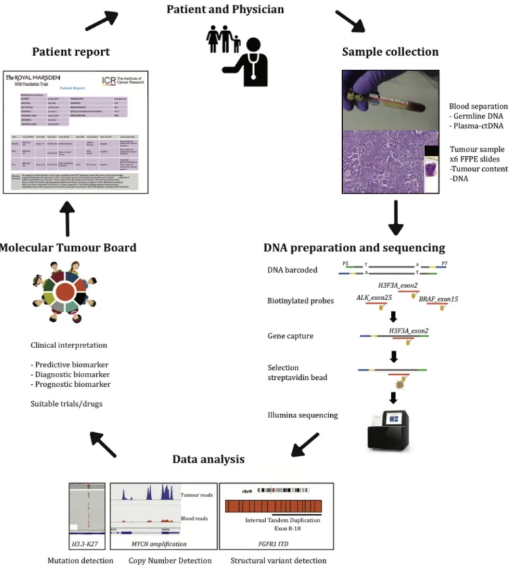
Fig. 1. Study overview. After obtaining informed consent, tumour and blood samples were collected. DNA was extracted, and sequence li-braries were prepared using the capture-based paediatric solid tumour panel. After sequencing, samples underwent an in-house data analysis pipeline that detects mutations, structural variants and copy number changes. Genomic alterations were manually reviewed by two independent scientists and then discussed in a molecular tumour board before a clinical report was issued. FFPE, formalin-fixed paraffin-embedded.
allele frequency. The correlation (r2) of VAF for SNVs and indels between droplet digital polymerase chain
reaction (ddPCR) and v2 was 0.9527 (Figure S1E) and
between v1 and v2 was 0.9301 (Figure S1F).
3.2. Patient samples and overall performance
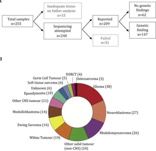
An overview of the study is given in Fig. 1. A total of
255 samples were submitted from 223 patients.
Although patients were eligible to enrol at any time, 90% of evaluable patients had at least one episode of progression/relapse before study enrolment. FFPE tis-sue from the most recent surgery was requested for all but 3 patients where fresh frozen tissue was used.
Adequate coverage for clinical reporting of results
was obtained in 82% of submitted samples (Fig. 2A).
Reasons for sample rejection or failure were as follows: tumour content less than 10%, DNA less than 20 ng and/or excessive DNA fragmentation. The median depth of coverage for all reported samples was 495
(interquartile range: 264e868). The most common
can-cers sequenced were glioma (38), neuroblastoma (27)
and rhabdomyosarcoma (26) (Fig. 2B).
3.3. Genetic findings
At least one genetic alteration was detected in 70% (145/
209) of samples at an allele frequency 5%. The
so-matic genetic alterations detected, grouped according to underlying diagnosis, are summarised inFig. 3,Table S3 andFig S2. In keeping with other studies[34], the most
frequently mutated gene was TP53in 36/209 (17%); in
addition high frequencies of alterations in genes known to be recurrently altered in paediatric malignancies such as ATRX, CDKN2A, CTNNB1 in 12/209 (5.7%),
MYCNin 11/209 (5.2%) andH3F3A, PIK3CAin 10/209
(4.3%) were detected.
3.4. Clinical actionability
Potentially targetable alterations, defined by OncoKB tiers of actionability in addition to predictive biomarkers for currently recruiting paediatric clinical trials, were detected in 51% of sequenced samples (Fig. 4A). Of the 107 tumour samples classified as potentially actionable, 42 (39%) had greater than one actionable alteration detected. For each tumour sample, only the alteration for which there was the highest tier of evidence for actionability was included.
Fig. 2. Tumour samples submitted for sequencing. Summary of sample flow and the total number of samples successfully sequenced (A). Distribution of tumour types among reported cases (B). DSRCT, desmoplastic small round cell tumour; CNS, central nervous system.
Glioma was the tumour type with more defined actionable alterations found, followed by osteosarcoma and rhab-domyosarcoma (Fig. 4B). No tier 1 alterations (US Food
and Drug Administration [FDA]erecognised biomarker
predictive of response to an FDA-approved drug) were detected, indicative of the lack of regulatory approvals for paediatric indications. Only one patient had a tier 2A alteration: a patient with an inflammatory myofibroblastic
tumour, harbouring anALK:SQSTM1translocation. The
patient had a complete surgical resection and did not require systemic therapy.
As a feasibility study, follow-up data were not routinely collected for all patients. Of the 57 patients with a tier 2B or 3 alteration and available follow-up data, only four (7%) received targeted therapies:
Three patients with BRAFV600E mutations were
treated with dabrafenib/trametinib combination therapy: patient 1 had a pleomorphic xanthoastrocytoma and was commenced on dabrafenib/trametinib after third disease progression. The patient remains on treatment with sta-ble disease after 9 months. Patient 2 had glioblastoma multiforme and was commenced on dabrafenib/trameti-nib after disease progression. The patient had stable disease for 13 months before further progression. Patient 3 had multiply relapsed metastatic ameloblastic
fibro-odontosarcoma[35]; by day 28 of treatment, there had
been a partial response but asymptomatic cardiac toxicity, required discontinuation of both drugs. On normalisation of the shortening and ejection fractions, the patient was recommenced on single-agent dabrafenib and had sustained partial response for 15 further months. A patient with multiply relapsed metastatic germinoma and PDGFRA/KIT amplification was given dasatinib, but progressed on treatment.
One patient with high-grade glioma (patient ID 045-T) had a total of 49 somatic mutations (Table S3) (in
w0.18 Mb) consistent with a hypermutator phenotype,
associated with mismatch repair deficiency and predic-tive of potential sensitivity to immune checkpoint
blockade [36]. However, the patient was not fit for
clinical trial enrolment by the time the sequencing re-sults were available.
Other patients had findings that informed prognosis:
a mutation inCTNNB1was found in a patient originally
diagnosed with supratentorial primitive
neuro-ectodermal tumour (PNET), biologically more in keep-ing with a WNT-activated medulloblastoma. Other
examples included an MYOD1 mutation in a patient
with embryonal rhabdomyosarcoma, associated with distinct clinical features and poor prognosis[37], and a
RELA-c11orf95 fusion in a patient with supratentorial ependymoma, associated with high-risk disease[38].
3.5. Analysis of paired samples
For eight patients, paired samples were sequenced at different stages of treatment (Fig. 5). In six of these,
there were differences between the variants detected at
different time points. Mutations in PTEN, NF1 and
TP53were observed in a patient with high-grade glioma
(patient 2) after dabrafenib/trametinib treatment but not in the pre-treatment sample. The patient subsequently received everolimus but progressed after 3 months on
treatment. The acquisition of NF1 mutations as a
resistance mechanism after BRAF inhibition is
consis-tent with findings in BRAFV600E-mutant melanoma
[39,40]. in another child with glioma sequenced at diagnosis and progression, the tumour harboured
shared alterations inH3F3AandTP53, whereasPTEN
was only present at diagnosis andPIK3CA at
progres-sion. In a patient with Wilms tumour, a potentially
targetableTSC2mutation was found in the 3rd relapse
sample, which was not present in the previous sample.
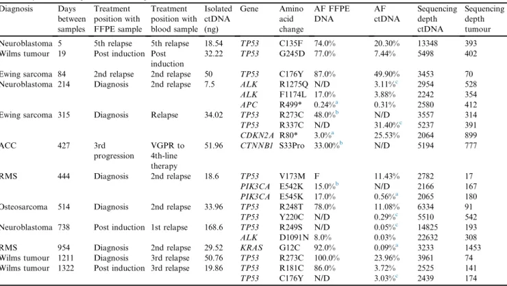
3.6. ctDNA analysis
ctDNA was sequenced in a cohort of 12 patients with extracranial tumours, in whom the tumour panel had detected a genetic alteration that was also covered by a commercially available ctDNA sequencing panel. In 3 patients, in whom ctDNA and FFPE were sequenced from the same time point, there was a direct concor-dance between findings. However, in 5 patients, from whom plasma was collected after at least one subsequent relapse, variants were detected in the plasma that were not detected in FFPE samples (Table 1). For example, in
a patient with neuroblastoma, an ALK F1174L
muta-tion was detected in both tumour and plasma; however,
an additionalALK hotspot mutation was also detected
in the plasma that was not present in the tumour sample. In addition, of note, in 2 cases, variants detected in plasma at relapse were only identified at very low levels in diagnostic tumour samples, below the predefined limit of detection for clinical reporting.
4. Discussion
Comprehensive molecular profiling strategies have been shown to be feasible in children with cancer[9e11]and show encouraging results. However, wide-scale imple-mentation is impractical in most health-care settings, and even if resources were unlimited, it is also restricted by the availability of biopsy material. We show that using as little as 50 ng of DNA, this assay is an accurate, reproducible and practical platform for molecular stratification and identification of actionable targets, required to accelerate precision medicine clinical trials in childhood tumours.
We are aware that although capture-based panel sequencing is an excellent tool, it has limitations. With our targeted panel approach, only a small portion of the genome is sequenced, and therefore, it is not always possible to distinguish between focal gains or deletions
and larger chromosomal gains or losses. Therefore, in version 3 of the panel, we are incorporating a new assay to determine this, which includes probes located across the chromosomes. In addition, as novel gene discoveries and/or targeted inhibitors become available, a wider approach is required for certain indications including a more extensive method for detection of structural vari-ants/translocations. Capture NGS panels are able to detect translocations in DNA with the ability to deter-mine the single-nucleotide breakpoint, so long as those breakpoints occur in or close to a targeted region. We used MANTA to detect spanning pair reads and split reads, thereby identifying fusion gene partners. However, detection of fusion genes is inevitably restricted. We are therefore currently validating a panel using anchored multiplex PCR-based enrichment to detect fusions from
RNA, removing the need to sequence long and complex intronic regions. Furthermore, methylation profiling is particularly relevant for precise diagnostic classification of central nervous system (CNS) tumours, many of which harbour few if any recurrent somatic alterations.
Therefore, in the Stratified Medicine Paediatrics (SMPaeds) national molecular profiling study for chil-dren with relapsed and refractory cancers, we will retain the practical advantages of panel sequencing and run this alongside other more comprehensive profiling mo-dalities including WES, RNA-seq, low-coverage WGS and methylation to support biomarker-driven clinical
trials in the UK, such as eSMART [41]. Furthermore,
where sufficient tissue is available, concurrent analysis via the National Health Service England WGS pro-gramme will be compared with SMPaeds genomic and
Table 1
Results of ctDNA panel sequencing of matched plasma samples and comparison with tumour panel sequencing for genes covered by both panels, ordered by the time elapsed between samples.
Diagnosis Days between samples Treatment position with FFPE sample Treatment position with blood sample Isolated ctDNA (ng) Gene Amino acid change AF FFPE DNA AF ctDNA Sequencing depth ctDNA Sequencing depth tumour Neuroblastoma 5 5th relapse 5th relapse 18.54 TP53 C135F 74.0% 20.30% 13348 393 Wilms tumour 19 Post induction Post
induction
32.22 TP53 G245D 77.0% 7.44% 5498 402
Ewing sarcoma 84 2nd relapse 2nd relapse 50 TP53 C176Y 87.0% 49.90% 3453 70 Neuroblastoma 214 Diagnosis 2nd relapse 7.5 ALK R1275Q N/D 3.11%c 2954 528
ALK F1174L 17.0% 3.88% 2242 354
APC R499* 0.24%a 0.31% 2580 412
Ewing sarcoma 315 Diagnosis Relapse 34.02 TP53 R273C 48.0%b N/D 3557 314
TP53 R337C N/D 31.40%c 5237 391 CDKN2A R80* 3.0%a 25.53% 2064 899 ACC 427 3rd progression VGPR to 4th-line therapy 51.96 CTNNB1 S33Pro 33.00%b N/D 5194 777 RMS 444 Diagnosis 2nd relapse 18.6 TP53 V173M F 11.43% 2782 17 PIK3CA E542K 15.0%b N/D 2166 167 PIK3CA E545K 17.0% 0.56%a 2065 180
Osteosarcoma 514 Diagnosis 2nd relapse 33.96 TP53 R248T 78.0% 11.08% 6334 91
TP53 Y220C N/D 0.29%c 5510 542
Neuroblastoma 738 Post induction 1st relapse 168.6 TP53 R249S N/D 0.05%c 14825 193
ALK D1091N 8.0% 0.03% 22632 308
RMS 954 Diagnosis 2nd relapse 29.52 KRAS G12C 92.0% 0.09%a 3233 1453
Wilms tumour 1211 Diagnosis 3rd relapse 50.76 TP53 R273C 100.0% 23.96% 3961 74 Wilms tumour 1322 Post induction 3rd relapse 19.86 TP53 R181C 86.0% 3.72% 2525 141
TP53 C176Y N/D 3.03%c 2439 174
FFPE, formalin-fixed paraffin-embedded; ctDNA, circulating tumour DNA; RMS, rhabdomyosarcoma; ACC, adrenocortical carcinoma; VGPR, very good partial response, postinduction, surgical resection after routine induction chemotherapy, AF, allele fraction; F, failed coverage; N/D, not detected.
a Below limit of detection. bDetected in tumour only. c
Detected in plasma only.
Fig. 3. Overview of sequencing results. Oncoprint represents somatic mutations and gains, amplification and deletions detected in genes that are covered by the targeted panel. Samples are grouped in columns with genes displayed along rows. Samples are arranged according to the tumour type and genes sorted by frequency. Panel version, sample type, molecular annotations and diagnosis are provided as bars according to the included key (A). Bar plot of most recurrent altered genes, sorted by frequency and colour coded according to the tumour type (B). FFPE, formalin-fixed paraffin-embedded; DSRCT, desmoplastic small round cell tumour; CNS, central nervous system; FF, fresh frozen.
clinical data. This approach will provide an unbiased assessment of the clinical utility and cost-effectiveness of multiple different modalities to enable formal recom-mendations for implementation into routine molecular diagnostics.
Despite the high detection rate of potentially actionable alterations, few patients received treatment with targeted agents. The reasons for this were multi-factorial and include the following: lack of available clinical trials, difficulties accessing novel drugs on a compassionate-use basis and/or clinical deterioration of
the patient. In addition, although many patients had relapsed/refractory disease, a considerable proportion of patients were still on either first-line therapy or proven standard relapse therapies at the time of sequencing. A number of patients were also enrolled in available phase I/II trials that did not require biomarker screening.
This was a pilot study, requiring retrieval of archival tissue, batching of samples for sequencing and infre-quent MTBs. However, for the prospective SMPaeds study, which mandates biopsy at relapse for molecular
Fig. 4. Clinical actionability. Somatic alterations were defined according to OncoKB levels of evidence. Actionability tiers are described in the key. Distribution of actionability tiers for the entire sequenced cohort (A). Distribution of actionability tiers across common tumours, colour coded according to the tumour type (B). DSRCT, desmoplastic small round cell tumour; CNS, central nervous system.
preselection for clinical trials, samples will be processed in a clinically relevant time frame, which after clinical feedback is currently 3e4 weeks, with the final goal of returning data in two weeks. For children with primary solid tumours (who are not enrolled in SMPaeds), as a result of this study, NGS panel sequencing on the paediatric solid tumour panel v2 is now offered in the UK as part of routine National Health Service diag-nostic testing with a turnaround time of 4 weeks from sample dispatch to reporting. Owing to ethical and consent constraints, we were not permitted to report germline findings in the present study. However, given the obvious clinical importance of predisposing muta-tions in paediatric cancer, we have now obtained suit-able consents to report germline mutations via an accredited genetics clinic at Great Ormond Street Hospital.
The sequencing of paired tumour samples at different times demonstrates the importance of tumour hetero-geneity and evolution, adding to the mounting literature in support of the clinical importance of biopsy at relapse for children with cancer[19,42]. Notably, many tumour
mutations emerging at the time of relapse (PTEN, NF1,
PIK3CA and TSC2) are recognised predictive bio-markers of a targeted therapeutic response.
Although sequencing tissue samples of patients is crucial, liquid biopsies offer the possibility of a non-invasive source for tumour genotyping and disease monitoring. Our preliminary findings from a small
number of children demonstrate that high-depth
sequencing of ctDNA can identify actionable somatic variants. We also identified some discrepancies between tumour and plasma, most likely a reflection of tumour heterogeneity and evolution. However, large-scale vali-dation studies comparing tumour and serial ctDNA findings in children with cancer are needed to define the clinical utility of ctDNA analysis, for which a bespoke ctDNA panel for paediatric solid tumours is currently being developed to be incorporated as part of the di-agnostics pipeline.
In summary, we demonstrate the value of targeted gene sequencing as a practical and cost-effective clinical tool to enable improved diagnosis, prognostication and therapeutic stratification for children with cancer.
Acknowledgements
This work was supported by Christopher’s Smile, the National Institute of Health Research (NIHR) Royal
Fig. 5. Comparison of results from paired samples, sequenced at different time points. Venn diagrams compare the genetic findings in eight patients. Shared alterations are illustrated by the intersection of the two ovals. Alterations detected at only the 1st time point are rep-resented in the pink oval, and alterations identified at the 2nd time point only are reprep-resented in the green oval. The size of the oval represents the number of variants identified in each patient.
Marsden Biomedical Research Centre (BRC), Children With Cancer UK (CWC UK) Cancer Research UK (CRUK), Abbie’s Fund, the Rosetree Trust and the KiCa Fund, managed by the King Baudouin Founda-tion. Roche provided support for Panel development. T.S.J. is funded by The Brain Tumour Charity, CWC UK, GOSH Children’s Charity (GOSH CC), CRUK, the Olivia Hodson Cancer Fund and the NIHR GOSH BRC. J.A. and D.H. are funded by the GOSH CC and NIHR GOSH BRC. L.V.M. is funded by the Oak Foundation. The authors thank all participants and the CCLG Tissue Bank for access to samples and contrib-uting CCLG Centres, including members of the ECMC Paediatric network. The CCLG Tissue Bank is funded by Cancer Research UK and CCLG.
Conflict of interest statement
There are no known conflicts of interest associated with this publication, and there has been no significant financial support for this work that could have influ-enced its outcome.
Appendix A. Supplementary data
Supplementary data to this article can be found online athttps://doi.org/10.1016/j.ejca.2019.07.027.
References
[1] Fiore RN, Goodman KW. Precision medicine ethics: selected is-sues and developments in next-generation sequencing, clinical oncology, and ethics. Curr Opin Oncol 2016;28(1):83e7. [2] Tuff-Lacey A, et al. A collaborative approach to enabling
strat-ified cancer medicine in the UK. Drug Discov Today 2015;20(12): 1414e8.
[3] Middleton G, et al. The National Lung Matrix Trial: translating the biology of stratification in advanced non-small-cell lung can-cer. Ann Oncol 2015;26(12):2464e9.
[4] Do K, O’Sullivan Coyne G, Chen AP. An overview of the NCI precision medicine trials-NCI MATCH and MPACT. Chin Clin Oncol 2015;4(3):31.
[5] Kim G, et al. FDA approval summary: olaparib monotherapy in patients with deleterious germline BRCA-mutated advanced ovarian cancer treated with three or more lines of chemotherapy. Clin Cancer Res 2015;21(19):4257e61.
[6] Loong HH, et al. Crizotinib in the management of advanced-stage non-small-cell lung cancer. Future Oncol 2015;11(5):735e45. [7] Stagno F, et al. Imatinib mesylate in chronic myeloid leukemia:
frontline treatment and long-term outcomes. Expert Rev Anti-cancer Ther 2016;16(3):273e8.
[8] Siegel R, Naishadham D, Jemal A. Cancer statistics. CA Cancer J Clin 2012;62(1):10e29. 2012.
[9] Mody RJ, et al. Integrative clinical sequencing in the management of refractory or relapsed cancer in youth. J Am Med Assoc 2015; 314(9):913e25.
[10] Worst BC, et al. Next-generation personalised medicine for high-risk paediatric cancer patients - the INFORM pilot study. Eur J Cancer 2016;65:91e101.
[11] Harttrampf AC, et al. Molecular screening for cancer treatment optimization (MOSCATO-01) in pediatric patients: a
single-institutional prospective molecular stratification trial. Clin Can-cer Res 2017;23(20):6101e12.
[12] Harris MH, et al. Multicenter feasibility study of tumor molecular profiling to inform therapeutic decisions in advanced pediatric solid tumors: the individualized cancer therapy (iCat) study. JAMA Oncol 2016;2(5):608e15.
[13] Ortiz MV, et al. Integrating genomics into clinical pediatric oncology using the molecular tumor board at the memorial sloan kettering cancer center. Pediatr Blood Cancer 2016;63(8): 1368e74.
[14] Castel D, et al. Histone H3F3A and HIST1H3B K27M mutations define two subgroups of diffuse intrinsic pontine gliomas with different prognosis and phenotypes. Acta Neuropathol 2015; 130(6):815e27.
[15] Schwartzentruber J, et al. Driver mutations in histone H3.3 and chromatin remodelling genes in paediatric glioblastoma. Nature 2012;482(7384):226e31.
[16] Peifer M, et al. Telomerase activation by genomic rearrangements in high-risk neuroblastoma. Nature 2015;526(7575):700e4. [17] Izquierdo E, et al. Development of a targeted sequencing
approach to identify prognostic, predictive and diagnostic markers in paediatric solid tumours. Oncotarget 2017;8(67): 112036e50.
[18] Padovan-Merhar OM, et al. Enrichment of targetable mutations in the relapsed neuroblastoma genome. PLoS Genet 2016;12(12): e1006501.
[19] Cohen B, et al. Pediatric oncology provider views on performing a biopsy of solid tumors in children with relapsed or refractory disease for the purpose of genomic profiling. Ann Surg Oncol 2016;23(Suppl 5):990e7.
[20] Thompson JC, et al. Detection of therapeutically targetable driver and resistance mutations in lung cancer patients by next-generation sequencing of cell-free circulating tumor DNA. Clin Cancer Res 2016;22(23):5772e82.
[21] Rothe F, et al. Plasma circulating tumor DNA as an alternative to metastatic biopsies for mutational analysis in breast cancer. Ann Oncol 2014;25(10):1959e65.
[22] Xu S, et al. Circulating tumor DNA identified by targeted sequencing in advanced-stage non-small cell lung cancer patients. Cancer Lett 2016;370(2):324e31.
[23] Bettegowda C, et al. Detection of circulating tumor DNA in early- and late-stage human malignancies. Sci Transl Med 2014; 6(224):224ra24.
[24] De Mattos-Arruda L, Caldas C. Cell-free circulating tumour DNA as a liquid biopsy in breast cancer. Mol Oncol 2016;10(3): 464e74.
[25] Alix-Panabieres C, Pantel K. Clinical applications of circulating tumor cells and circulating tumor DNA as liquid biopsy. Cancer Discov 2016;6(5):479e91.
[26] Kurihara S, et al. Circulating free DNA as non-invasive diag-nostic biomarker for childhood solid tumors. J Pediatr Surg 2015; 50(12):2094e7.
[27] Combaret V, et al. Detection of tumor ALK status in neuroblastoma patients using peripheral blood. Cancer Med 2015;4(4):540e50. [28] Panditharatna E, et al. Clinically relevant and minimally invasive
tumor surveillance of pediatric diffuse midline gliomas using patient-derived liquid biopsy. Clin Cancer Res 2018;24(23):5850e9. [29] Jimenez I, et al. Circulating tumor DNA analysis enables mo-lecular characterization of pediatric renal tumors at diagnosis. Int J Cancer 2019;144(1):68e79.
[30] Chicard M, et al. Whole-exome sequencing of cell-free DNA reveals temporo-spatial heterogeneity and identifies treatment-resistant clones in neuroblastoma. Clin Cancer Res 2018;24(4):939e49. [31] Allin DM, et al. Circulating tumour DNA is a potential
biomarker for disease progression and response to targeted ther-apy in advanced thyroid cancer. Eur J Cancer 2018;103:165e75. [32] Chakravarty D, et al. OncoKB: a precision oncology knowledge
[33] Forbes SA, et al. COSMIC: somatic cancer genetics at high-res-olution. Nucleic Acids Res 2017;45(D1):D777e83.
[34] Grobner SN, et al. The landscape of genomic alterations across childhood cancers. Nature 2018;555(7696):321e7.
[35] Gatz SA, et al. Chemotherapy responsiveness in a patient with multiply relapsed ameloblastic fibro-odontosarcoma of the maxilla. Pediatr Blood Cancer 2015;62(11):2029e32.
[36] Lee L, Gupta M, Sahasranaman S. Immune Checkpoint in-hibitors: an introduction to the next-generation cancer immuno-therapy. J Clin Pharmacol 2016;56(2):157e69.
[37] Kohsaka S, et al. A recurrent neomorphic mutation in MYOD1 defines a clinically aggressive subset of embryonal rhabdomyo-sarcoma associated with PI3K-AKT pathway mutations. Nat Genet 2014;46(6):595e600.
[38] Pajtler KW, et al. Molecular classification of ependymal tumors across all CNS compartments, histopathological grades, and age groups. Cancer Cell 2015;27(5):728e43.
[39] Whittaker SR, et al. A genome-scale RNA interference screen implicates NF1 loss in resistance to RAF inhibition. Cancer Discov 2013;3(3):350e62.
[40] Van Allen EM, et al. The genetic landscape of clinical resistance to RAF inhibition in metastatic melanoma. Cancer Discov 2014; 4(1):94e109.
[41] European Proof-of Concept Therapeutic Stratification Trial of Molecular Anomalies in Relapsed or Refractory Tumours.https:// clinicaltrials.gov/ct2/show/NCT02813135?termZesmart&rankZ5. [42] Eleveld TF, et al. Relapsed neuroblastomas show frequent