Psychology Faculty Research and Publications
Psychology, Department of
1-1-2016
Exercise Training and Functional Connectivity
Changes in Mild Cognitive Empairment and
Healthy Elders
Theresa J. Chirles
University of Maryland at College Park
Katherine Reiter
Marquette University, [email protected]
Lauren R. Weiss
University of Maryland at College Park
Alfonso J. Alfini
University of Maryland at College Park
Kristy A. Nielson
Marquette University, [email protected]
See next page for additional authors
Accepted version.
Journal of Alzheimer's Disease,
Vol. 57 (2017): 845-856.
DOI
. © 2017 IOS Press.
Used with permission.
Journal of Alzheimer’s Disease, Vol 57 (2017): pg. 845-856. DOI. This article is © IOS Press and permission has been granted for this version to appear in e-Publications@Marquette. IOS Press does not grant permission for this article to be further copied/distributed or hosted elsewhere without the express permission from IOS Press.
1
Exercise Training and Functional
Connectivity Changes in MCI and
Healthy Elders
Theresa J. Chirles
University of Maryland
College Park, MD
Katherine Reiter
Marquette University
Milwaukee, WI
Lauren R. Weiss
University of Maryland
College Park, MD
Alfonso J. Alfini
University of Maryland
College Park, MD
Kristy A. Nielson
Department of Psychology, Marquette University
Medical College of Wisconin
Milwaukee, WI
J. Carson Smith
University of Maryland
Journal of Alzheimer’s Disease, Vol 57 (2017): pg. 845-856. DOI. This article is © IOS Press and permission has been granted for this version to appear in e-Publications@Marquette. IOS Press does not grant permission for this article to be further copied/distributed or hosted elsewhere without the express permission from IOS Press.
2
Abstract:
Background: Effective interventions are needed to improve brain function in
Mild Cognitive Impairment (MCI), an early stage of Alzheimer’s disease (AD).
The posterior cingulate cortex (PCC)/precuneus is a hub of the Default Mode Network (DMN) and is preferentially vulnerable to disruption of functional connectivity in MCI and AD.
Objective: We investigated whether 12 weeks of aerobic exercise could
enhance functional connectivity of the PCC/precuneus in MCI and healthy elders.
Methods: Sixteen MCI and 16 healthy elders (age range = 60-88) engaged
in a supervised 12-week walking exercise intervention. Functional MRI (fMRI) was acquired at rest; the PCC/precuneus was used as a seed for correlated brain activity maps.
Results: A linear mixed effects model revealed a significant interaction in the right parietal lobe: the MCI group showed increased connectivity while the healthy elders showed decreased connectivity. In addition, both groups showed increased connectivity with the left postcentral gyrus. Comparing pre to post intervention changes within each group, the MCI group showed increased connectivity in 10 regions spanning frontal, parietal, temporal and insular lobes and the cerebellum. Healthy elders did not demonstrate any significant connectivity changes.
Conclusion: The observed results show increased functional connectivity of
the PCC/precuneus in individuals with MCI after 12 weeks of moderate intensity walking exercise training. The protective effects of exercise training on cognition may be realized through the enhancement of neural recruitment mechanisms, which may possibly increase cognitive reserve. Whether these effects of exercise training may delay further cognitive decline in patients diagnosed with MCI remains to be demonstrated.
Keywords:Alzheimer’s disease, connectivity, cognitive disorders, aging, default mode network, posterior cingulate, precuneus, exercise interventions, resting state fMRI
Introduction
Neuropathological changes associated with Alzheimer’s disease (AD) occur many years before the onset of clinical symptoms.1 Older
adults who have declined cognitively but who do not meet criteria for a diagnosis of AD are often classified as having Mild Cognitive
Impairment (MCI),2,3 and more than half of these individuals progress
to an AD diagnosis within five years.4 There is an urgency to identify
biomarkers for preclinical detection of neuropathology prior to the onset of symptoms in order to inform treatment strategies and to aid in the understanding of AD progression.5 Resting state functional
connectivity is emerging as a viable biomarker and predictor of future conversion to AD6-9 and as an indicator of treatment efficacy.10,11
Journal of Alzheimer’s Disease, Vol 57 (2017): pg. 845-856. DOI. This article is © IOS Press and permission has been granted for this version to appear in e-Publications@Marquette. IOS Press does not grant permission for this article to be further copied/distributed or hosted elsewhere without the express permission from IOS Press.
3 Resting state functional connectivity in this paper is based on the correlations of spontaneous blood oxygenation level-dependent (BOLD) functional magnetic resonance imaging (fMRI) signals during the absence of an external task. It is assumed to reflect the underlying anatomy of the neuronal architecture12 through direct and indirect
neural networks consisting of monosynaptic and polysynaptic connections.13-15 Temporal correlations of spatially distinct brain
regions indicate either direct or indirect neuronal connections, and resting state functional connectivity has been found to predict
performance on higher order cognitive tests.16-18 Higher order cognitive
processes require the integration of several segregated, domain-specific neural processing pathways,12 and these diverse pathways
intersect in regions of the brain called ‘hubs’, characterized by a disproportionately high number of functional, and often concurrently anatomical, connections.19 These hubs, while few in number, may limit
the large metabolic cost of neural communication by integrating otherwise disparate networks20 and play an important role in
information flow.21 The posterior cingulate (PCC) and precuneus
regions together constitute a key hub of the default mode network (DMN). This hub fosters efficient communication between the DMN and the medial temporal lobe (MTL) network, a network with an important role in memory processes22 that is highly vulnerable to AD pathology.23
The PCC/precuneus is also an area associated with the accumulation of amyloid-β (Aβ) plaque, a hallmark of AD pathology.12 The
PCC/precuneus exhibits reduced functional connectivity in MCI, early
AD,24,25 and in clinically normal older adults that test positive for brain
amyloid burden.26,27 Changes in the functional connectivity of the
PCC/precuneus have also been associated with accelerated atrophy and other preclinical pathological changes associated with AD,24,28,29
underscoring its potential role as a predictive biomarker. Thus,
alterations in resting state functional connectivity, while concurrently associated with cognitive decline, may also precede measureable cognitive changes.
Prior to evidence of cognitive decline in AD, the PCC/precuneus exhibits increased connectivity with frontal and parietal brain regions that do not show AD pathology until very late disease stages,30 and
Journal of Alzheimer’s Disease, Vol 57 (2017): pg. 845-856. DOI. This article is © IOS Press and permission has been granted for this version to appear in e-Publications@Marquette. IOS Press does not grant permission for this article to be further copied/distributed or hosted elsewhere without the express permission from IOS Press.
4 regions in older adults is a compensatory response to aging.31
Although this compensatory response is associated with neuronal damage,32 it is also thought to be indicative of maintaining cognitive
function.33,34 In individuals diagnosed with MCI and AD, declines in
connectivity have been noted in brain areas affected early in the
progression of AD, such as the MTL.23 Interventions or treatments that
preserve and/or increase the connectivity of the PCC/precuneus with available frontal and parietal resources in older adults may help promote cognitive stability.
It is well established that both leisure time physical activity and exercise training help to improve and maintain cognitive function in healthy older adults,35,36 even in those at increased risk for AD.37-40
Aerobic training in healthy elders appears to increase the functional connectivity within the DMN41 and hippocampal networks.42
Furthermore, although local neuronal networks exhibit deterioration in healthy elders, high levels of physical activity have been shown to protect these networks.43 Exercise training in individuals diagnosed
with MCI has been shown to improve cognition4,44-46 and increase
neuronal efficiency during a semantic memory retrieval task,46 but it is
not known if exercise training results in changes to neural network functional connectivity in older adults diagnosed with MCI. If exercise training does increase the connectivity of hubs enhancing network recruitment in this population, it may indicate a gain of cognitive reserve that would help to preserve cognitive abilities and possibly delay cognitive decline. Cognitive reserve is a concept that explains the typically nonlinear relationship between cognitive performance and neuronal damage or brain disruption,31 and is associated with the
ability to recruit additional brain networks when the primary networks are disrupted47 as in MCI. Greater levels of cognitive reserve proxies,
such as education, have been associated with increased functional connectivity,48 and physical activity (PA) also has been implicated as
one of the factors that increases cognitive reserve.49
The current study extends this literature with evidence that aerobic exercise training may stimulate functional connectivity of the PCC/precuneus in individuals diagnosed with MCI. This is a
continuation of our previously published paper that reported a 12-week walking intervention resulted in decreased fMRI activation in
Journal of Alzheimer’s Disease, Vol 57 (2017): pg. 845-856. DOI. This article is © IOS Press and permission has been granted for this version to appear in e-Publications@Marquette. IOS Press does not grant permission for this article to be further copied/distributed or hosted elsewhere without the express permission from IOS Press.
5 several cortical regions during a semantic memory related task.46 We
hypothesized that healthy elders and individuals diagnosed with MCI would demonstrate increased connectivity between the PCC/precuneus and frontal-parietal cortices from before to after the intervention, indicating enhanced network recruitment capabilities. We expected increased connectivity to the MTL in the MCI group, as the MTL is particularly vulnerable to AD progression.23
Materials and Methods
Participants and pre-screening
This study used resting state fMRI data from participants (17 MCI and 18 healthy elders) described in previous work,46 except that
resting state fMRI data were missing for one MCI and two healthy elder participants. Community dwelling older adults, ages 60 to 88, were recruited through physician referrals, local newspaper
advertisements, and in-person informational sessions at retirement communities and recreational centers. Interested volunteers who were still eligible after a phone interview met face-to-face with a study team member to review all procedures, expectations, possible risks, and a physician approval form for moderate intensity exercise was obtained. A neurological evaluation completed the eligibility evaluation. Informed consent was obtained from all individual participants included in the study. All procedures were in accordance with the ethical standards of the institutional and/or national research committee and with the 1964 Helsinki declaration and its later amendments or comparable ethical standards.
Inclusion and exclusion criteria
In order to maximize the effect of exercise training, all study participants indicated they engaged in only light physical activity two or fewer days/week for the past six months. Participants were
excluded if they reported a history or evidence of: 1) medical illnesses or conditions that may affect brain function (including glaucoma, chronic obstructive pulmonary disease, and untreated hypertension); 2) neurological illnesses or conditions (including cerebral ischemia,
Journal of Alzheimer’s Disease, Vol 57 (2017): pg. 845-856. DOI. This article is © IOS Press and permission has been granted for this version to appear in e-Publications@Marquette. IOS Press does not grant permission for this article to be further copied/distributed or hosted elsewhere without the express permission from IOS Press.
6 vascular headache, head trauma with loss of consciousness (>30 min), epilepsy, carotid artery disease, cerebral palsy, brain tumor, normal-pressure hydrocephalus, chronic meningitis, pernicious anemia, multiple sclerosis, Huntington’s disease, Parkinson’s disease or HIV infection); 3) current untreated Axis I psychiatric disturbance meeting DSM-IV Axis I criteria (including substance abuse or dependence and severe depressive symptoms); 4) exclusion criteria specific to MR scanning (such as pregnancy, history of claustrophobia, weight
inappropriate for height, and ferrous objects within the body); 5) any unstable or severe cardiovascular disease or asthmatic condition; 6) left-handedness (laterality quotient [LQ] <50);50 6) current use of
psychoactive medications, except stable doses of antidepressants; and 7) history of transient ischemic attack or >4 on the modified Hachinski ischemic scale. Participants were also excluded if they scored >15 on the Geriatric Depression Scale (GDS)51 or showed relatively impaired
activities of daily living (ADL) using the Lawton and Brody Self-Maintaining and Instrumental Activities of Daily Living Scale.
Neuropsychological test battery and clinical criteria for
MCI
The Neuropsychological test battery included the Mini-Mental State Exam,52 Mattis Dementia Rating Scale-2 (DRS),53 Rey Auditory
Verbal Learning Test (AVLT),54 Logical Memory and Letter-Number
Sequencing subtests of the Wechsler memory Scale-III,55 Symbol-Digit
Modalities Test,56 Controlled Oral Word Association Test,57 animal
fluency, and the Clock Drawing Test.58 This comprehensive battery was
administered before and after the exercise intervention, and alternate forms of the AVLT and DRS were used at each testing session.
Cognitive status of the participants was determined using the core clinical criteria set by the NIH-Alzheimer’s association workgroup on MCI due to AD.7 MCI was defined as a subjective concern regarding
a change in cognition supported by an informant, impairment in one or more cognitive domains (defined as 1.5 standard deviations below age and education matched means on delayed recall on the AVLT), and intact activities of daily living. Three neuropsychologists (including K.A.N.) reached a consensus on impairment. A neurologist ruled out all other possible etiologies. Healthy elders had no specific cognitive
Journal of Alzheimer’s Disease, Vol 57 (2017): pg. 845-856. DOI. This article is © IOS Press and permission has been granted for this version to appear in e-Publications@Marquette. IOS Press does not grant permission for this article to be further copied/distributed or hosted elsewhere without the express permission from IOS Press.
7 complaint, intact cognitive performance in all domains, and intact activities of daily living.
Exercise test
Participants completed a submaximal exercise test on a
motorized treadmill (General Electric, Milwaukee, WI) to estimate peak aerobic capacity (
V̇̇O
2peak) before and after the exercise intervention. The exercise test used a modified Balke-Ware protocol of 2.0 miles/hr beginning with a 00 grade and increasing 10 per minute.59Concentrations of oxygen and carbon dioxide in expired air were collected every 15 seconds by a metabolic measurement system (ParvoMedics, Sandy, UT). Each test included measurements of heart rate, blood pressure (every 2 minutes), and ratings of perceived exertion (RPE; each minute). Test termination criteria included reaching 85% of age-predicted heart rate max, a diastolic blood pressure greater than 110 mmHg, or the participant’s desire to stop. The peak rate of oxygen uptake (
V̇̇O
2peak) was estimated from the highestV̇̇O
2 value achieved during the test (expressed as ml/kg/min at STPD).59 Additional details have been described by Smith et al.46Exercise intervention
A qualified personal fitness trainer or an exercise physiologist supervised the participants in the 12-week intervention at fitness centers located near the participants’ homes or within their
communities. The exercise intensity, session duration, and weekly frequency were increased during the first four weeks until the participants were walking for 30 minutes, four times a week, at approximately 50-60% of HRR (heart rate reserve). Each session began and ended with 10 minutes of light walking and flexibility
exercises. Participants wore a Polar® heart rate monitor and provided subjective RPE’s using the Borg 6-20 RPE scale throughout each exercise session at minutes 5, 15, and 30.60,61 The treadmill grade
and/or speed were modified to moderately challenge the participant based on the heart rate and perception of effort (not more than 15 on the Borg scale). This is considered a moderate intensity exercise for older adults.62
Journal of Alzheimer’s Disease, Vol 57 (2017): pg. 845-856. DOI. This article is © IOS Press and permission has been granted for this version to appear in e-Publications@Marquette. IOS Press does not grant permission for this article to be further copied/distributed or hosted elsewhere without the express permission from IOS Press.
8
MRI acquisition procedures
Prior to the first MRI acquisition using a General Electric
(Waukesha, WI) 3.0 Tesla scanner, participants were familiarized with the MRI environment by lying in a mock scanner. During MRI
acquisition, participants were instructed to lie as still as possible and foam padding was used to limit movement and improve comfort. Anatomical and resting state sequences were run during the scanning session. High-resolution, three-dimensional spoiled gradient recalled at steady-state (SPGR) anatomic images were acquired (TE = 3.9ms; TR = 9.6ms; inversion recovery (IR) preparation time = 450ms; flip angle = 12°; number of excitations (NEX) = 1; slice thickness = 1.0mm; FOV = 240mm; resolution = 256 x 224). During the resting state scan, participants were instructed to keep their eyes open and to look at a fixation cross. A gradient-echo, echo-planar pulse sequence sensitive to blood oxygenation level-dependent (BOLD) contrast were acquired (TE = 25ms; TR = 2000ms; flip angle = 77°; NEX = 1; 36 axial slices; 4.0 mm isotropic voxels; FOV = 240mm; resolution = 64 x 64;
duration 6 minutes).
MRI preprocessing
Preprocessing steps were done using tools from the Analysis of Functional NeuroImages (AFNI) software package.63 During the initial
preprocessing and analysis, the researcher (TC) was blind to each participant’s group classification. Time series and anatomical images were aligned and skull-stripping, slice time correction, and motion correction procedures were performed. The first 3 TRs were removed, a 0.005 to 0.10 Hz bandpass filter was applied, and the following sources of noise were regressed out: six-parameter rigid body head motion, ventricle signal, white matter signal, mean global signal, and the derivatives of the motion parameters, white matter signal, and ventricle signal. The time series data were smoothed using a 4mm full-width at half-maximum Gaussian blur and normalized to Montreal Neurological Institute (MNI) space.
Journal of Alzheimer’s Disease, Vol 57 (2017): pg. 845-856. DOI. This article is © IOS Press and permission has been granted for this version to appear in e-Publications@Marquette. IOS Press does not grant permission for this article to be further copied/distributed or hosted elsewhere without the express permission from IOS Press.
9
Seed based analysis
The seed based analysis was also conducted using AFNI. The seed region of interest (ROI) was defined using a 5mm spherical mask surrounding the MNI coordinates -2, -50, 36, the peak voxel
coordinates of the PCC/precuneus reported by Buckner and colleagues.12 The time course in the ROI was extracted, and seed
correlation maps for all participants at each testing session were
formed by correlating the seed ROI with all other voxels in the brain. A Fisher’s r to z transformation was implemented to normalize the
correlation coefficients. Group and Time differences were analyzed using linear mixed effects64 and cluster-based analysis (after
interpolation to 2 mm3 voxels, 105 voxels or more with primary
threshold = 0.01; cluster based statistic p < 0.05, FWER controlled). This allows for sensitivity (minimizing Type II errors), while
maintaining some spatial specificity. The AFNI 3dLME command was used to run the linear mixed effects model using age as a covariate. This is an ideal analysis for repeated measures analyses because it allows for random intercepts; thus initial variability in the correlations are taken into account. F-statistics indicate main effects and
interactions, and we also conducted post hoc paired sample t-tests within each group to assess changes from baseline to
post-intervention.
Results
Participant and baseline characteristics
Usable resting state fMRI data were available for 16 healthy elders and 16 participants diagnosed with MCI. As shown in Table 1, the healthy elders and individuals diagnosed with MCI did not
significantly differ in sex, age, education, depression or activities of daily living. As expected, the MCI group exhibited poorer baseline performance than the healthy group on all but two neuropsychological subtests (DRS Attention and DRS Construction). Participants’ baseline characteristics are presented in Table 1.
Journal of Alzheimer’s Disease, Vol 57 (2017): pg. 845-856. DOI. This article is © IOS Press and permission has been granted for this version to appear in e-Publications@Marquette. IOS Press does not grant permission for this article to be further copied/distributed or hosted elsewhere without the express permission from IOS Press.
10
Exercise intervention fidelity
The mean (SD) number of exercise sessions attended,
adherence rate, exercise intensity and perceived exertion over the first 4 weeks and during weeks 5-12 of the intervention did not differ
between groups. Out of a total of 44 sessions, 42.3 (2.2) were completed resulting in a 96.1 (5.0) % adherence rate. During weeks 1-4 and weeks 5-12, the mean intensity was 46.9 (7.1) %HRR and 54.7 (11.0) %HRR, respectively. RPEs were most closely associated with the verbal descriptor “light” at 10.6 (1.8) and 10.8 (2.0) during the first 4 weeks and last 8 weeks, respectively. There was also a mean increase over the 12-week intervention in
V̇̇O
2peak by2.0ml/kg/min – an approximate 10.6% increase in cardiorespiratory fitness. More details regarding the change in cardiorespiratory fitness can be found in Smith et al.46
Neuropsychological test performance
Neuropsychological test results for the entire sample (35 participants) have been previously reported.46 The results reported
here are consistent with those reported by Smith et al. (2013), and reflect the slightly smaller sample size (32 participants) in the current study. A repeated measures ANOVA revealed a significant effect of Time in Trial 1 of AVLT (p = .013), where both groups demonstrated improvement from baseline to post-intervention (mean (SD): MCI pre: 3.75 (2.02), MCI post: 4.81 (1.97)); healthy elders pre: 5.50 (2.00) healthy elders post: 6.38 (1.57)). The Group by Time interaction for Trial 1 was not significant (p = 0.800), and there were no significant changes in the other Rey AVLT indices.
Seed based functional connectivity: Group by Time
interaction
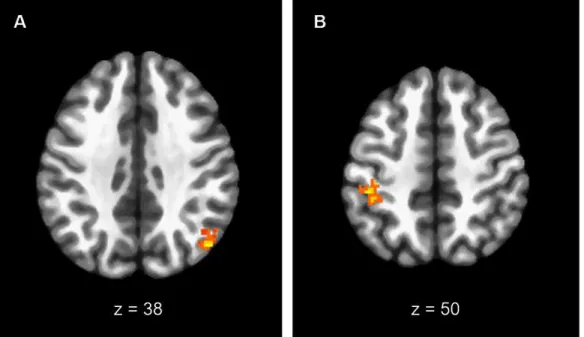
Connectivity results are based on the correlation maps of the mean BOLD time course from the PCC/precuneus seed ROI and the remaining voxels in the brain. One significant Group by Time
interaction was observed and is presented in Figure 1A and Table 2. The MCI group showed an increased correlation between the
Journal of Alzheimer’s Disease, Vol 57 (2017): pg. 845-856. DOI. This article is © IOS Press and permission has been granted for this version to appear in e-Publications@Marquette. IOS Press does not grant permission for this article to be further copied/distributed or hosted elsewhere without the express permission from IOS Press.
11 PCC/precuneus and right inferior parietal lobe (IPL). The healthy elders showed a decreased correlation with the cluster in the right IPL.
Seed based functional connectivity: Time main effects
A Time main effect, reflecting significant changes from before to after the intervention on average collapsed across both groups, was found in the left postcentral gyrus. There was an increased correlation of the PCC/precuneus with this cluster, and the region is presented inFigure 1B and Table 2.
Seed based functional connectivity: Post-hoc t-tests:
Changes within each group
Significant changes in the MCI group are identified in Table 3
and Figure 2A. The MCI group exhibited increased correlations after the exercise intervention in ten regions. Clusters had peak voxels in the right MFG, superior frontal gyrus, postcentral gyrus, PHG, and claustrum. Clusters were also found in the left IPL and bilateral precentral gyrus (two clusters in the left precentral gryrus) and
culmen. No significant clusters demonstrating changes in connectivity across time were found in the healthy elders group.
Discussion
We investigated the effects of a 12-week walking intervention on the functional connectivity of the PCC/precuneus in individuals diagnosed with MCI and healthy elders. We hypothesized that both the MCI group and healthy elders would show increased connectivity with frontal and parietal regions, suggestive of enhanced recruitment of preserved brain regions. We also hypothesized that the intervention would increase PCC/precuneus connectivity with MTL regions in the MCI group. We did find the hypothesized changes in functional
connectivity of the PCC/precuneus with frontal, temporal, and parietal brain regions in the MCI group, but only one region, the left
postcentral gyrus, showed increased connectivity in both groups. Additionally, a group by time interaction in the right IPL revealed that the MCI group showed the expected increased connectivity while the
Journal of Alzheimer’s Disease, Vol 57 (2017): pg. 845-856. DOI. This article is © IOS Press and permission has been granted for this version to appear in e-Publications@Marquette. IOS Press does not grant permission for this article to be further copied/distributed or hosted elsewhere without the express permission from IOS Press.
12 healthy elders demonstrated decreased connectivity in this region. These results, in conjunction with the findings from our previously published paper on the same subjects,46 may indicate that exercise
training has divergent effects on neural compensation and neural efficiency in the MCI group compared to healthy elders.
In developing our hypotheses for this study, we focused primarily on neural compensation as demonstrated by increased connectivity of the PCC/precuneus with preserved brain regions as a mechanism to increase cognitive reserve. After exercise training, the MCI group, presumed to be on the AD continuum, did demonstrate increased synchrony between compensatory networks and the
PCC/precuneus, a region preferentially targeted in AD pathology. We expected, but did not observe, a similar increase in connectivity after exercise training in the healthy elders. This prediction was based on a previous report by Voss and colleagues,41 which found trends of
connectivity changes in the DMN after 6 months of exercise training. However, these effects did not reach statistical significance in their study until after the 12-month intervention. As our intervention lasted only 3 months, it is possible that a longer exercise intervention is needed to observe connectivity changes in cognitively healthy older adults.
Another hypothesized mechanism to increase cognitive reserve is augmenting neural reserve – an indicator of neural efficiency.65 In
our previous paper utilizing the same subjects,46 we found group
differences in activation changes in the precuneus and PCC during a famous name discrimination task. While both groups maintained equal task performance, the activation intensity decreased in the healthy elders after the exercise intervention, while there were no changes in the MCI group. This suggests increased neural efficiency in the healthy elders (as found in several other regions of the semantic memory network), and a need to maintain a compensatory response in the MCI group, as this region is targeted by AD pathology. Unfortunately, we were not able to measure levels of amyloid deposition or neurological damage in either group, so this possibility needs to be further
explored. However, our current results are consistent with this interpretation and suggest that the MCI group demonstrated neural compensation through increased connectivity of the PCC/precuneus,
Journal of Alzheimer’s Disease, Vol 57 (2017): pg. 845-856. DOI. This article is © IOS Press and permission has been granted for this version to appear in e-Publications@Marquette. IOS Press does not grant permission for this article to be further copied/distributed or hosted elsewhere without the express permission from IOS Press.
13 while the healthy elders, who appear to not have reached a critical threshold for age related changes, did not require neural
compensation.65 Rather, exercise training may have resulted in
increased neural reserve (or efficiency) of the PCC/precuneus, as indicated by our previously published findings of reduced activation during memory retrieval.46 While the participants diagnosed with MCI
did not differ in education from the healthy elders group (both were highly educated), the presence of cognitive impairment indicates a critical threshold was reached through combined age-related and AD processes. Thus, in those diagnosed with MCI, our findings support the idea that exercise training may stimulate increased cognitive reserve through enhanced recruitment of compensatory networks, such as increased function connectivity with a key neural hub in the
PCC/precuneus.
As a first attempt to examine changes in resting state functional connectivity in MCI after an exercise intervention, we conducted a priori post hoc analyses to further explore the functional connectivity changes within this group. Results showed that the MCI group
demonstrated increased connectivity of the PCC/precuneus with frontal and parietal regions from pre to post intervention. These effects
suggest improved coordination of intrinsic activity of PCC/precuneus and several network regions including the fronto-parietal network (bilateral precentral gyrus and right middle frontal gyrus), the somatosensory network (right postcentral gyrus), and DMN regions (right superior frontal gyrus and left IPL). There was also increased connectivity between the PCC and the right parahippocampal gyrus, a region that links the DMN to the medial temporal lobe system.66 The
increased connectivity suggests possible enhancement of posterior-anterior connections vulnerable to aging.18,31 Our findings raise the
possibility that these increased compensatory connections across networks connected to the PCC/precuneus may in part explain the neural protective effects of physical activity in MCI. This pattern of stronger connectivity with the PCC/precuneus after exercise training is a stark contrast to the typical progression of connectivity disruptions with the emergence of clinical symptoms. As AD progresses there has been shown to be an initial decrease in functional connectivity within the DMN, followed by compensatory hyperconnectivity in the
Journal of Alzheimer’s Disease, Vol 57 (2017): pg. 845-856. DOI. This article is © IOS Press and permission has been granted for this version to appear in e-Publications@Marquette. IOS Press does not grant permission for this article to be further copied/distributed or hosted elsewhere without the express permission from IOS Press.
14 an overall loss of connectivity.32 The increased functional connectivity
after exercise training observed in the MCI group between the
PCC/precuneus and the other 10 regions suggests a possible reversal of the expected progression of connectivity decrements in MCI and, furthermore, enhancement of recruitment mechanisms that would increase cognitive reserve and possibly delay cognitive decline. Future randomized controlled trials should test this hypothesis.
Interestingly, the MCI group showed increased connectivity with regions in the insular lobe and cerebellum after the walking
intervention. Connectivity in insular networks are reported to
correspond to cognitive performance in individuals with amnesic MCI and cognitively healthy older adults.67 Additionally, while we focused
on the recruitment of frontal and parietal regions by the
PCC/precuneus as an example of increased compensation to protect the DMN, the cerebellar region identified in our study corresponds to regions identified to be functionally related to the DMN.68 Our results
may indicate that the insular lobe and cerebellum are additional resources of reserve for the DMN. These results should be interpreted with some caution due to lower signal to noise ratio in these regions and particularly in the cerebellum.
Future research on the effects of an exercise intervention on functional connectivity in MCI and healthy elders should address
potential mechanisms for these effects. Candidate measures that have been linked to exercise training would be BDNF, which has been found to modify functional connectivity,69 cerebral blood flow,42 hippocampal
brain volume70,71 white matter integrity,72 and Aβ plaque burden.73
Given the evidence that exercise may oppose the actions of
acetylcholinesterase in the hippocampus and cerebral cortex of rats,74
the effects of exercise training on the cholinergic system should also be considered. We have also recently reported, in this same cohort, that increased cardiorespiratory fitness after the exercise intervention was positively correlated in both groups with increased cortical
thickness in regions including the precuneus, posterior cingulate cortex, pre-central and post-central gyri, and the medial and middle frontal gyri;75 regions that partly overlap with the areas that showed
changes in resting state connectivity in the current analysis. Future studies should focus on multimodal imaging techniques to understand
Journal of Alzheimer’s Disease, Vol 57 (2017): pg. 845-856. DOI. This article is © IOS Press and permission has been granted for this version to appear in e-Publications@Marquette. IOS Press does not grant permission for this article to be further copied/distributed or hosted elsewhere without the express permission from IOS Press.
15 the mechanisms of exercise on neural plasticity in older adults and if this changes by disease state.
The lack of a non-exercise control group is a limitation of this study, and some caution is warranted in the interpretation of these effects. We cannot rule out the possibility that the walking intervention and its social context (most participants exercised alone under the supervision of a certified personal trainer) combined to produce
changes in connectivity. However, the passage of time does not seem to be a plausible explanation for the changes we observed, as a
longitudinal study in healthy older adults (ages 49 to 79) found
functional connectivity within the DMN to be stable over a period of six years.76 We observed that several regions showed bilateral increases
in functional connectivity, and the fact that the effects were more pronounced in those diagnosed with MCI argues against a
generalizable influence of the experimental context.
Many longitudinal studies have shown that the risk of cognitive decline is reduced in older adults who are physically active77,78 and
cognition is protected in individuals with MCI who have greater
physical activity.44,45 Our results indicate that these protective effects
may manifest in individuals with MCI through the enhanced
recruitment of the PCC/precuneus, an important hub for higher order cognitive processes, and the preservation of posterior-anterior resting state functional connections. These connections are vulnerable in normal aging, and when these aging effects are combined with AD, the results are even more devastating to cognition.31 The pathological
processes of AD and normal aging have divergent effects on brain networks,79,80 and the differential effects of exercise training on
functional connectivity in our study suggest that exercise-induced neural plasticity may vary based on AD progression and available cognitive reserve. While it remains to be conclusively demonstrated that exercise training may delay the conversion of individuals
diagnosed with MCI to AD, or delay the onset of MCI among the
cognitively intact, these results further underscore the complexity and pleiotropic nature of exercise as a potential intervention to modify neural network connectivity along the AD continuum.
Journal of Alzheimer’s Disease, Vol 57 (2017): pg. 845-856. DOI. This article is © IOS Press and permission has been granted for this version to appear in e-Publications@Marquette. IOS Press does not grant permission for this article to be further copied/distributed or hosted elsewhere without the express permission from IOS Press.
16
Acknowledgements
We thank the participants for their time and dedication to this project. The authors also thank Piero Antuono, Alissa Butts, Ryan Hanson, Nathan Hantke, Mahshid Najafi, Karen Outzen, Hyuk Oh, Mihai Sirbu, and Matthew Verber for assistance with various phases of this study.
This study was supported by the University of Wisconsin-Milwaukee Graduate School Research Growth Initiative; and the National Center for Advancing Translational Sciences, National Institutes of Health grant numbers 8UL1TR000055 and
8KL2TR000056. Its contents are solely the responsibility of the authors and do not necessarily represent the official views of the NIH.
References
1Jack CR, Knopman DS, Jagust WJ, Shaw LM, Aisen PS, Weiner MW, Petersen
RC, Trojanowski JQ (2010) Hypothetical model of dynamic biomarkers
of the Alzheimer's pathological cascade. Lancet Neurol 9, 119-128.
2Petersen RC, Smith GE, Waring SC, Ivnik RJ, Tangalos EG, Kokmen E (1999)
Mild cognitive impairment: clinical characterization and outcome. Arch
Neurol 56, 303-308.
3Petersen RC (2004) Mild cognitive impairment as a diagnostic entity. J Intern
Med 256, 183-194.
4Gauthier S, Reisberg B, Zaudig M, Petersen RC, Ritchie K, Broich K, Belleville
S, Brodaty H, Bennett D, Chertkow H, Cummings JL, de Leon M, Feldman H, Ganguli M, Hampel H, Scheltens P, Tierney MC,
Whitehouse P, Winblad B, impairment IPAEComc (2006) Mild cognitive
impairment. Lancet 367, 1262-1270.
5Pievani M, de Haan W, Wu T, Seeley WW, Frisoni GB (2011) Functional
network disruption in the degenerative dementias. Lancet Neurology
10, 829-843.
6Greicius M (2008) Resting-state functional connectivity in neuropsychiatric
disorders. Curr Opin Neurol 21, 424-430.
7Albert MS, DeKosky ST, Dickson D, Dubois B, Feldman HH, Fox NC, Gamst A,
Holtzman DM, Jagust WJ, Petersen RC, Snyder PJ, Carrillo MC, Thies B, Phelps CH (2011) The diagnosis of mild cognitive impairment due to Alzheimer's disease: recommendations from the National Institute on Aging-Alzheimer's Association workgroups on diagnostic guidelines for
Journal of Alzheimer’s Disease, Vol 57 (2017): pg. 845-856. DOI. This article is © IOS Press and permission has been granted for this version to appear in e-Publications@Marquette. IOS Press does not grant permission for this article to be further copied/distributed or hosted elsewhere without the express permission from IOS Press.
17
8Pievani M, Filippini N, van den Heuvel MP, Cappa SF, Frisoni GB (2014) Brain
connectivity in neurodegenerative diseases--from phenotype to
proteinopathy. Nat Rev Neurol 10, 620-633.
9Yamasaki T, Muranaka H, Kaseda Y, Mimori Y, Tobimatsu S (2012)
Understanding the Pathophysiology of Alzheimer's Disease and Mild
Cognitive Impairment: A Mini Review on fMRI and ERP Studies. Neurol
Res Int 2012, 719056.
10Goveas JS, Xie C, Ward BD, Wu Z, Li W, Franczak M, Jones JL, Antuono PG,
Li SJ (2011) Recovery of hippocampal network connectivity correlates with cognitive improvement in mild Alzheimer's disease patients
treated with donepezil assessed by resting-state fMRI. J Magn Reson
Imaging 34, 764-773.
11Li W, Antuono PG, Xie C, Chen G, Jones JL, Ward BD, Franczak MB, Goveas
JS, Li SJ (2012) Changes in regional cerebral blood flow and functional connectivity in the cholinergic pathway associated with cognitive performance in subjects with mild Alzheimer's disease after 12-week
donepezil treatment. Neuroimage 60, 1083-1091.
12Buckner RL, Sepulcre J, Talukdar T, Krienen FM, Liu H, Hedden T,
Andrews-Hanna JR, Sperling RA, Johnson KA (2009) Cortical hubs revealed by intrinsic functional connectivity: mapping, assessment of stability, and
relation to Alzheimer's disease. J Neurosci 29, 1860-1873.
13Allen G, McColl R, Barnard H, Ringe WK, Fleckenstein J, Cullum CM (2005)
Magnetic resonance imaging of prefrontal and
cerebellar-parietal functional connectivity. Neuroimage 28, 39-48.
14Vincent JL, Patel GH, Fox MD, Snyder AZ, Baker JT, Van Essen DC, Zempel
JM, Snyder LH, Corbetta M, Raichle ME (2007) Intrinsic functional
architecture in the anaesthetized monkey brain. Nature 447, 83-86.
15Vincent JL, Kahn I, Snyder AZ, Raichle ME, Buckner RL (2008) Evidence for
a frontoparietal control system revealed by intrinsic functional
connectivity. J Neurophysiol 100, 3328-3342.
16Wang L, Laviolette P, O'Keefe K, Putcha D, Bakkour A, Van Dijk KR,
Pihlajamäki M, Dickerson BC, Sperling RA (2010) Intrinsic connectivity between the hippocampus and posteromedial cortex predicts memory
performance in cognitively intact older individuals. Neuroimage 51,
910-917.
17Wang L, Negreira A, LaViolette P, Bakkour A, Sperling RA, Dickerson BC
(2010) Intrinsic interhemispheric hippocampal functional connectivity predicts individual differences in memory performance ability.
Hippocampus 20, 345-351.
18Andrews-Hanna JR, Snyder AZ, Vincent JL, Lustig C, Head D, Raichle ME,
Buckner RL (2007) Disruption of large-scale brain systems in advanced
Journal of Alzheimer’s Disease, Vol 57 (2017): pg. 845-856. DOI. This article is © IOS Press and permission has been granted for this version to appear in e-Publications@Marquette. IOS Press does not grant permission for this article to be further copied/distributed or hosted elsewhere without the express permission from IOS Press.
18
19Sporns O, Honey CJ, Kötter R (2007) Identification and classification of hubs
in brain networks. PLoS One 2, e1049.
20Bassett DS, Bullmore E (2006) Small-world brain networks. Neuroscientist
12, 512-523.
21Power JD, Fair DA, Schlaggar BL, Petersen SE (2010) The development of
human functional brain networks. Neuron 67, 735-748.
22Alvarez P, Squire LR (1994) Memory consolidation and the medial temporal
lobe: a simple network model. Proc Natl Acad Sci U S A 91,
7041-7045.
23Albert MS (2011) Changes in cognition. Neurobiol Aging 32 Suppl 1,
S58-63.
24Zhou Y, Dougherty JH, Hubner KF, Bai B, Cannon RL, Hutson RK (2008)
Abnormal connectivity in the posterior cingulate and hippocampus in
early Alzheimer's disease and mild cognitive impairment. Alzheimers
Dement 4, 265-270.
25Greicius MD, Menon V (2004) Default-mode activity during a passive
sensory task: uncoupled from deactivation but impacting activation. J
Cogn Neurosci 16, 1484-1492.
26Hedden T, Van Dijk KR, Becker JA, Mehta A, Sperling RA, Johnson KA,
Buckner RL (2009) Disruption of functional connectivity in clinically
normal older adults harboring amyloid burden. J Neurosci 29,
12686-12694.
27Drzezga A, Becker JA, Van Dijk KRA, Sreenivasan A, Talukdar T, Sullivan C,
Schultz AP, Sepulcre J, Putcha D, Greve D, Johnson KA, Sperling RA (2011) Neuronal dysfunction and disconnection of cortical hubs in
non-demented subjects with elevated amyloid burden. Brain 134,
1635-1646.
28Buckner RL, Snyder AZ, Shannon BJ, LaRossa G, Sachs R, Fotenos AF,
Sheline YI, Klunk WE, Mathis CA, Morris JC, Mintun MA (2005) Molecular, structural, and functional characterization of Alzheimer's disease: evidence for a relationship between default activity, amyloid,
and memory. J Neurosci 25, 7709-7717.
29Huang C, Wahlund LO, Svensson L, Winblad B, Julin P (2002) Cingulate
cortex hypoperfusion predicts Alzheimer's disease in mild cognitive
impairment. BMC Neurol 2, 9.
30Zhang HY, Wang SJ, Xing J, Liu B, Ma ZL, Yang M, Zhang ZJ, Teng GJ
(2009) Detection of PCC functional connectivity characteristics in
resting-state fMRI in mild Alzheimer's disease. Behavioural Brain
Research 197, 103-108.
31Buckner RL (2004) Memory and executive function in aging and AD:
multiple factors that cause decline and reserve factors that
Journal of Alzheimer’s Disease, Vol 57 (2017): pg. 845-856. DOI. This article is © IOS Press and permission has been granted for this version to appear in e-Publications@Marquette. IOS Press does not grant permission for this article to be further copied/distributed or hosted elsewhere without the express permission from IOS Press.
19
32Hillary FG, Roman CA, Venkatesan U, Rajtmajer SM, Bajo R, Castellanos ND
(2015) Hyperconnectivity is a fundamental response to neurological
disruption. Neuropsychology 29, 59-75.
33Reuter-Lorenz PA, Park DC (2010) Human neuroscience and the aging
mind: a new look at old problems. J Gerontol B Psychol Sci Soc Sci 65,
405-415.
34Reuter-Lorenz PA, Park DC (2014) How does it STAC up? Revisiting the
scaffolding theory of aging and cognition. Neuropsychol Rev 24,
355-370.
35Kramer AF, Erickson KI, Colcombe SJ (2006) Exercise, cognition, and the
aging brain. J Appl Physiol (1985) 101, 1237-1242.
36Cotman CW, Berchtold NC (2002) Exercise: a behavioral intervention to
enhance brain health and plasticity. Trends Neurosci 25, 295-301.
37Smith JC, Nielson KA, Woodard JL, Seidenberg M, Rao SM (2013) Physical
activity and brain function in older adults at increased risk for
Alzheimer's disease. Brain Sci 3, 54-83.
38Heyn P, Abreu BC, Ottenbacher KJ (2004) The effects of exercise training on
elderly persons with cognitive impairment and dementia: a
meta-analysis. Arch Phys Med Rehabil 85, 1694-1704.
39Gates N, Fiatarone Singh MA, Sachdev PS, Valenzuela M (2013) The effect
of exercise training on cognitive function in older adults with mild cognitive impairment: a metaanalysis of randomized controlled trials. Am J Geriatr Psychiatry 21, 1086-1097.
40Woodard JL, Sugarman MA, Nielson KA, Smith JC, Seidenberg M, Durgerian
S, Butts A, Hantke N, Lancaster M, Matthews MA, Rao SM (2012) Lifestyle and genetic contributions to cognitive decline and
hippocampal structure and function in healthyaging. Curr Alzheimer
Res 9, 436-446.
41Voss MW, Prakash RS, Erickson KI, Basak C, Chaddock L, Kim JS, Alves H,
Heo S, Szabo AN, White SM, Wójcicki TR, Mailey EL, Gothe N, Olson EA, McAuley E, Kramer AF (2010) Plasticity of brain networks in a
randomized intervention trial of exercise training in older adults. Front
Aging Neurosci 2.
42Burdette JH, Laurienti PJ, Espeland MA, Morgan A, Telesford Q, Vechlekar
CD, Hayasaka S, Jennings JM, Katula JA, Kraft RA, Rejeski WJ (2010) Using network science to evaluate exercise-associated brain changes
in older adults. Front Aging Neurosci 2, 23.
43Heisz JJ, Gould M, McIntosh AR (2015) Age-related shift in neural
complexity related to task performance and physical activity. J Cogn
Neurosci 27, 605-613.
44Baker LD, Frank LL, Foster-Schubert K, Green PS, Wilkinson CW, McTiernan
Journal of Alzheimer’s Disease, Vol 57 (2017): pg. 845-856. DOI. This article is © IOS Press and permission has been granted for this version to appear in e-Publications@Marquette. IOS Press does not grant permission for this article to be further copied/distributed or hosted elsewhere without the express permission from IOS Press.
20 Mehta PD, Craft S (2010) Effects of aerobic exercise on mild cognitive
impairment: a controlled trial. Arch Neurol 67, 71-79.
45Lautenschlager NT, Cox KL, Flicker L, Foster JK, van Bockxmeer FM, Xiao J,
Greenop KR, Almeida OP (2008) Effect of physical activity on cognitive function in older adults at risk for Alzheimer disease: a randomized
trial. JAMA 300, 1027-1037.
46Smith JC, Nielson KA, Antuono P, Lyons JA, Hanson RJ, Butts AM, Hantke
NC, Verber MD (2013) Semantic memory functional MRI and cognitive
function after exercise intervention in mild cognitive impairment. J
Alzheimers Dis 37, 197-215.
47Stern Y (2009) Cognitive reserve. Neuropsychologia 47, 2015-2028.
48Arenaza-Urquijo EM, Landeau B, La Joie R, Mevel K, Mézenge F, Perrotin A,
Desgranges B, Bartrés-Faz D, Eustache F, Chételat G (2013) Relationships between years of education and gray matter volume,
metabolism and functional connectivity in healthy elders. Neuroimage
83, 450-457.
49Fratiglioni L, Paillard-Borg S, Winblad B (2004) An active and socially
integrated lifestyle in late life might protect against dementia. Lancet
Neurol 3, 343-353.
50Oldfield RC (1971) The assessment and analysis of handedness: the
Edinburgh inventory. Neuropsychologia 9, 97-113.
51Yesavage JA (1988) Geriatric Depression Scale. Psychopharmacol Bull 24,
709-711.
52Folstein MF, Folstein SE, McHugh PR (1975) "Mini-mental state". A practical
method for grading the cognitive state of patients for the clinician. J
Psychiatr Res 12, 189-198.
53Jurica P, Leitten C, Mattis S (2001) Dementia Rating Scale-2 Professional
Manual, Psychological Assessment Resources, Lutz, Florida.
54Rey A (1964) L'examen clinique en psychologic, Presses Universitaires de
France Paris, Paris.
55Wechsler D (1997) WAIS-III/WMS-III technical manual, Psychological
Corporation, San Antonio.
56Smith A (1991) Symbol Digit Modalities Test, Western Psychological
Services, Los Angeles.
57Benton A, Hamsher K (1978) Multilingual Aphasia Examination: Manual of
Instructions, Associates A, Iowa City.
58Cosentino S, Jefferson A, Chute DL, Kaplan E, Libon DJ (2004) Clock
drawing errors in dementia: neuropsychological and neuroanatomical
considerations. Cogn Behav Neurol 17, 74-84.
59American College of Sports Medicine (2010) ACSM's guidelines for exercise
testing and prescription, Lippincott Williams & Wilkins, Philadelphia.
60Borg G (1998) Borg's Perceived Exertion and Pain Scales, Human Kinetics,
Journal of Alzheimer’s Disease, Vol 57 (2017): pg. 845-856. DOI. This article is © IOS Press and permission has been granted for this version to appear in e-Publications@Marquette. IOS Press does not grant permission for this article to be further copied/distributed or hosted elsewhere without the express permission from IOS Press.
21
61Cook D, O'Connor P, Eubanks S, Smith J, Lee M (1997) Naturally occurring
muscle pain during exercise: Assessment and experimental evidence. Medicine & Science in Sports & Exercise 29, 999-1012.
62Mazzeo RS, Tanaka H (2001) Exercise prescription for the elderly: current
recommendations. Sports Med 31, 809-818.
63Cox RW (1996) AFNI: software for analysis and visualization of functional
magnetic resonance neuroimages. Comput Biomed Res 29, 162-173.
64Chen G, Saad ZS, Britton JC, Pine DS, Cox RW (2013) Linear mixed-effects
modeling approach to FMRI group analysis. Neuroimage 73, 176-190.
65Barulli D, Stern Y (2013) Efficiency, capacity, compensation, maintenance,
plasticity: emerging concepts in cognitive reserve. Trends Cogn Sci
17, 502-509.
66Ward AM, Schultz AP, Huijbers W, Van Dijk KR, Hedden T, Sperling RA
(2014) The parahippocampal gyrus links the default-mode cortical
network with the medial temporal lobe memory system. Hum Brain
Mapp 35, 1061-1073.
67Xie C, Bai F, Yu H, Shi Y, Yuan Y, Chen G, Li W, Zhang Z, Li SJ (2012)
Abnormal insula functional network is associated with episodic memory
decline in amnestic mild cognitive impairment. Neuroimage 63,
320-327.
68Buckner RL, Krienen FM, Castellanos A, Diaz JC, Yeo BT (2011) The
organization of the human cerebellum estimated by intrinsic functional
connectivity. J Neurophysiol 106, 2322-2345.
69Foster PP (2015) Role of physical and mental training in brain network
configuration. Front Aging Neurosci 7, 117.
70Erickson KI, Voss MW, Prakash RS, Basak C, Szabo A, Chaddock L, Kim JS,
Heo S, Alves H, White SM, Wojcicki TR, Mailey E, Vieira VJ, Martin SA, Pence BD, Woods JA, McAuley E, Kramer AF (2011) Exercise training
increases size of hippocampus and improves memory. Proc Natl Acad
Sci U S A 108, 3017-3022.
71Smith JC, Nielson KA, Woodard JL, Seidenberg M, Durgerian S, Hazlett KE,
Figueroa CM, Kandah CC, Kay CD, Matthews MA, Rao SM (2014) Physical activity reduces hippocampal atrophy in elders at genetic risk
for Alzheimer's disease. Front Aging Neurosci 6, 61.
72Smith JC, Lancaster MA, Nielson KA, Woodard JL, Seidenberg M, Durgerian
S, Sakaie K, Rao SM (2016) Interactive effects of physical activity and
APOE-ε4 on white matter tract diffusivity in healthy elders.
Neuroimage 131, 102-112.
73Head D, Bugg JM, Goate AM, Fagan AM, Mintun MA, Benzinger T, Holtzman
DM, Morris JC (2012) Exercise Engagement as a Moderator of the
Effects of APOE Genotype on Amyloid Deposition. Arch Neurol 69,
Journal of Alzheimer’s Disease, Vol 57 (2017): pg. 845-856. DOI. This article is © IOS Press and permission has been granted for this version to appear in e-Publications@Marquette. IOS Press does not grant permission for this article to be further copied/distributed or hosted elsewhere without the express permission from IOS Press.
22
74Pihlajamaki M, Sperling RA (2009) Functional MRI assessment of
task-induced deactivation of the default mode network in Alzheimer's
disease and at-risk older individuals. Behavioural Neurology 21,
77-91.
75Reiter K, Nielson KA, Smith TJ, Weiss LR, Alfini AJ, Smith JC (2015)
Improved Cardiorespiratory Fitness Is Associated with Increased
Cortical Thickness in Mild Cognitive Impairment. J Int Neuropsychol
Soc 21, 757-767.
76Persson J, Pudas S, Nilsson LG, Nyberg L (2014) Longitudinal assessment of
defaultmode brain function in aging. Neurobiol Aging 35, 2107-2117.
77Middleton LE, Manini TM, Simonsick EM, Harris TB, Barnes DE, Tylavsky F,
Brach JS, Everhart JE, Yaffe K (2011) Activity energy expenditure and
incident cognitive impairment in older adults. Arch Intern Med 171,
1251-1257.
78Etgen T, Sander D, Huntgeburth U, Poppert H, Förstl H, Bickel H (2010)
Physical activity and incident cognitive impairment in elderly persons:
the INVADE study. Arch Intern Med 170, 186-193.
79Hedden T, Gabrieli JD (2004) Insights into the ageing mind: a view from
cognitive neuroscience. Nat Rev Neurosci 5, 87-96.
80Small SA, Schobel SA, Buxton RB, Witter MP, Barnes CA (2011) A
pathophysiological framework of hippocampal dysfunction in ageing
Journal of Alzheimer’s Disease, Vol 57 (2017): pg. 845-856. DOI. This article is © IOS Press and permission has been granted for this version to appear in e-Publications@Marquette. IOS Press does not grant permission for this article to be further copied/distributed or hosted elsewhere without the express permission from IOS Press.
23 Running head: EFFECTS OF A 12-WEEK EXERCISE INTERVENTION ON CONNECTIVTY OF THE POSTERIOR CINGULATE/PRECUNEUS IN MCI AND HEALTHY ELDERS.
Table 1. Demographic data and baseline characteristics of the participants diagnosed with mild cognitive impairment (MCI) and the healthy elders (HE).
Logical Memory, Wechsler Memory Scale-III subtest: IR, immediate recall; DR,
delayed recall; DRS, Mattis Dementia Rating Scale-2; LNS, Wechsler Adult Intelligence Scale-III Letter Number Sequencing; BDS, Behavioral Dyscontrol Scale; COWA, Controlled Oral Word Association Test; GDS, Geriatric Depression Scale (scores were available for 14 MCI, 15 HE); ADL, activities of daily living. *p-value based on chi-sq.
Journal of Alzheimer’s Disease, Vol 57 (2017): pg. 845-856. DOI. This article is © IOS Press and permission has been granted for this version to appear in e-Publications@Marquette. IOS Press does not grant permission for this article to be further copied/distributed or hosted elsewhere without the express permission from IOS Press.
24
Table 2. Regions that showed a significant Group by Time interaction and
Time main effect for functional connectivity changes with the PCC/Precuneus from before to after a 12-week exercise training intervention in older adults diagnosed with mild cognitive impairment (MCI; n = 16) and healthy elders (HE; n = 16).
Common superscript within region indicates significant difference, p<0.01. BA: Brodmann Area; k: cluster size; r and F: correlation and statistic of the peak,
respectively; MCI: mild cognitive impairment; HE: healthy elders; L: left hemisphere; R: right hemisphere; IPL: inferior parietal lobule. xyz: MNI coordinates. Pre indicates baseline; Post indicates after exercise intervention. Shown in Figure 1.
Table 3. Regions that showed a significant change in functional connectivity with the PCC/precuneus from before to after a 12-week exercise training intervention in older adults diagnosed with mild cognitive impairment (MCI; n = 16).
BA: Brodmann Area including the peak voxel; k: cluster size; xyz: peak voxel MNI coordinates; MCI: mild cognitive impairment. L: left hemisphere; R: right hemisphere;
Journal of Alzheimer’s Disease, Vol 57 (2017): pg. 845-856. DOI. This article is © IOS Press and permission has been granted for this version to appear in e-Publications@Marquette. IOS Press does not grant permission for this article to be further copied/distributed or hosted elsewhere without the express permission from IOS Press.
25
IPL: inferior parietal lobule; HP: hippocampus; PHG: parahippocampal gyrus. Pre indicates baseline; Post indicates after exercise intervention. Shown in Figure 2.
Figure Legends
Figure 1. Statistically significant (family-wise error corrected, p < .05) Group by Time
interactions and main effects of Time for functional connectivity of the PCC/precuneus in response to a 12-week walking exercise intervention in older adults diagnosed with mild cognitive impairment (MCI; n = 16) and healthy elders (HE; n = 16). The mean correlation coefficients, MNI coordinates, and cluster size for each region are shown in Table 2. Panel A: A significant Group by Time interaction was found in the right inferior parietal lobule (IPL), where functional connectivity with the PCC/precuneus increased after exercise training in the MCI group and decreased in the HE group. Panel B: Functional connectivity significantly increased between the PCC/precuneus and the postcentral gyrus after exercise training in both the MCI and HE groups.
Journal of Alzheimer’s Disease, Vol 57 (2017): pg. 845-856. DOI. This article is © IOS Press and permission has been granted for this version to appear in e-Publications@Marquette. IOS Press does not grant permission for this article to be further copied/distributed or hosted elsewhere without the express permission from IOS Press.
26
Figure 2. Resting state functional connectivity changes with the PCC/precuneus seed
region in response to a 12-week walking exercise intervention in older adults diagnosed with mild cognitive impairment (MCI; n = 16).. All 10 highlighted brain regions indicate increased functional connectivity with the PCC/precuneus from before to after exercise training (familywise error corrected, p < .05). The MNI coordinates and cluster sizes for each region are shown in Table 3.