Cryptococcus
species identification by multiplex PCR
ANA LUSIA LEAL*, JOSIANE FAGANELLO*, MARIA CRISTINA BASSANESI
$& MARILENE H. VAINSTEIN*
*
Centro de Biotecnologia, Universidade Federal do Rio Grande do Sul, and
$Instituto de Pesquisas Biolo´gicas, Laborato´rio
Central do Estado do Rio Grande do Sul, Porto Alegre, Rio Grande do Sul, Brazil
Members of the
Cryptococcus
species complex are encapsulated basidiomycetous
yeasts, which can affect the central nervous system (CNS) and if untreated may
cause meningitis.
Cryptococcus neoformans
is an opportunistic pathogen causing
infections mainly in immunocompromised individuals.
Cryptococcus gattii
is a
primary pathogen responsible for a high incidence of cryptococcomas in the lung
and brain and shows a delayed response to antifungal therapy. The differentiation
between the two species is primarily based on their growth on and color change of
canavanine-glycine-bromothymol blue agar (CGB). Since this test is not always
reliable, a multiplex PCR to identify both
Cryptococcus
species using more than
130 samples was standardized and the results obtained compared to those with the
CGB test, using the Crypto Check serotyping kit as the standard. The multiplex
PCR was shown to be more specific than the CGB test, in that results obtained
with it were in agreement with those from serotyping all the samples, while the data
from the CGB test disagreed with 6 out of 131 samples.
Keywords
Cryptococcus neoformans
,
Cryptococcus gattii
, multiplex PCR, CGB
Introduction
Cryptococcus neoformans
and
Cryptococcus gattii
are
pathogenic yeasts that infect both
immunocompro-mised and healthy individuals.
C. neoformans
var.
grubii
(serotype A) and
C. neoformans
var.
neoformans
(serotype D) are cosmopolitan, while
C. gattii
, recently
raised to species status, is considered to be restricted to
tropical and subtropical regions [1,2]. However, more
recently,
C. gattii
has been isolated from humans, many
different animal species and several environmental
locations on Vancouver Island and its surrounding
areas of the Canadian and USA mainland [3
6].
Differences between the diseases caused by these two
species have been reported, such as the delayed
response of
C. gattii
to antifungal therapy and a high
frequency of cryptococcomas in the lung and the brain
caused by this species [7]. Moreover,
C. gattii
has been
shown to be less susceptible to antifungal agents than
C. neoformans
[8]. These characteristics make it
im-portant for clinicians to obtain a fast and reliable
identification of the two
Cryptococcus
species. The
canavanine-glycine-bromothymol blue agar test (CGB)
was proposed by Kwon-Chung
et al
. [9] to distinguish
C. neoformans
from
C. gattii
and since its introduction
has been used in many laboratories [10
12]. However,
some results suggest that a CGB-positive reaction alone
is not enough to reliably discriminate the two species
[13,14]. In this report we propose a multiplex PCR
method to differentiate the species based on primers
constructed earlier [15]. The method was tested on 141
samples isolated in Brazil and 19 recovered from
several countries around the world. The Crypto Check
serotyping kit (Iatron, Tokyo, Japan) was used as
standard to compare the PCR and the CGB results.
The multiplex PCR presented in this work appears to
be more specific than the CGB agar method.
Materials and methods
Strains
All strains used in this study are listed in Table 1 and
include 131 isolates recovered and previously identified
by subculturing on CGB media (LACEN-Laborato´rio
Correspondence: Marilene H. Vainstein, Centro de Biotecnologia, Universidade Federal do Rio Grande do Sul, PO Box 15005, 91501-970, Porto Alegre, RS, Brazil. Tel: 55 51 33166060; Fax:55 51 33167309; E-mail: [email protected]
Received 6 June 2007; Accepted 24 November 2007
Central do Estado do Rio Grande do Sul, Porto
Alegre, RS, Brazil) as
C. neoformans
(121 isolates) and
as
C. gattii
(10 isolates) [9]. As the number of
C. gattii
isolates was much smaller than
C. neoformans
, we also
used 10 strains which had been described in previous
reports from our laboratory [10,16] and additional 15
global strains from Canada, Mexico, Australia,
Thai-land, New ZeaThai-land, USA, Paraguay, South Africa,
India, Columbia and Greece (kindly provided by Dr
Wieland Meyer) [5,17
19]. The isolates were serotyped
by the Crypto Check kit (Iatron, Tokyo, Japan)
according to the manufacturer’s instructions [20].
DNA extraction
DNA was extracted as previously described [21].
Multiplex PCR
PCR reactions were carried out in a volume of 25
m
l,
containing; 10
20 ng of DNA, reaction buffer (10 mM
Tris HCl pH 8.3; 50 mM KCl; 2.3 mm MgCl
2), 200
m
M
of deoxynucleotide triphosphates, 25 pmol of each
primer and 1 U Taq DNA polymerase (CENBIOT
Enzimas, Porto Alegre, Brazil). Primers were CNa-70S
(5
?
- ATTGCGTCCACCAAGGAGCTC -3
?
) and
CNa-70A (5
?
- ATTGCGTCCATGTTACGTGGC -3
?
) for
C. neoformans
; and CNb-49S (5
?
-
ATTGCGTCCAA-GGTGTTGTTG -3
?
) and CNb-49A (5
?
-
ATTGCGTC-CATCCAACCGTTATC -3
?
) for
C. gattii
[15]. An
initial denaturation at 94
8
C for 8 min was followed
by 35 cycles at 94
8
C for 1 min, annealing at 65
8
C for 1
min, elongation at 72
8
C for 2 min, and a final
elongation at 72
8
C for 8 min. The PCR amplicons
were electrophoresed on 1% agarose gels in 1X
Tris-borate-EDTA (TBE) buffer at 100 V for 40 min and
then stained in a solution of 0.5
m
g/ml ethidium
bromide.
Sensitivity and specificity tests
To evaluate the sensitivity of the method, dilutions of a
control DNA (HC5
serotype A, previously
character-ized [16]) were performed and subjected to the
multi-plex PCR. To test the primer specificity, the DNA from
Mycobacterium tuberculosis
,
Candida albicans
,
Candida
dubliniensis
,
Candida parapsilosis
,
Candida tropicalis
,
Candida guilliermondii
,
Candida krusei
,
Sporothrix
schenckii
,
Rhodotorula
mucilaginosa
,
Cryptococcus
luteolus
,
Neisseria meningitidis
,
Staphylococcus aureus
,
Escherichia coli
and human DNA were subjected to
multiplex PCR. The amplicons obtained for
C.
neofor-mans
and
C. gattii
were compared with the sequences
from the microorganisms listed above and with the
human sequence using the GenBank database and the
program BLASTN, to verify their specificity.
RFLP analysis
To confirm the species-specificity of the obtained PCR
products six
C. gattii
and six
C. neoformans
amplicons
were digested with the restriction endonuclease
Eco
RI
(Invitrogen) according to the manufacturer’s
instruc-tions.
Results
Multiplex PCR
The primer sequences were submitted to a BLASTN
search in GenBank against the
C. neoformans
JEC21
complete genome. The sequence retrieved from the
search flanked by the CNa-70S and CNa-70A primers
corresponded to a region on chromosome 3 that
includes the coding sequence of a putative
aminotrans-ferase gene. The sequence retrieved from the blast
search flanked by the CNb-49S and CNb-49A primers
corresponded to a region on chromosome 2, which
includes the coding sequence of a putative polymerase
gene. The multiplex PCR amplified DNA fragments of
695 and 448 bp for
C. neoformans
and
C. gattii
,
respectively. According to the size of the fragments,
6 of the 131 isolates from LACEN were identified as
C. gattii
(4.58%) and remaining 125 were found to be
C. neoformans
(95.42%) (Fig. 1). All PCR results were
in accordance with the results obtained by serotyping.
Comparing the serotype data with results with the CGB
agar test, we found the latter gave 5 false positives and 1
false negative. The multiplex PCR method also
con-firmed the previous identification of the 24
C. gattii
strains from our previous works and those provided as
global isolates (see Table 1).
Sensitivity and specificity tests
Among the DNA dilutions tested we found the minimal
amount of DNA which resulted in an amplification
product with the proposed multiplex PCR was 1.25 ng
(data not shown). None of the tested DNAs, from
other
microorganisms
or
humans,
resulted
in
an amplification product using the multiplex PCR
(Fig. 2 A, B). This was confirmed by blasting the
obtained PCR products for
C. neoformans
and
C. gattii
against GenBank. According to
in silico
analysis,
no significant identity among the amplicons from
C. neoformans
or
C. gattii
and sequences from other
microorganisms or humans was found.
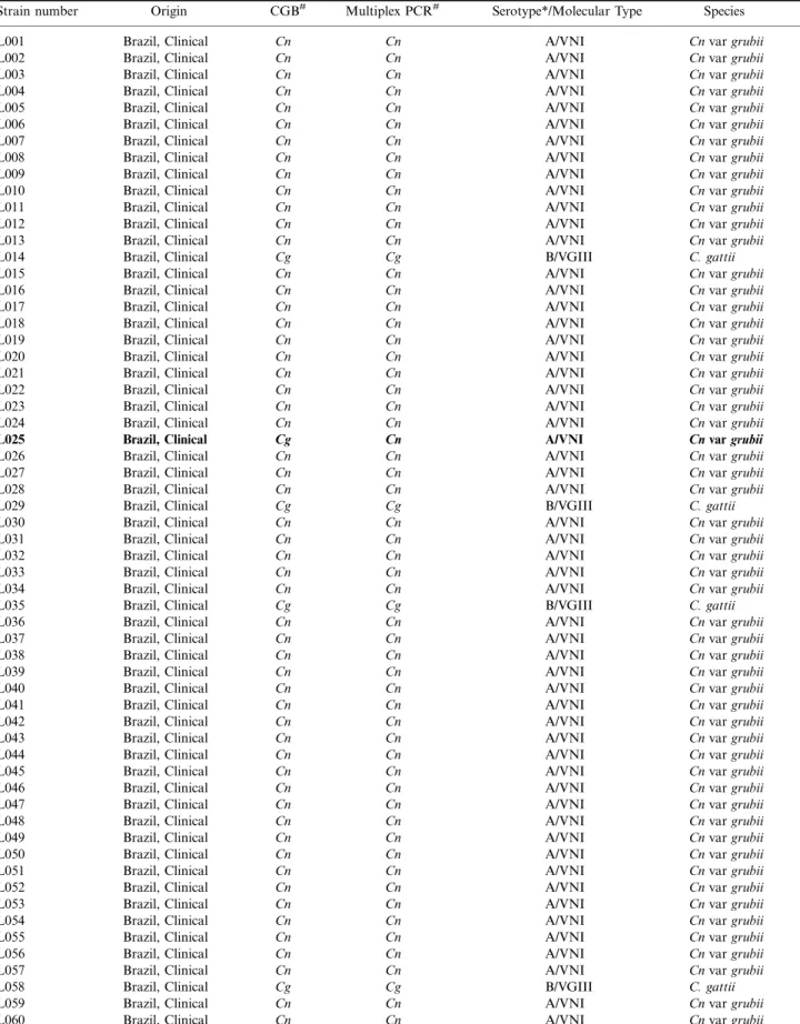
Table 1 Isolates used in this work.
Strain number Origin CGB# Multiplex PCR# Serotype*/Molecular Type Species L001 Brazil, Clinical Cn Cn A/VNI Cnvargrubii
L002 Brazil, Clinical Cn Cn A/VNI Cnvargrubii
L003 Brazil, Clinical Cn Cn A/VNI Cnvargrubii
L004 Brazil, Clinical Cn Cn A/VNI Cnvargrubii
L005 Brazil, Clinical Cn Cn A/VNI Cnvargrubii
L006 Brazil, Clinical Cn Cn A/VNI Cnvargrubii
L007 Brazil, Clinical Cn Cn A/VNI Cnvargrubii
L008 Brazil, Clinical Cn Cn A/VNI Cnvargrubii
L009 Brazil, Clinical Cn Cn A/VNI Cnvargrubii
L010 Brazil, Clinical Cn Cn A/VNI Cnvargrubii
L011 Brazil, Clinical Cn Cn A/VNI Cnvargrubii
L012 Brazil, Clinical Cn Cn A/VNI Cnvargrubii
L013 Brazil, Clinical Cn Cn A/VNI Cnvargrubii
L014 Brazil, Clinical Cg Cg B/VGIII C. gattii
L015 Brazil, Clinical Cn Cn A/VNI Cnvargrubii
L016 Brazil, Clinical Cn Cn A/VNI Cnvargrubii
L017 Brazil, Clinical Cn Cn A/VNI Cnvargrubii
L018 Brazil, Clinical Cn Cn A/VNI Cnvargrubii
L019 Brazil, Clinical Cn Cn A/VNI Cnvargrubii
L020 Brazil, Clinical Cn Cn A/VNI Cnvargrubii
L021 Brazil, Clinical Cn Cn A/VNI Cnvargrubii
L022 Brazil, Clinical Cn Cn A/VNI Cnvargrubii
L023 Brazil, Clinical Cn Cn A/VNI Cnvargrubii
L024 Brazil, Clinical Cn Cn A/VNI Cnvargrubii
L025 Brazil, Clinical Cg Cn A/VNI Cnvargrubii
L026 Brazil, Clinical Cn Cn A/VNI Cnvargrubii
L027 Brazil, Clinical Cn Cn A/VNI Cnvargrubii
L028 Brazil, Clinical Cn Cn A/VNI Cnvargrubii
L029 Brazil, Clinical Cg Cg B/VGIII C. gattii
L030 Brazil, Clinical Cn Cn A/VNI Cnvargrubii
L031 Brazil, Clinical Cn Cn A/VNI Cnvargrubii
L032 Brazil, Clinical Cn Cn A/VNI Cnvargrubii
L033 Brazil, Clinical Cn Cn A/VNI Cnvargrubii
L034 Brazil, Clinical Cn Cn A/VNI Cnvargrubii
L035 Brazil, Clinical Cg Cg B/VGIII C. gattii
L036 Brazil, Clinical Cn Cn A/VNI Cnvargrubii
L037 Brazil, Clinical Cn Cn A/VNI Cnvargrubii
L038 Brazil, Clinical Cn Cn A/VNI Cnvargrubii
L039 Brazil, Clinical Cn Cn A/VNI Cnvargrubii
L040 Brazil, Clinical Cn Cn A/VNI Cnvargrubii
L041 Brazil, Clinical Cn Cn A/VNI Cnvargrubii
L042 Brazil, Clinical Cn Cn A/VNI Cnvargrubii
L043 Brazil, Clinical Cn Cn A/VNI Cnvargrubii
L044 Brazil, Clinical Cn Cn A/VNI Cnvargrubii
L045 Brazil, Clinical Cn Cn A/VNI Cnvargrubii
L046 Brazil, Clinical Cn Cn A/VNI Cnvargrubii
L047 Brazil, Clinical Cn Cn A/VNI Cnvargrubii
L048 Brazil, Clinical Cn Cn A/VNI Cnvargrubii
L049 Brazil, Clinical Cn Cn A/VNI Cnvargrubii
L050 Brazil, Clinical Cn Cn A/VNI Cnvargrubii
L051 Brazil, Clinical Cn Cn A/VNI Cnvargrubii
L052 Brazil, Clinical Cn Cn A/VNI Cnvargrubii
L053 Brazil, Clinical Cn Cn A/VNI Cnvargrubii
L054 Brazil, Clinical Cn Cn A/VNI Cnvargrubii
L055 Brazil, Clinical Cn Cn A/VNI Cnvargrubii
L056 Brazil, Clinical Cn Cn A/VNI Cnvargrubii
L057 Brazil, Clinical Cn Cn A/VNI Cnvargrubii
L058 Brazil, Clinical Cg Cg B/VGIII C. gattii
L059 Brazil, Clinical Cn Cn A/VNI Cnvargrubii
L060 Brazil, Clinical Cn Cn A/VNI Cnvargrubii
Table 1(Continued)
Strain number Origin CGB# Multiplex PCR# Serotype*/Molecular Type Species
L061 Brazil, Clinical Cn Cn A/VNI Cnvargrubii
L062 Brazil, Clinical Cn Cn A/VNI Cnvargrubii
L063 Brazil, Clinical Cn Cn A/VNI Cnvargrubii
L064 Brazil, Clinical Cn Cn A/VNI Cnvargrubii
L065 Brazil, Clinical Cn Cn A/VNI Cnvargrubii
L066 Brazil, Clinical Cn Cn A/VNI Cnvargrubii
L067 Brazil, Clinical Cn Cn A/VNI Cnvargrubii
L068 Brazil, Clinical Cn Cn A/VNI Cnvargrubii
L069 Brazil, Clinical Cn Cn A/VNI Cnvargrubii
L070 Brazil, Clinical Cn Cn A/VNI Cnvargrubii
L071 Brazil, Clinical Cn Cn A/VNI Cnvargrubii
L072 Brazil, Clinical Cn Cn A/VNI Cnvargrubii
L073 Brazil, Clinical Cn Cn A/VNI Cnvargrubii
L074 Brazil, Clinical Cg Cn A/VNI Cnvargrubii
L075 Brazil, Clinical Cg Cn A/VNI Cnvargrubii
L076 Brazil, Clinical Cg Cn A/VNI Cnvargrubii
L077 Brazil, Clinical Cg Cn A/VNI Cnvargrubii
L078 Brazil, Clinical Cn Cn A/VNI Cnvargrubii
L079 Brazil, Clinical Cn Cn A/VNI Cnvargrubii
L080 Brazil, Clinical Cn Cn A/VNI Cnvargrubii
L081 Brazil, Clinical Cn Cn A/VNI Cnvargrubii
L082 Brazil, Clinical Cn Cn A/VNI Cnvargrubii
L083 Brazil, Clinical Cn Cn A/VNI Cnvargrubii
L084 Brazil, Clinical Cn Cn A/VNI Cnvargrubii
L085 Brazil, Clinical Cn Cn A/VNI Cnvargrubii
L086 Brazil, Clinical Cn Cn A/VNI Cnvargrubii
L087 Brazil, Clinical Cn Cg B/VGIII C. gattii
L088 Brazil, Clinical Cn Cn A/VNI Cnvargrubii
L089 Brazil, Clinical Cn Cn A/VNI Cnvargrubii
L090 Brazil, Clinical Cn Cn A/VNI Cnvargrubii
L091 Brazil, Clinical Cn Cn A/VNI Cnvargrubii
L092 Brazil, Clinical Cn Cn A/VNI Cnvargrubii
L093 Brazil, Clinical Cn Cn A/VNI Cnvargrubii
L094 Brazil, Clinical Cn Cn A/VNI Cnvargrubii
L095 Brazil, Clinical Cn Cn A/VNI Cnvargrubii
L096 Brazil, Clinical Cn Cn A/VNI Cnvargrubii
L097 Brazil, Clinical Cn Cn A/VNI Cnvargrubii
L098 Brazil, Clinical Cn Cn A/VNI Cnvargrubii
L099 Brazil, Clinical Cn Cn A/VNI Cnvargrubii
L100 Brazil, Clinical Cn Cn A/VNI Cnvargrubii
L101 Brazil, Clinical Cn Cn A/VNI Cnvargrubii
L102 Brazil, Clinical Cn Cn A/VNI Cnvargrubii
L103 Brazil, Clinical Cn Cn A/VNI Cnvargrubii
L104 Brazil, Clinical Cn Cn A/VNI Cnvargrubii
L105 Brazil, Clinical Cn Cn A/VNI Cnvargrubii
L106 Brazil, Clinical Cn Cn A/VNI Cnvargrubii
L107 Brazil, Clinical Cn Cn A/VNI Cnvargrubii
L108 Brazil, Clinical Cn Cn A/VNI Cnvargrubii
L109 Brazil, Clinical Cn Cn A/VNI Cnvargrubii
L110 Brazil, Clinical Cn Cn A/VNI Cnvargrubii
L111 Brazil, Clinical Cn Cn A/VNI Cnvargrubii
L112 Brazil, Clinical Cn Cn A/VNI Cnvargrubii
L113 Brazil, Clinical Cn Cn A/VNI Cnvargrubii
L114 Brazil, Clinical Cn Cn A/VNI Cnvargrubii
L115 Brazil, Clinical Cn Cn A/VNI Cnvargrubii
L116 Brazil, Clinical Cn Cn A/VNI Cnvargrubii
L117 Brazil, Clinical Cn Cn A/VNI Cnvargrubii
L118 Brazil, Clinical Cn Cn A/VNI Cnvargrubii
L119 Brazil, Clinical Cn Cn A/VNI Cnvargrubii
L120 Brazil, Clinical Cn Cn A/VNI Cnvargrubii
RFLP analysis
Digestion with the restriction endonuclease
Eco
RI
resulted in fragments of 447 and 248 bp for
C.
neoformans
and 324 and 124 bp for
C. gattii
(Fig. 3).
These results confirmed the specificity of the amplified
fragments for the respective species.
Discussion
The CGB test has become a traditional method to
differentiate
C. neoformans
from
C. gattii
ever since it
was proposed in 1982 [9,10,22]. One problem with this
test is the possibility of erroneous results, creating
non-reliable diagnostic data in laboratories where no
other identification methods are available. Moreover,
false positive reactions have been reported, suggesting
that CGB alone is not sufficient to accurately
discriminate between the two species [13,14]. The
results of the CGB test in the current work were
found to be 95.4% in agreement (125 out of 131
samples) with the serotyping data. Five false positive
and one false negative were noted, confirming the
need for other procedures to obtain a trustworthy
identification.
Table 1(Continued)
Strain number Origin CGB# Multiplex PCR# Serotype*/Molecular Type Species
L121 Brazil, Clinical Cn Cn A/VNI Cnvargrubii
L122 Brazil, Clinical Cn Cn A/VNI Cnvargrubii
L123 Brazil, Clinical Cn Cn A/VNI Cnvargrubii
L124 Brazil, Clinical Cn Cn A/VNI Cnvargrubii
L125 Brazil, Clinical Cn Cn A/VNI Cnvargrubii
L126 Brazil, Clinical Cn Cn A/VNI Cnvargrubii
L127 Brazil, Clinical Cg Cg B/VGIII C. gattii
L128 Brazil, Clinical Cn Cn A/VNI Cnvargrubii
L129 Brazil, Clinical Cn Cn A/VNI Cnvargrubii
L130 Brazil, Clinical Cn Cn A/VNI Cnvargrubii
L131 Brazil, Clinical Cn Cn A/VNI Cnvargrubii
AL6 Brazil, Clinical Cg Cg B/VGIII C. gattii
AL10 Brazil, Clinical Cg Cg B/VGIII C. gattii
AL13 Brazil, Clinical Cg Cg B/VGIII C. gattii
AL14 Brazil, Clinical Cg Cg B/VGIII C. gattii
AL32 Brazil, Clinical Cg Cg B/VGIII C. gattii
AL33 Brazil, Clinical Cg Cg B/VGIII C. gattii
AL34 Brazil, Clinical Cg Cg B/VGIII C. gattii
HSL3 Brazil, Clinical Cg Cg B/VGIII C. gattii
C5 Brazil, Clinical Cg Cg B/VGIII C. gattii
C43 Brazil, Clinical Cg Cg B/VGIII C. gattii
WM179 Australia, Clinical Cg Cg B/VGI C. gattii
WM178 Australia, Clinical Cg Cg B/VGII C. gattii
WM175 Australia, Clinical Cg Cg B/VGIII C. gattii
WM779 South Africa, Veterinary Cg Cg C/VGIV C. gattii
WM148 Australia, Clinical Cn Cn A/VNI Cnvargrubii
WM626 Australia, Clinical Cn Cn A/VNII Cnvargrubii
WM628 Australia, Clinical Cn Cn AD/VNIII Cnhybrid AD ATCC28957 Cn Cn D/VNIV Cnvarneoformans
LA1 Mexico, Clinical Cg Cg B/VGI C. gattii
AV55 Greece, Clinical Cg Cg B/VGII C. gattii
MC-S-239 Thailand, Clinical Cg Cg B/VGII C. gattii
CN043 New Zealand, Clinical Cg Cg C/VGIII C. gattii
WM728 USA, Environmental Cg Cg B/VGIII C. gattii
LA290 Paraguay, Clinical Cg Cg -/VGIII C. gattii
B5748 India, Clinical Cg Cg B/VGIV C. gattii
LA568 Columbia, Clinical Cg Cg B/VGIV C. gattii
LA390 Mexico, Clinical Cg Cg -/VGIV C. gattii
R265 Canada, Clinical Cg Cg B/VGII C. gattii
R272 Canada, Clinical Cg Cg B/VGII C. gattii
#CnC. neoformans;CgC. gattii.
Samples in bold indicates disagreements in identification among the tests used. *Crypto Check serotyping kit (Iatron Labs, Tokyo, Japan).
The proposed multiplex PCR is a simple, fast and
species-specific test, which did not amplify DNA from
other microorganisms or humans, and showed
poten-tial applicability for correctly identifying
Cryptococcus
species within the tested samples. Serotyping using the
Crypto Check kit was used as a gold standard in this
study. The multiplex PCR identifications were in 100%
agreement with those obtained through serotyping. The
PCR was more accurate than the CGB test in six
(4.58%) of the samples, proving it to be an efficient
method. Since the Crypto Check kit is no longer
produced and no similar kits are commercially available
there is a rising need for the development of new
methods for use in clinical diagnostic laboratory to
identify the two
Cryptococcus
species which are more
reliable than the CGB test alone. It is also important to
point out that the application of PCR is about six days
faster than that of the CGB test. Speed is important
due to the severity of illness and urgency in beginning
the treatment as early as possible. The results suggest
that the multiplex PCR is more reliable and faster than
the CGB test, providing a trustworthy species
identifi-cation which can be used as a complementary method
to confirm the CGB results and avoiding possible
diagnostic mistakes caused by false positive and
negative which may occasionally be generated by the
CGB test. Moreover, the multiplex PCR proved to be
species-specific and is sufficiently sensitive to be applied
in clinical laboratories.
Acknowledgements
This work was supported by Conselho Nacional de
Desenvolvimento Cientı´fico e Tecnolo´gico (CNPq),
Fig. 1 Cryptococcus species identification by multiplex PCR. Agarose gel electrophoresis of products amplified by the multiplex PCR using the primer pairs CNA70A CNA70S and CNB49A
CNB49S. (A) Lanes 1 to 3,Cryptococcus neoformansvar.grubiiand lanes 4 to 6, Cryptococcus gattii; lane 7, negative control. (B) Amplification of the major molecular types of this species complex as identified byURA5-RFLP and PCR-fingerprinting [23]. Lanes 1 to 8, WM148 (VNI, serotype A), WM626 (VNII, serotype A), WM628 (VNIII, serotype AD) and ATCC28957 (VNIV, serotype D), WM179 (VGI, serotype B), WM178 (VGII, serotype B), WM175 (VGIII, serotype B) and WM779 (VGIV, serotype C). The DNA marker (100bp DNA ladder, Invitrogen) is indicated on the left side (M).
Fig. 2 Specificity test of the multiplex PCR. Agarose gel electro-phoresis of products amplified by the multiplex PCR using the primer pairs CNA70ACNA70S and CNB49ACNB49S from different microorganisms and human. Cryptococcus neoformans var. grubii
(lane 1) andCryptococcus gattii(lane 2) were used as internal control. (A) Lanes (3)S. aureus, (4)M. tuberculosis, (5)C. albicans, (6)E. coli, (7)N. meningitidis, (8) human DNA, (9) negative control. (B) Lanes (3) C. dubliniensis, (4) C. parapsilosis, (5) C. tropicalis, (6) C. guilliermondii, (7) C. krusei, (8) S. schenckii, (9) R. mucilaginosa, (10) C. luteolus, (11) negative control. The DNA marker (100 bp DNA ladder, Invitrogen) is indicated on the left side (M).
Fig. 3 RFLP analysis of multiplex PCR products. Agarose gel electrophoresis of products digested with the restriction endonuclease
EcoRI. Lanes 1, 3 and 5, Cryptococcus neoformans var. grubii,
Cryptococcus neoformans var. neoformans and Cryptococcus gattii
PCR products, respectively; lanes 2, 4 and 6, EcoRI digested
Cryptococcus neoformansvar.grubii, Cryptococcus neoformansvar.
neoformansandCryptococcus gattiiPCR products, respectively. The DNA marker (100 bp DNA ladder, Invitrogen) is indicated on the left side (M).
Fundac¸a˜o Estadual de Produc¸a˜o e Pesquisa em Sau´de
(FEPPS) and Fundac¸a˜o de Amparo a Pesquisa do
Estado do Rio Grande do Sul (FAPERGS). The
authors wish to thank Dr Irene Schrank for critically
contributing to this work and Dr Wieland Meyer
(Molecular Mycology Research Laboratory, The
Uni-versity of Sydney at Westmead Hospital, Sydney,
Australia) for providing the global strains.
References
1 Idnurm A, Bahn YS, Nielsen K,et al. Deciphering the model pathogenic fungusCryptococcus neoformans. Nat Rev Microbiol
2005;3: 753764.
2 Kwon-chung KJ, Boekout T, Fell JW, Diaz M. Proposal to conserve the nameCryptococcus gattiagainstC. hondurianusand
C. bacillisporus.Taxon2002;51: 804806.
3 Kidd SE, Bach PJ, Hingston AO, et al. Cryptococcus gattii
dispersal mechanism, British Columbia, Canada. Emerg Infect Dis2007;13: 5157.
4 MacDougall L, Fyfe M. Emergence ofCryptococcus gattiiin a novel environment provides clues to its incubation period.J Clin Microbiol2006;44: 18511852.
5 Kidd SE, Hagen F, Tscharke RL, et al. A rare genotype of
Cryptococcus gattii caused the cryptococcosis outbreak on Vancouver Island (British Columbia, Canada). Proc Natl Acad Sci USA2004;101: 1725817263.
6 Stephen C. Multispecies outbreak of cryptococcosis on southern Vancouver Island, British Columbia.Can Vet J2002;43: 792794. 7 Sorrel TC. Cryptococcus neoformans varietygattii. Med Mycol
2001;39: 155168.
8 Trilles L, Fernandez-Torres B, Lazera MR, Wanke B, Guarro J.In
vitro antifungal susceptibility of Cryptococcus gattii. J Clin Microbiol2004;42: 48154817.
9 Kwon-Chung KJ, Polacheck I, Bennett J. Improved diagnostic medium for separation ofCryptococcus neoformans var. neofor-mans(serotypes A and D) andCryptococcus neoformansvar.gattii
(serotypes B and C).J Clin Microbiol1982;15: 535537. 10 Abegg MA, Cella FL, Faganello J,et al.Cryptococcus neoformans
andCryptococcus gattiiisolated from the excreta of Psittaciformes in a southern brazilian zoological garden.Mycopathologia2006;
161: 8391.
11 Horta JA, Staats CC, Casali AK,et al. Epidemiological aspects of clinical and environmental Cryptococcus neoformans isolates in
the Brazilian state Rio Grande do Sul.Med Mycol2002;40: 565
571.
12 Canelo C, Navarro A, Guevara M,et al. Determinacio´n de la variedad de cepas de Cryptococcus neoformans aisladas de pacientes con SIDA.RevMed Exp1999;15: 4447.
13 Nakamura Y, Kano R, Sato H,et al. Isolates ofCryptococcus neoformansserotype A and D developed on canavanine-glycine-bromothymol blue medium.Mycoses1998;41: 3540.
14 Khan ZU, Al-Alnezi AA, Chandy R, Xu J. Disseminated cryptococcosis in an AIDS patient caused by a canavanine-resistant strain of Cryptococcus neoformans var. grubii. J Med Microbiol2003;52: 271275.
15 Aoki FH, Amai T, Tanaka R,et al. New primer pairs specific for
Cryptococcus neoformansserotype A or B prepared on the basis of random amplified polymorphic DNA fingerprinting pattern analyses.J Clin Microbiol1999;37: 315320.
16 Casali AK, Goulart L, Rosa e Silva LK,et al. Molecular typing of clinical and environmental Cryptococcus neoformans isolates in the Brazilian state Rio Grande do Sul.FEMS Yeast Res2003;3: 405415.
17 Latouche GN, Huynh M, Sorrel TC, Meyer W. PCR-Restriction Fragment Length Polymorphism analysis of the phospholipase B (PLB1) gene for subtyping ofCryptococcus neoformansisolates.
Appl Environ Microbiol2003;69: 20802085.
18 Fraser JA, Giles SS, Wenink EC,et al. Same-sex mating and the origin of the Vancouver Island Cryptococcus gattii outbreak.
Nature2005;437: 13601364.
19 Diaz MR, Boekhout T, Kiesling T, Fell JW. Comparative analysis of the intergenic spacer regions and population structure of the species complex of the pathogenic yeastCryptococcus neoformans.
FEMS Yeast Res2005;5: 11291140.
20 Kabasawa K, Itagaki H, Ikeda R, et al. Evaluation of a new method for identification ofCryptococcus neoformanswhich uses serologic tests aided by selected biological tests.J Clin Microbiol
1991;29: 28732876.
21 Zhang D, Yang Y, Castlebury LA, Cerniglia CE. A method for the large scale isolation of high transformation.FEMS Microbiol Lett
1996;145: 261265.
22 Chaturvedi S, Dyavaiah M, Larsen RA, Chaturvedi V. Crypto-coccus gattiiin AIDS patients, Southern California.Emerg Infect Dis2005;11: 16861692.
23 Meyer W, Castan˜eda A, Jackson S,et al. Molecular typing of IberoAmerican Cryptococcus neoformans isolates. Emerg Infect Dis2003;9: 189195.