ORIGINAL C O N T R I B U T I O N
asthma, magnesium; magnesium, asthma
Intravenous Magnesium for Acute Asthma:
Failure to Decrease Emergency Treatment Duration
or Need for Hospitalization
From the Departments of Emergency Medicine, Riverside General Hospital, Riverside, California, and Loma Linda Uaiversity Medical Center, Loma Linda, California; and California Emergency Physi- cians Medical Group, Oakland, California.
Received for publication April 24, 1991. Revision received August 21, 1991. Accepted for publication November 5, 1991.
Presented at the Scientific Assembly of the American College of Emergency Physicians in Boston, October 1991.
Steven M Green, MD Steven G Rothrock, MD
Study objective: To evaluate the efficacy of routine early administration of IV magnesium to patients with acute asthma. Design: Prospective, randomized clinical trial.
Setting: Urban teaching hospital emergency department.
Type of p a r t i c i p a n t s : One hundred twenty consecutive patients aged 18 to 65 years with acute asthma unresponsive to a single albuterol treatment.
I n t e r v e n t i o n s : All patients received oxygen, 125 mg IV
methylprednisolone, and hourly albuterol inhalation therapy. The study group also received 2 g IV magnesium sulfate infused over 20 minutes. Measurements and main results: Demographic and clinical characteristics were similar in both groups. Hospitalization was necessary in 13 of 58 patients who received magnesium (22%; 95% confidence intervals [01L 13% to 32%) and 11 of 62 control patients (17%; 95%01, 10% to 26%; P= .523). Duration of ED treatment in discharged patients was 224 + 75 minutes in the magnesium group (95% CI, 208 to 240 minutes) and 228 + 90 minutes in the control group (95% Cl, 209 to 247 minutes, P=. 832). In addition, changes in peak expiratory flow were not statistically different.
Conclusion: Routine early administration of IV magnesium in acute asthma does not alter treatment outcome. [Green SM, Rothrock SG: Intravenous magnesium for acute asthma: Failure to decrease emergency treatment duration or need for hospitalization. Ann Emerg Med March 1992;21:260-265.]
IV M A G N E S I U M Green & Rothrock
INTRODUCTION
Research regarding magnesium has undergone a renaissance in recent years, with striking reports of the ability of this ion to reduce mortality and dysrhythmias in acute myocardial infarc- tion, 1"4 to diminish digoxin cardiotoxicity, s-v and to pharmaco- logically convert ventricular tachycardia 7-9 and torsade de pointes. 7'1° Case reports 11-13 and limited series 14-19 have simi- larly described varying degrees of improvement in patients with acute asthma who were treated with IV magnesium.
In 1989, Skobeloff et al reported a placebo-controlled trial of 1.2 g magnesium IV in 38 patients with moderate-to-severe asthma they considered "refractory to inhaled albuterol. ''2° Significant improvement in peak expiratory flow rates as well as markedly lower hospital admission rates (37% vs 79%, P < .01) were noted in patients treated with IV magnesium.
These findings suggest that routine early administration of magnesium to patients with asthma might add to or synergize with conventional therapy (eg, oxygen, J3-agonists, and steroids). Accordingly, we performed a randomized clinical trial to com- pare the addition of magnesium with conventional therapy alone. The null hypothesis of this investigation was that the early addi- tion of magnesium would not diminish the duration of emergency department treatment or rate of hospitalization.
MATERIALS AND METHODS
Patients 18 to 65 years old who presented to our urban teaching hospital ED with an acute exacerbation of asthma were eligible for the study. The American Thoracic Society definition of asth- ma was used: "A disease characterized by an increased respon- siveness of the trachea and bronchi to various stimuli and mani- fested by a widespread narrowing of the airways that changes in severity either spontaneously or as a result of therapy. ''21 Patients with atheroselerotie heart disease, angina, chest pain, uncontrolled hypertension, congestive heart failure, heart block, metastatic cancer, renal disease, temperature above 38.3 C, sys- tolic blood pressure less than 120 mm Hg, or pregnancy (known or suspected) were excluded. Patients with radiographic evidence of confounding pulmonary disease (eg, pneumonia, pneumothorax, lung cancer, or congestive heart failure) or those requiring mechanical ventilation at any point during their ED visit or subsequent hospitalization also were excluded. Only the first visit for each individual patient was analyzed to avoid sampling unit confusion.
On arrival, all patients were treated with oxygen and inhaled albuterol. The latter was administered by respiratory therapists
either as a nebulized aerosol (0.5 mL in 2.5 mL saline) or through supervised inhalations of a metered-dose inhaler with spacer titrated to a therapeutic effective dose. If dyspnea and wheezing had not improved such that the patient could be dis- charged after this initial inhalation therapy, the patient received 125 mg IV methylprednisolone. Hourly albuterol treatments were repeated according to hospital protocol until the patient was either admitted or discharged. Consecutive patients lacking exclusion criteria and receiving IV methylprednisolone were entered into the study.
Patients presenting on odd days were given 2 g magnesium sulfate diluted in 50 mL DsW administered intravenously over 20 minutes. This infusion was begun immediately after methyl- prednisolone administration and within 45 minutes of treatment initiation. Patients presenting on even days did not receive mag- nesium. Peak expiratory flow measurements were obtained before and after each albuterol treatment by respiratory therapists using a Mini-Wright Peak Flow Meter (Armstrong Medical Industries, Inc, Lincolnshire, Illinois). Three attempts were made at each measurement, and the highest value was recorded. Physicians were not blinded to patient randomization; however, patients and respiratory therapists were unaware that a study was being performed.
Other therapy (eg, theophylline, injectable J3-agonists, or epinephrine) was left to the discretion of the treating physician. Theophylline was administered only after a subtherapeutic serum concentration was obtained. Serum magnesium levels were not obtained. All care was given or supervised by attending physi- cians board certified in emergency medicine.
Patients were discharged only if dyspnea was relieved and aus- cultation demonstrated either clear breath sounds or minimal wheezing. ED treatment duration for discharged patients was defined as the time from initial physician contact to actual patient sign-out. All patients were instructed to return to the study hospital if their condition worsened. Relapse was defined as a return visit to our ED within 72 hours of discharge. Major complications were defined as events requiring discontinuation of the magnesium infusion or other interventions for patient safe- ty (eg, hypotension, bradycardia). Minor reactions such as flush- ing sensations or malaise were not quantified. Radiographic chronic obstructive pulmonary disease (COPD) was defined as comments indicating emphysema or chronic bronchitis on the typed report of the most recent chest radiograph (either at the ED visit or within the prior two years). Initial peak expiratory flow was defined as the best of three attempts before the first albuterol inhalation treatment. Final peak expiratory flow was •
V MAGNESIUM ;reen& Rothrock
defined as the best of three attempts immediately after the final ED albuterol inhalation treatment. Absolute change in peak expiratory flow was defined as the final measurement minus the initial measurement. Relative change in peak expiratory flow was defined as the absolute change divided by the initial measure- ment and multiplied by 100.
Before the investigation, sample size calculations were performed assuming an c~ of .05 and a [3 of .20. 22 No prior stud- ies have indicated that magnesium worsens asthma, so a one- tailed statistical approach was used. Approximately 15% of asth-
Table 1,
Characteristics of study and control groups
Magnesium Control Characteristic (N = 58) (N = 62) Statistic P Demographics Age 40.9 (14.5%) 39.8 (14.8%1 t = ,023 .982 Sex Female 46 (79%) 46 (74%) X 2 = .439 .508 Male 12 (21%) 16 (26%1 Ethnicity White 34 (59%) 40 (64%) X z = 2.68 .443 Hispanic 19 {33%) 13 (21%) Black 3 (5%) 6 (10%) Oriental 2 (3%) 3 (5%)
Vital Signs and Clinical Information
Temperature (C) 36.£ ± 0.6 36.8 ± 0.7 t = /.067 288
Pulse 108 ± 20 107 _+ I7 t = .117 .907
Respirations 28 ± 7 29 ± 8 t = 1.198 .233
Sputum production 30 (52%) 29 (47%) X 2 = .2£4 .588 Symptom duration (hr] 32 ± 24 28 +_ 24 t = 259 .449 Medications at Time of Study Entry
Inhaled ~-agonist 49 {84%) 53 (65%) X 2 = .024 .878
inhaled anticholinergic 4 (7%) 2 (3%) Fisher's .428
Inhaled steroid 3 (5%) 1 {2%1 Fisher's .352
Home oxygen 0 (0%) 1 (2%1 Fisher's 1.000
Terbutaline 10 (17%) 9 (15%) X 2 = .167 .683
Theophylline 43 (74%) 37 (60%) x 2 = 2.82 .093
0ral steroid 14 (24%) 12 (19%) X 2 = AO4 .525
Diuretic 3 (5%) 3 (5%) Fisher's 1.000 Emergency Treatment Albuterel treatments 2.72 +_ 1.12 2.61 ± 1.03 t = .565 .573 Terbutaline SC 0.57 +- 0.70 0.63 + 0.79 t = .439 .661 Epinephrine SC 0.02 -+ 0.13 0.03 ± 0.18 t = .528 .599 Theophyl/ine IV (mg) 104 +_ 156 137 ± 177 t = 1.081 .262 Diagnostic Data* WBC count 10.9 ± 4.0 11.6 + 3.8 t = .658 .514
Theophylline level (rag/L) 8.8 ± 6.2 6.8 ± 5.9 t = 1.35 .180
Radiographic COPO 8 (16%1 8 (f6%1 X ~ = .621 .886
Hypoxia (Po 2 < 60) 1 (6%) 3 (17%) Fisher's .619 Hypercarbia (Pco2 > 46) 1 (6%) 4 (24%) Fisher's .366 *Not all diagnostic items were obtained in all patients. WBC counts were obtained in 24 magnesium and
22 control cases; thaophylline levels were ohtaioecl in 35 magnesium and 32 control cases; chest radiographs (at visit or within two years of visit) were available in 51 magnesium and 51 control cases; and arterial bqood gases were obtained in 17 magnesium and 18 control eases.
matic patients require hospital admission; 23"24 thus, to detect a 20% difference in hospital admission rates with 80% power, 57 patients each were required in the magnesium and control groups. 22 For patients with asthma who eventually are
discharged, the average El) treatment time is approximately four hours with an SD of one hour; 23"24 thus, to detect a 30-minute difference in ED treatment time with 80% powei, 49 patients each were required in the magnesimn and control groups, z2 Thus, a minimum of 57 patients were required in each group.
Statistical comparisons were made using the unpaired t test, Z 2 analysis, and Fisher's exact test. Analyses were performed with statistical software (SYSTAT 5 . 0 , Systat, Inc, Evanston, Illinois). P < .05 was considered significant. All confidence intervals (CI) were 95%. No corrections were made for multiple comparisons, as the exact comparisons to be made were predetermined. The study was approved by the hospital institutional review board.
RESULTS
Two hundred seventeen consecutive patient visits met study entry criteria between March 29, 1990, and March 21, 1991, and were enrolled. Eighty of these visits were repeats and were excluded; only the first visit for eaeh patient was included in the data analysis to avoid sampling unit confusion. Seventeen other patients were removed from data analysis because of misplaced medical records (two), missing peak expiratory flow data (seven), respiratory failure requiring mechanical ventilation (one), pneu- monia (four), lung cancer (one), or congestive heart failure (two). This left 120 patients for data analysis - - 58 in the magnesium group (48%) and 62 in the control group (52%). The characteris- tics of the study and eontrol groups are given; none was statisti- cally significant (Table 1).
Twenty-four of the 120 visits (20%) resulted in hospital admis- sion ('Fable 2). Hospitalization was necessary in 22% of the mag- nesium group (95% CI, 13% to 32%) and 17% of the control group (95% CI, 10% to 26%; P = .523). There was no significant difference between mean ED treatment time in discharged mag- nesium patients (224 + 75 minutes; 95% CI, 208 to 240 minutes) and discharged control patients (228 +_ 90 minutes; 95% CI, 209 to 247 minutes; P = .832). Absolute change in peak expiratory flow was 122 + 75 L/rain in the magnesium group (95% CI, 106 to 138 L/rain) and 13.3 4-- 82 L/rain in the control group (95% CI, 116 to ]50 L/min; P = .419); relative change in peak expiratory flow was 103 _+ 91% in the magnesium group (95% CI, 83% to 123%) and 123 +_ 26% in the control group (9.5% CI, 101% to 145%; P = .272). ~.
I V M A G N E S I U M Green & Rothrock
The relapse rate for discharged patients and the length of hos- pitalization for admitted patients also were not significantly dif- ferent, although the limited numbers of patients in each of these categories limit the power of these comparisons.
No major complications were noted at any time. Minor compli- cations such as flushing sensations occurred occasionally but were not quantified.
D I S C U S S I O N
The therapy of greatest efficacy fox acute asthma exacerbations is clearly oxygen and inhaled [~-agonists. Early administration of methylprednisolone appears to be a helpful adjunct, 23'25 although this is disputed, z4,26 Theophylline administration remains ubiquitous despite frequent toxicity and ample evidence of marginal efficacy, z7-29 If beneficial for asthma, magnesium therapy would have tremendous appeal due to its widespread availability, low cost, and minimal adverse effects.
Magnesium has been postulated to produce bronehodilation through the counteraction of calcium-mediated smooth muscle constriction. 13'14'16'18'3° Magnesium depletion occurs in periods of adrenergic excess such as asthma exacerbations, as well as from diuretic use and alcohol a b u s e ) ' n Hypermagnesemia is rare in the absence of impaired renal function. 31
A wide range of magnesium doses has been used for asthma, with reported amounts varying from 1.2 to 2.5 g IV over 20 min- utes. 14"19'2° Two grams (8.1 retool) has been described as a safe dose with minimal side effects, 1A9'31 so it was chosen for our investigation. This quantity raises the serum magnesium level to approximately twice the original level with equilibration into the intracellular space occurring over approximately 24 hours. 14
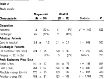
Table 2, Study results Magnesium Control Characteristic (N = 58) (N = 62) Statistic P Disposition Admitted 13 (22%) 11 (18%) X 2 = .409 .523 Discharged 45 (78%) 51 (82%) Admitted Patients Days in hospital 2.4 ± 1.5 2.1 ± 0.7 t = .648 .525 Discharged Patients
ED treatment time (min) 224 + 75 228 ± 90 t = .212 .832 Relapse < 72 hr (%) 1 (2%) 5 (8%) Fisher's .208 Peak Expiratory Flow Data
Initial (L/min) 141 ± 77 144 ± 79 t = .198 .844 Final (Umin) 263 _+ 122 278 '-+ 104 t = ,699 .486 Absolute change (L/rain) 122 + 75 133 ± 82 t - .811 .419 Relative change (%) 103 ± 91 123 + 102 t = 1.104 .272
Side effects of magnesium infusions are mild and include tran- sient sensations of facial warmth, flushing, or malaise; significant adverse events have not been repotted. 1"14"15'19"2° Alterations of pulse and blood pressure are not seen when magnesium is given as an infusion; 14'15'19'2°'32 however, hypotension and bradycar- dia can occur with rapid IV administration. 33 At extremely high serum levels (5 to 7.5 retool/L, 10 to 15 mEq/L), absent reflexes, muscle weakness, respiratory depression, and cardiac conduction abnormalities c a n o c c u r . 1 4 " 1 5 ' 3 1 ' 3 3 However, to produce these
levels, doses of approximately 15 g or more are necessary. 33 Treatment of acute exacerbations of asthma with parenteral magnesium was first described in the late 1930s. 11A2 Recent case reports and limited series have revived interest in this ther- apy; 13"16-19 in one instance, magnesium was thought to obviate the need for intubation) 3 In a small series of ten mild asthmat- ics, Okayama et al noted that 2.5 g IV magnesium improved peak expiratory flow rates in a dose-dependent manner with concur- rent relief of d y s p n e a ) 4 Unfortunately, no controls were used. In a second uncontrolled study of six inpatient asthmatics, Noppen et al noted mild improvements in pulmonary function testing after magnesium infusion. 15
In 1989, Skobeloff et al reported the only controlled trial of magnesium for asthma other than the current report. 2° In their study, patients with moderate-to-severe asthma considered "refractory" to inhaled albuterol (ie, asthmatics unable to double their peak expiratory flow measurements after two inhaled albuterol treatments and IV methylprednisolone) were given 1.2 g IV magnesium in a randomized double-blind manner. Significant improvement in peak expiratory flow rates was noted in the treatment group as well as a markedly lower rate of hospi- tal admission (seven of 19 in the treatment group [37%] vs 15 of 19 in the control group [79%], P < .01).
Unfortunately, there are several limitations to this report. The number of patients in this investigation was small (38), and no sample size calculations or other rationale for early study temai- nation are given. The authors do not provide data comparing the number of total ~-agonist inhalation treatments each group received, making it unclear Whether one group received more of this fundamental therapy. In addition, patients in the placebo group had lower initial peak expiratory flow rates, suggesting that the control group might have comprised "sicker" patients with a pre-existing higher likelihood of admission or poor response to [3-agonists. Finally, the admission rate of the control group (79%) is surprisingly high, raising doubts about the applicability of these findings to other ED settings. ~.
IV M A G N E S I U M Green & Rothrock
We have been unable to demonstrate any benefit from the rou- tine early administration of 2 g IV magnesium sulfate in our gen- eral asthmatic population, contradicting the findings of Skobeloff et al, who used a lower dose of 1.2 g. Several possible reasons for this contradiction are hypothesized; most are related to differ- ences in the patient population studied.
Skobeloff et al included only patients poorly responsive to two inhalational treatments, whereas we investigated those poorly responsive to a single treatment. It is possible that our "healthi- er" patients were more responsive to ]3-agonist therapy and that this may have overshadowed a smaller bronchodilatory effect of magnesium. Skobeloff et al enrolled only 38 patients over a seven-month period in a major urban ED, suggesting that their "poor responders" comprised a quite limited subset of all avail- able asthmatics. The patients in our investigation, however, were consecutive and therefore a more reliable cross section of the general asthmatic population. Skobeloff et al used an average of three peak expiratory flow attempts at each measurement, where- as we reported the best of three. Peak expiratory flow assessment is highly dependent on patient effort, and we feel that our method better depicts true ventilatory function.
Our population also received magnesium therapy earlier than that of Skobeloff et al; one might speculate that the bronchodila- tion of magnesium is transient and had diminished by the time our disposition decisions and final peak expiratory flow measure- ments were made. This is supported by the results of Rolla et al, 19 who noted that the bronchodilation resulting from 2 g IV magnesium had disappeared 90 minutes after the infusion. If the effect of magnesium is short lived, then the "late" use of magne- sium to improve patients "refractory" to ~-agonists as advocated by Skobeloff et al 2° is fraught with hazard, as rebound broncho- spasm might result soon after discharge.
We chose to use the definition of asthma put forth by the American Thoracic Society 21 because of its widespread use in studies regarding asthma. 14,2°oa4 A clear distinction between COPD and asthma cannot always be made, and the society's defi- nition includes patients with elements of COPD. We chose to compare the relative proportions of COPD within our study (as defined by radiographic criteria) instead of attempting to exclude these patients through arbitrary criteria. Radiographic COPD was noted to be present in 16% of both treatment and control groups (Table 1). An age cutoff of 18 to 65 years was considered appropriate to avoid potential confounding effects of pediatric and geriatric populations.
We chose to use relief of dyspnea and auscuhatory findings of either clear breath sounds or minimal wheezing as our criteria for
ED discharge. This end point is that most commonly used by practicing physicians and most often described in clinical trials
of a s t h m a . 2°'23-27 An alternative criterion based on peak expira- tory flow assessment was rejected, as these measurements often val~¢ greatly with patient cooperation and effort.
A limitation of our investigation is that it was not blinded to physicians. Because outcome was measured by relatively objec- tive criteria (ie, duration of treatment, hospitalization rate, peak expiratory flow), we postulate that potential physician biases would have limited impact, if any, on the results. In our opinion, the improved study validity inherent to a consecutive series outweighed the potential influence of physician knowledge, Physicians were enthusiastic about magnesium, and we postulate that any bias, if present, would favor magnesium. If physicians discharged magnesium patients sooner or more frequently than control patients, our results do not indicate that this difference was significant.
Patients were unaware that data on their treatment were being collected and rarely asked what medication was being adminis- tered. Patient blinding was thought to be ethically acceptable due to the extreme safety of magnesium and the widespread use of both treatment regimens within our hospital and others. Respiratory therapists also were unaware that a study was being conducted; they routinely performed peak expiratory flow mea- surements before and after each inhalational treatment according to hospital protocol. Thus, measurements of peak expiratory flow were essentially double-blinded to patient and respiratory therapist.
Our method of calculating relapses included only return visits to our hospital, potentially underestimating actual relapse rates. We believe this limitation to be minor, as for financial reasons our largely indigent population has no alternative facility at which to seek care. We observed only one relapse in the magne- sium group compared with five relapses in the control group. This trend generates speculation that magnesium might exert a sustained bronchodilatory effect after discharge with subsequent protection against relapse. Our sample size was inadequate to assess this hypothesis; based on a control relapse rate of 8%, we calculate that a total sample of more than 700 patients would be necessary to detect a 4% decrease in relapse rate with 80%
power. 22
We allowed albuterol inhalational therapy to be performed either as a traditional nebulization or a metered-dose inhaler with spacer supervised by a respiratory- therapist. The decision as to which of these modalities would be used for each treatment was made by a blinded therapist with occasional input ~,
iV M A G N E S I U M Green, & Rothrock
from physicians. Supervised use of a metered-dose inhaler has been shown to be as effective as traditional nebulization. 35
Based on prospective sample size calculations, this study had 80% power to detect a 20% difference in admission rates and a 30-minute difference in ED treatment times. Sample size calcu- lations were not performed before study initiation on peak expi- ratory flow parameters; however, if study data are applied retro- spectively, the sample size used would have 80% power to detect a difference in absolute improvement of 37 L/min and relative improvement of 46% .22
CONCLUSION
A prospective, randomized clinical trial was conducted to evalu- ate the efficacy of routine early administration of IV magnesium to patients with acute asthma. No significant differences were noted in rate of hospitalization, peak expiratory flow measure- ments, or ED treatment time for discharged patients. We conclude that routine early administration of IV magnesium to ED patients with acute asthma does not alter treatment outcome. •
The authors thank Tim Nesper, MD; John Naflel, MD; Victor Levine, MD; David Joss, MD; and Tami Thomas, MD, for their participation and enthusiasm and the nurses and respiratory therapists of Riverside General Hospital for their help in carry- ing out the
study.
REFERENCES
1. Abraham AS, Rosenmann D, Kramer M, et ah Magnesium in the prevention of lethal arrhythmias in acute myocardial infarction. Arch Intern Mad 1987;147:753-755.
2. Rasmussen HS, Norregard P, Lindeneg O, et ah Intravenous magnesium in acute myocardial infarction. Lancet 1986;1:234-236.
3. Rasmussen HS: Magnesium infusion reduces the incidence of arrhythmias in acute myocardial infarction. Clin Cordial 1987;10:351.
4. Shechter M: Beneficial effect of magnesium sulfate in acute myocardial infarction. Am J Cardiot 1990;66:271.
5. French JH, Thomas RG, Siskind AP, et at: Magnesium therapy in massive digoxin intoxication.
Ann Emerg Mef11984;13:562-566.
6. Green SM, Naftel J: Antiarrhythmic efficacy of magnesium in the setting of life-threatening digoxin toxicity. Am J Emerg Med 1989;7:347-348.
7. Iseri LT: Magnesium and cardiac arrhythmias. Magnesium 1986;5:111-126.
8. Iseri LT, Chung P, Tobis J: Magnesium therapy for intractable ventricular tachyarrhythmias in normomagnesemic patients. West J Med 1983;138:823-828.
9. Alien B J: Magnesium sulfate therapy for sustained monomorphic ventricular tachycardia. Arn J Cordial 1989;64:1202.
10, Tzivoni D, }(eren A, Cohen AM: Magnesium therapy for torsades de pointes. Am J Cordial
1964;53:528-530.
11. Haury VG: Blood serum magnesium in bronchial asthma and its treatment by the administration of magnesium sulfate. J Lab Clin Med 1940!26:340-341.
12. RoseIla JC, Pla JC: Magnesium sulfate in crisis of asthma. Prensa MedArgent 1936;23:1677. 13. McNamara RM, Spivey WH, Skobeloff E, et ah Intravenous magnesium sulfate in the
management of acute respiratory failure complicating asthma. Ann Emerg Mad 1989;2:197-199. 14. Okayama H, Aikawa T, Okayama M, et ah Bronchodilating effect of intravenous magnesium sulfate in bronchial asthma. JAMA 1987;257:1876-1078.
15. Noppen M, Vanmaele L, Impens N, et ah Bronchodilating effect of intravenous magnesium sulfate in acute severe bronchial asthma. Chest 1990;97:373-376.
16. Rolla G, Bucca C, Carla E: Dose-related effect of inhaled magnesium sulfate on histamine bronchial challenge in asthmatics. Drugs Exp Clin Res 1988;14:609-6t2.
17. Ralla G, Bucca C, Bugiani M, et al: Reduction of histamine-induced bronchoconstriction by magnesium in asthmatic subjects. Aflergy1987;42:186-188.
18. Rolla G, Bucca C, Arossa W, et at: Magnesium a;tenuates methacholine-induced bronchoconstriction in asthmatics. Magnesium 1987;6:201-204.
19. Rella G, Bucca C, Coda E, et ah Acute effect of intravenous magnesium sulfate on airway obstruction of asthmatic patients. Ann Allergy 1988;61:388-391.
20. Skobeloff EM, Spivey WH, McNamara RM, et ah Intravenous magnesium sulfate for the treatment of acute asthma in the emergency department. JAMA 1989;262:1210-1213. 21. Committee on Diagnostic Standards for Nontuberculous Respiratory Diseases, American Thoracic Society: Chronic bronchitis, asthma, and pulmonary emphysema. Am Rev Respir Dis
1962;85:762-768.
22. Hulley SB, Cummings SR: Designing Clinical Research: An EpidemiologicalApprnach.
Baltimore, Williams & Wilkins, 1988, p 215-217.
23. Littenberg B, Gluck EH: A controlled trial of methylprednisolone in the emergency treatment of acute asthma. N Engl J Med 1986;3t4:150-152.
24. Stein LM, Cole RP: Early administration of corticosteroids in emergency room treatment of acute asthma. Ann Intern Meal 1990;112:822-827.
25. Murata GH, Gorby MS, Chick TW, et ah Intravenous and oral corticosteroids for the prevention of relapse after treatment of decompensated COPD. Chest 1990;98:845-849. 26. Emerman CL: A randomized controlled trial of methylprednisolone in the emergency treatment of acute exacerbations of COPD. Chest 1989;95:563.
27. Rice KL, Leatherman JW, Duane PG, et ah Aminophylline for acute exacerbations of chronic obstructive pulmonary disease. Ann Intern Med 1987;107:305-309.
28. Litternberg B: Aminophylline treatment in severe, acute asthma: A meta-analysis. JAMA
1988;259:1678-1684.
29. Lam A, Newhouse MT: Management of asthma and chronic airflow limitation: Are methylxanthines obsolete? Chest 1990;98:44-52.
30. Spivey WH, Skobeloff EM, Levin RM: Effect of magnesium cNoride on rabbit bronchial smooth muscle. Ann Emerg Mad 1990;19:1107-1112.
31. Goodman LS, Gilman A: The Pharmacological Basis of Therapeutics, ed 7. New York, Macmillan Publishing Co, 1985.
32. Mroczek W J, Lee WR, Davidov ME: Effect of magnesium sulfate on cardiovascular hemodynamics. Angiology 1977;28:720-724.
33. LyphoMed, Inc: Magnesium sulfate injection, product insert, 1984.
34. Chapman KR, Verbeek PR, White JG, et ah Effect of a short course of prednisone in the prevention of early relapse after the emergency room treatment of acute asthma. N Engl J Meal
1991 ;324:788-794.
35. Mestitz H, Copland JM, McDonald CF: Comparison of outpatient nebulized vs metered dose inhaler terbutaline in chronic outflow obstruction. Chest 1989;96:1237-1240.
Address for reprints: Steven M Green, MD, Department of Emergency Medicine, Riverside General Hospital, 9851 Magnolia Avenue, Riverside, California.