Virtual reality to improve lower extremity function, kinematic
parameters, and walking speed post-stroke: A case series.
Carlos Luque-Moreno
I.R.C.C.S. Fondazione Ospedale SanCamillo, Venezia, Italy Via Alberoni 70 30126 Venice Italy 0039 041 220 7111 [email protected]
Cleofás Rodríguez-Blanco
Departamento de Fisioterapia Universidad de Sevilla C/Avicena s/n. 41009 Sevilla Spain 0034 954486528 [email protected]Ángel Oliva-Pascual-Vaca
Departamento de Fisioterapia Universidad de Sevilla C/Avicena s/n. 41009 Sevilla Spain 0034 954486528 [email protected]Michela Agostini
I.R.C.C.S. Fondazione Ospedale San Camillo, Venezia, Italy
Via Alberoni 70 30126 Venice Italy 0039 041 220 7111
Pawel Kiper
I.R.C.C.S. Fondazione Ospedale San Camillo, Venezia, Italy
Via Alberoni 70 30126 Venice Italy 0039 041 220 7111
Andrea Turolla
I.R.C.C.S. Fondazione Ospedale San Camillo, Venezia, Italy
Via Alberoni 70 30126 Venice Italy 0039 041 220 7111
ABSTRACT
Introduction: Virtual reality (VR) is an innovative tool that can enrich physiotherapy treatment in individuals with stroke. The increased use of feedback provides them with useful additional information to improve walking speed, kinematics, and functionality of the lower limb. Our aim is to evaluate these changes in two individuals with stroke. Case description: A 58-year-old man (4.5 months post-stroke) and a 49-58-year-old man (3 months post-stroke) followed a virtual reality training to improve kinematics, functionality, and gait speed. Each participant underwent 15 sessions (three weeks) of conventional physiotherapy combined with training and virtual reality. Outcomes: The lower extremity Fugl-Meyer scale (FM) improved in both participants; in motor evaluation, participant 1 increased 4 points and patient 2 increased 6 points. Participant 1 was highly functional but had difficulty in the race at baseline, while participant 2 improved on the Ambulatory Functional Scale (FAC) from 3/5 to 4/5 and the Berg Balance Scale (BBS) from 50 to 53, with a constant permanent score of 122/126 on the Functional Independence Measure scale (FIM). Both participants improved the kinematic parameters in leg stance on plegic lower extremity and walking speed > Minimally Clinically Important Difference (MCID) (participant 1: improvement of 0.16m/s, participant 2: 0.34m/s). Discussion: Our results suggest that VR can contribute to improving kinematics and functionality which are linked to significant clinical increases in gait speed.
Categories and Subject Descriptors
I.2.1.[Applications and Expert Systems]: Medicine and Science. H.5.1.[Multimedia Information Systems]: Artificial, Augmented, and Virtual Reality.
General Terms
Experimentation.Keywords
Virtual reality, stroke, gait speed, feedback, physiotherapy.
1.
INTRODUCTION
The Virtual reality (VR) and interactive video games are an innovative therapy in the field of stroke rehabilitation. Treatment includes the use of computer-based programs designed to simulate real-life events [1], among other uses. Deficits in gait remarkably limited functionality in individuals who suffered a stroke and many of them who recovered ability to walk without physical assistance are still disabled by their slow walking speed and can only walk short distances [2]. Several authors have addressed the use of VR systems [3-10] in recovering the function of the plegic lower limb (obtaining satisfactory results in the increase of gait speed [3-5,7-10], cortical re-organisation [5], balance [8,10] and kinematic parameters [7,8], most of which were based on an immersive virtual reality system. This type of system gives the user a more authentic perception of the environment, enhancing the 3 dimensions and the sensation of actually being there. Nevertheless, many immersive systems have not only resulted in practical difficulties but also brought on a set of symptoms defined as “cybersickness” [11] (nausea, vomiting, drowsiness, loss of balance, etc). Reinforced VR systems, such as the one used in this study, allow individuals to simultaneously see the images generated by the computer and the physical world around them, providing mixed solutions and a reduced level of immersion.
Reinforced feedback (RF) is an important resource in enhancing motor learning in stroke individuals [12]; therefore, its integration in VR systems is an additional benefit to conventional physiotherapy. Our system provided auditory and visual feedback, knowledge of performance (KP), and knowledge of results (KR). It has been shown that the use of visual and proprioceptive feedback can be used to improve spatial parameters and running speed in this disorder [13]. Moreover, to improve gait patterns, an assistance-as-needed paradigm may promote greater gains than with locomotor training without assistance [13,14].
A kinematic evaluation is important in assessing improvements in the quality and precision of movement. Satisfactory results were obtained by using a VR-based system with free software coupled to a motion tracking in the assessment and treatment of arm motor deficiency after stroke [15-19]. During this period, we experimented with the use of a system of this kind in order to study the kinematics of lower extremity movement in the restorative process after stroke. The physiotherapist is able to continuously modify the virtual environment where individuals with stroke interact, assisting in their recovery process, helping them overcoming their difficulties and enhancing progress in motor learning. In this case series, we describe the intervention with reinforced feedback in virtual environment (RFVE), aimed at improving kinematics and motor function (especially gait speed) by analyzing the results of the two participants described below.
2.
CASE DESCRIPTION
Participant 1:
The first participant was a 58-year-old man with left hemiparesis diagnosed from an ischemic stroke (posterior limb of the right internal capsule, corona radiata) 4.5 months prior to evaluation. Some authors suggest that most neurological recovery occurs in the first three months, with the possibility of extending this period of greater plasticity to six months [20,21]. After suffering from the stroke, he recovered in a rehabilitation hospital where he was reintroduced to gait and basic daily activities. His past medical history was insignificant. He obtained a high level of functionality working with conventional physiotherapy techniques. However, before the stroke he exercised regularly and after being discharged his physical limitations became evident. Moreover, a slight deficit in the left body hemisphere was shown, particularly in the precision of fine movements of the left hand and slight asymmetry caused in the gait speed which was lower than before. He came to our hospital for specialized physiotherapy using robotics to improve the quality of movement in his hand and upper limb and VR to improve lower limb motricity and gait. He could be classified as ambulator-independent, scoring 5/5 on the Functional Ambulation Classification (FAC): the patient can ambulate independently on uneven and even surfaces, stairs, and inclines. He also showed very good functionality, reaching the maximum score of 56/56 on the Berg Balance Scale (BBS) and 126/126 on the Functional Independence Measure (FIM) scale. He scored 106/112 points on the total lower extremity scale of the Fugl-Meyer assessment of motor recovery after stroke (FM), with a maximum pain score range of motion and sensitivity (no restriction of joint range), 29/34 in motor evaluation and 13/14 in balance. We measured spasticity in the hip, knee and ankle and found no proximal spasticity. However, we aimed for a spasticity of 2/5 in the modified Ashworth scale (MAS) at the ankle plantarflexors, which exhibited muscular weakness as well as
spasticity. We measured gait speed and obtained 259.4 m in the 3-minute-walking test (3MWT) [Table 1].
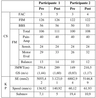
Table 1: Pre-post outcomes: Clinical evaluation (gait speed and scores on scales) and kinematics of exercises created in leg stance on plegic side starting with the knee extended and ending with controlled knee flexion.
Participante 1 Participante 2 Pre Post Pre Post
CS FAC 5 5 3 4 FIM 126 126 122 122 BBS 56 56 50 53 FM Total 106 111 100 108 Pain-Amp 40 40 40 40 Sensit. 24 24 24 24 Motor Eval 29 33 26 32 Balance 13 14 10 12 3MWT(m) GS (m/s) 259,4 (1,44) 289 (1,60) 149 (0,83) 210,5 (1,17) K P SE (mm2) 5055,4 2 3.123,0 1 6882,9 4 5146,8 9 Speed (mm/s) 136,92 140,92 60,12 61,93 Submov 7,1 5 19,4 10,9
CS: Clinical Scales; KP: Kinematic Parameters; Amp: amplitude; Sensit: Sensitivity; Motor Eval: Motor Evaluation; GS: Gait Speed; SE: Spatial error; Submov: submovements.
Participant 2:
The second participant was a 49-year-old man with hemiparesis after a hemorrhagic stroke (left cerebellar intraparenchymal and paraventricular) 3 months prior to evaluation. His medical records were insignificant. He completed a physical therapy program before recovering good functionality in activities of daily living (ADL), but used a wheelchair for long trips. Our program involves increasing walking speed and stability, improving kinematics and avoiding the misuse of compensation. He could be classified as an ambulator-dependent for supervision, scoring 3/5 in the FAC: person requires supervision or verbal stand-by help from one person without physical contact. It was shown that he was very functional with a high score on the FIM almost 122/126, with good balance and independence as the BBS (50/56). He reached a score of 100/112 on the FM, with top marks in pain-joint range and sensitivity (no restriction of joint range). He obtained 26/34 points in the functional assessment of the lower limb and 10/14 in the balance evaluation. As the data demonstrates, patient no. 2 has a greater balance deficit than patient no. 1. We measured the hip, knee and ankle spasticity. We didn´t find spasticity at proximal level but we discovered the same level of spasticity in the same location (ankle plantar flexors) as in patient no. 1. We took different measures of walking speed, obtaining 140 m in the 3MWT.
3.
INTERVENTION
Our equipment included a computer workstation connected to a 3D motion-tracking system (Polhemus FasTrak 3Space, Vermont, USA) and a high-resolution LCD projector which displayed the virtual scenarios on a large wall screen. The electromagnetic sensor was positioned at different locations on the patient's leg. The physical therapist could create numerous virtual tasks for the leg through the use of flexible software called Virtual Reality Rehabilitation System (VRRS – Khymeia Group, Itlaly), originally developed at the Massachusetts Institute of Technology (Cambridge, MA, U.S.), which processes data coming from the motion of the end-effector. The physiotherapist could select the characteristics and the complexity of the motor tasks according to each participant's lower limb deficit. In the virtual scenario, the physiotherapist determined the starting position and the different paths of movement the participant was asked to perform. VRRS enables us to visualize additional virtual objects to increase the complexity of motion.
During the RFVE therapy, participants were asked to perform tasks according to constraints previously specified by the physiotherapist. They were given information about their leg movements during the performance of motor skills (KP) based on the movement of the end-effector virtual representation. The physiotherapist's movement and trajectory could also be displayed in the background of the virtual scene in order to facilitate the subject's perception and adjustment to motion errors (learning by imitation) [22].
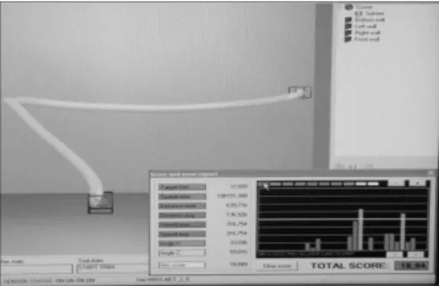
Moreover, the KR regarding the achievement of a requested motor task was given to participants in the form of standardized scores along with an augmented sensory feedback when the score surpassed a predetermined threshold [Fig 1 and 2]. Initially the abovementioned scores were KP and KR provided at a frequency of more than 90% and gradually decreased as performance improved [15].
Fig. 1: Representation in the virtual environment of the task created by the physiotherapist showing the performance feedback. Patient 1 performs a motion path with the sensor placed in the healthy foot (right) leg stance and the plegic lower limb with the aim of improving the proprioception in the plegic foot.
Participants received VR treatment one hour daily (Monday to Friday) in addition to the one-hour conventional physiotherapy program, for a total of three weeks (15 sessions). Conventional physiotherapy did not include specific programs using robotics or complex systems for the treatment of lower limb and balance (gait trainer, smart balance master, etc. to avoid biased results). None of the participants had aphasia, apraxia or cognitive impairments; therefore, we should ensure an adequate understanding so that a proper assimilation of motor tasks is carried out. Both therapies were focused on lower extremity motor rehabilitation. In the
RFVE program, the subject was asked to perform various motor tasks. The movement of the entire leg was simultaneously represented in a virtual scenario by means of motion-tracking equipment. As an example, trajectories were designed with a starting point and an ending point (two different colored squares with a slim trajectory line between them) that participants were to follow during their workout. If they did not complete the entire circuit, they did not receive auditory feedback on arrival. In addition, if during this practice the distance of the trajectory diverged from the range of position marked by the physiotherapist, the sound became louder as it got further away (auditory feedback) and a ball which simulated sensor location changed color [Fig.1].
Fig. 2: Representative trajectories reaching patient performed by touching two points above the ground with the sound foot while supported by standing on the plegic foot, from the starting position to the target that consists of two bottles resting on the ground. Trajectories were scattered at the baseline (a), but became more regular after training sessions (b).
Both participants received virtual reality treatment to improve the stability of monopodal support on the plegic lower limb and the quality of distal movement therein. The closed-chain work helped us reduce spasticity at ankle level, achieving an eccentric muscle work at triceps surae level. Therefore, we proposed basic exercises that could progress in difficulty as the patient advanced. For instance, one of the exercises participant 1 completed was an analytical exercise in leg stance on the plegic side. The participant was asked to lift his heel (triceps surae concentric work) to improve the generation of power needed in the final support stage and to achieve an adequate step length. A sensor was placed at the level of the participant's heel and an initial trajectory was recorded allowing him to lower part of his weight while leaning on a stick he was holding in the opposite hand. Thus, the center of gravity was centered and the weight that fell on the plegic lower limb was lower, allowing maximum elevation of the heel (greater amplitude in the range of dorsal flexion). When the participant arrived at the peak, the system provided a sound and knowledge of results feedback. This was repeated and as the exercise progressed less weight was placed on the stick and more weight fell on the plegic leg. Finally, the participant managed to achieve this task without using the stick [Fig 3].
As the participant was able to meet the objective, the therapist could add on to the exercise by placing a sensor on the top part of the torso. This sensor was simultaneously connected to the heel sensor so that if the patient tried to compensate by flexing his torso during leg movement, the ball deviated from the marked path. This last point was important in order to improve the performance of selective movements while avoiding the typical compensation that individuals with stroke rely on when walking (during the final stage of support, the hip has to stay extended. At the same time, it is important that we aim to improve the strength of the ankle. There must be a system to guide the participant on how to position the rest of the body so a more global sequence of movement is obtained). The physiotherapist continuously interacted with the
system and modified all the above parameters based on the participant's potential to make progress.
Participant 2 was asked to use his healthy foot to reach reference points on the ground in order to work on the proprioception of the supporting plegic foot. The distance between the points was progressively increased. To avoid visual compensation, the participant was asked to look at the screen and not his foot after several repetitions, making it harder to locate points exponentially high off the ground. Different elements were introduced in the real world and these were reflected in the virtual surroundings to partially modify the exercise so that the participant would be able to face new challenges (high steps, etc.). Trajectories were plotted in such a way so that the participant had to touch different objects located at different heights. In addition, tactile feedback was obtained when touched with the healthy toe. Therefore, when moving from one point to another the time of support on the plegic side was increased and the distribution of weight on the supporting foot was more balanced, thus improving proprioception. This mechanism prevented the participant from looking at his leg while performing the task so as to draw greater attention to the intrinsic proprioception process. Exercises in this line were also carried out with participant 1 using more complex methods. In our case, the advantage of working with flexible software is that it allowed the physiotherapist to program the most suitable performance and to provide appropriate feedback to improve individual performance.
Fig. 3: In the image the patient nº. 1 it is seen leaning on the plegic side, the sensor is placed at the plegic calcaneus level and
ask him to rise the heel increasing ankle plantarflexion,
significant movement on toe-off in the pre-swing phase. While this task is being performed, the screen shows a down square as a starting point, an up square as a point of arrival, a gray line and a ball that shows the movement of the sensor. The patient gets continuous feedback, more accentuated on the arrival so that the range of movement is completed.
4.
OUTCOMES
Despite the fact that no FIM pre-postraining differences were observed due to the ceiling effect in patients which is based on a certain functionality [23], both participants reported an improvement in functionality in difficult tasks. Participant 1 was able to run at a higher speed and started to play sports that he used to play such as tennis. Participant 2 managed to leave the wheelchair that he used for long journeys on stable ground. In the BBS, participant 1 continued to obtain the highest score, an improvement of 3 points over participant 2.
Spasticity decreased in both patients by two points in the MAS at the ankle to a score of 1.
In the FM scale, both participants improved lower limb skills. Participant 1 increased motricity by 5 points (4 points in motor evaluation and 1 in balance) and participant 2 by 8 points (6 points in motor evaluation and 2 in balance). This indicates an improvement of the plegic lower limb in both participants. Participant 1 increased his walking speed by 10.24% (0.16 m/s) in the 3MWT. With participant 2 there was an increase of 29.21% (0.34 m/s) in the 3MWT.
In the kinematic parameters, we can see that the speed reached in exercise did not vary significantly in both participants; however, variations were present in spatial error (SE), thus indicating how much the participants deviated from the trajectory and the mean number of submovements (or speed peaks, the greater amount of submovements the choppier and less fluid the movement became). Patient 1 experienced a decrease of 38.22% in the SE and 29.58% in submovements. Patient 2 also showed a decrease in both parameters, 25.22% in SE and 43.81% in submovements. Execution speeds remained more or less constant in both participants, being a kinematic parameter we ignore. That is, an increase in speed does not imply improved kinematics as it could create a lack of control in monopodal support with a rapid collapse of the plegic limb due to gravity. However, the reduction of spatial error and submovements has positive results and leads to greater precision in performing the task.
5.
DISCUSSION
The average spontaneous speed in adults is 1.37m /s in women and 1.43m/s in men [24]; participant 1 initially walked at a speed of 1.44m/s, which would normally be acceptable if it was a comfortable speed. However, our participants were asked to walk at the fastest speed possible during the 3MWT. Thus, the increase by more than 0.16m/s in the maximum speed obtained by the participant is clinically significant as it coincides with the MCID (minimally clinically important difference) referring to acute stroke [25]. This increase is significant, considering participant 1 was in a subacute state and the starting score was high (and as a result more difficult to improve), in order to reach our functional objective of increasing gait speed and facilitating the race, an activity that the participant was able to perform prior to the ictus (the 3MWT not only measures gait speed but also the subject’s resistance while walking thus the application of this method is comparable to the data obtained with the 6MWT) [26]. As for participant 2, who initiated from a much more inferior gait speed, the increase of 0.34m/s is also clinically significant as it is synonymous with functional improvement. It is difficult to compare these results with those from other authors who have used other virtual reality systems since the participants’ conditions differed from those of our participants [27]. The walking speed has been related to a recovery rate evaluated by FM. The result of Harro et al. (1987) shows that patients with <90% score had difficulty increasing their walking speed, comparing them to subjects with higher recovery level scores> 90% [28]. Our two cases obtained values over 90% in FM, hence there is potential for increasing walking speed. However, this increased capability also depends on other functional parameters. The increase in gait functionality obtained in FAC by participant 2 is outstanding. This indicates that aside from increasing walking speed, functional independence increased.
Regarding kinematic results, it has been impossible for us to compare results during walking with those from other authors [27] because our kinematic evaluation was in analytical sequences and was not during walking itself. However, the fact is that our system allows us to create open and individualized evaluation templates for specific deficits and to evaluate not only the range of motion but also the accuracy of it. To that extent, results are very satisfactory taking into account that this assessment focuses on one leg, which was the most deficient item for both participants as shown in their results from clinical scales. Thus, the decrease of submovements and SE indicates that motor control improved, which carries clinical importance, since an increase in gait speed without this control would imply compensatory mechanisms [29]. Results are positive so we can deduce that the combined treatment of conventional physiotherapy and virtual reality treatment could be effective in improving the performance of motor tasks and stability in leg stance on the plegic side, with improvement of functionality during walking. However, a kinematic evaluation of gait by means of a gait analysis could enhance our results. Controlled studies are needed to determine the role of VR in these improvements.
6.
REFERENCES
[1] Laver K, George S, Thomas S, et al. Cochrane review: virtual reality for stroke rehabilitation. Eur J Phys Rehabil
Med. 2012;48(3):1-2.
[2] Hassid E, Rose D, Commisarow J, et al. Improved gait symmetry in hemiparetic stroke patients induced during body weight-supported treadmill stepping. J Neurol Rehab. 1997;11:21-6.
[3] Jaffe DL, Brown DA, Pierson-Carey CD, et al. Stepping over obstacles to improve walking in individuals with poststroke hemiplegia. J Rehabil Res Dev. 2004;41(3A):283-92.
[4] Moreira MC, De Amorim Lima AM, Ferraz KM, et al. Use of virtual reality in gait recovery among post stroke patients – a systematic literature review. Disabil Rehabil Assist
Technol. 2013;8(5):357-62.
[5] You SH, Jang SH, Kim YH, et al. Virtual reality-induced cortical reorganization and associated locomotor recovery in chronic stroke: an experimenter-blind randomized study.
Stroke. 2005;36(6):1166-71.
[6] Fung J, Richards CL, Malouin F, et al. A treadmill and motion coupled virtual reality system for gait training post-stroke. Cyberpsychol Behav. 2006(2);9:157-62.
[7] Dunning K, Levine P, Schmitt L, et al. An ankle to computer virtual reality system for improving gait and function in a person 9 months poststroke. Top Stroke Rehabil. 2008;15(6):602-10.
[8] Yang YR, Tsai MP, Chuang TY, et al. Virtual reality-based training improves community ambulation in individuals with stroke: a randomized controlled trial. Gait Posture. 2008;28(2):201-6.
[9] Mirelman A, Bonato P, Deust JE. Effects of training with a robot-virtual reality system compared with a robot alone on the gait of individuals after stroke. Stroke. 2008;40:169-74. [10] Kim JH, Jang SH, Kim CS, Jung JH, You JH. Use of virtual
reality to enhance balance and ambulation in chronic stroke: A double-blind randomized controlled study, Am J Phys
Med Rehabil. 2009;88(9):693-701.
[11] Holden M. Virtual environments for motor rehabilitation: review. Cyberpsichology Behav. 2005;8(3):187-211.
[12] Kiper P, Piron L, Turolla A, et al. The effectiveness of reinforced feedback in virtual environment in the first 12 months after stroke. Neurol Neurochir Pol. 2011;45(5):436-44.
[13] Lewek MD, Feasel J, Wentz E, et al. Use of Visual and Propioceptive Feedback to improve gait speed and spatiotemporal symmetry following chronic stroke: a case series. Phys Ther. 2012;92(5):748-56.
[14] Lewek MD, Cruz TH, Moore JL, et al. Allowing intralimb kinematic variability during locomotor training poststroke improves kinematic consistency: A subgroup analysis from a randomized clinical trial. Phys Ther. 2009;89(9):829-39. [15] Piron L, Tonin P, Piccione F, et al. Virtual environment
training therapy for arm motor rehabilitation. Presence. 2005;14(6):732-40.
[16] Piron L, Turolla A, Agostini M, et al. Assessment and treatment of the upper limb by means of virtual reality in post-stroke patients. Stud Health Technol Inform.
2009;145:55-62.
[17] Piron L, Turolla A, Agostini M, et al. Exercises for paretic upper limb after stroke: a combined virtual-reality and telemedicine approach. J Rehabil Med. 2009;41(12):1016-102.
[18] Piron L, Tonin P, Atzori AM, et al. The augmented-feedback rehabilitation technique facilitates the arm motor recovery in patients after a recent stroke. Stud Health Technol Inform. 2003;94:265-7.
[19] Holden M, Todorov E, Callahan J, Bizzi E. Virtual environment training improves motor performance in two patients with stroke: Case report. Neurology report. 1999;23(2):57-67.
[20] Wade DT, Wood VA, Langton Hewer R. Recovery after stroke: the first 3 months. J Neurol Neurosurg Psychiatry. 1985(1);48:7-13.
[21] Bagg SD. Outcome predictors and the effectiveness of stroke rehabilitation. Phys Med Rehabil: state of the art reviews. 1988;12:581-92.
[22] Byrne RW, Russon AE. Learning by imitation: a hierarchical approach. Behav Brain Sci. 1998;21(5):667-84.
[23] Chang YH, Cong CS, Eng JY, Wong BP, Chai CP, Chen ZZ et al. Functional outcome in stroke patients enrolled in early supported discharge program not reaching minimal clinically important change in Functional Independence Measure.
Stroke. 2012;43:A3310.
[24] Perry J. Gait analysis. Normal and pathological function. New York: Slack Incorporated, 1992.
[25] Tilson JK, Sullivan KJ, Cen SY, Rose DK, Koradla CH, Azen SP, et al. Meaningful gait speed improvement during the first 60 days poststroke: Minimal clinically important difference. Phys Ther. 2010;90(2):196-208.
[26] Iriberri M, Gáldiz JB, Gorostiza A, Ansola P, Jaca C. Comparison of the distances covered during 3 and 6 min walking test. Respir Med. 2002;96(10):812-6.
[27] Mirelman A, Patritti BL, Bonato P, et al. Effects of virtual reality training on gait biomechanics of individuals post-stroke. Gait Posture. 2010;31(4):433-37.
[28] Harro CC, Giuliani CA. Kinematics and EMG analysis of hemiplegic gait patterns during free and fast walking speeds.
Neurol Rep. 1987;11:57-62.
[29] Bowden MC, Behrman AL, Neptune RR, Gregory CM, Kautz SA. Locomotor rehabilitation of individuals with chronic stroke: difference between responders and nonresponders. Arch Phys Med Rehabil. 2013;94(5):856-62.