Scholarship@Western
Scholarship@Western
Electronic Thesis and Dissertation Repository
4-19-2013 12:00 AM
Improving Cardiac Repair by Stem Cell Factor Post-Myocardial
Improving Cardiac Repair by Stem Cell Factor Post-Myocardial
Infarction
Infarction
Fuli Xiang
The University of Western Ontario
Supervisor Dr Qingping Feng
The University of Western Ontario Graduate Program in Physiology
A thesis submitted in partial fulfillment of the requirements for the degree in Doctor of Philosophy
© Fuli Xiang 2013
Follow this and additional works at: https://ir.lib.uwo.ca/etd
Part of the Circulatory and Respiratory Physiology Commons
Recommended Citation Recommended Citation
Xiang, Fuli, "Improving Cardiac Repair by Stem Cell Factor Post-Myocardial Infarction" (2013). Electronic Thesis and Dissertation Repository. 1204.
https://ir.lib.uwo.ca/etd/1204
This Dissertation/Thesis is brought to you for free and open access by Scholarship@Western. It has been accepted for inclusion in Electronic Thesis and Dissertation Repository by an authorized administrator of
i
POST-MYOCARDIAL INFARCTION
(Thesis format: Integrated Article)
by
Fu-Li Xiang
Graduate Program in Physiology
A thesis submitted in partial fulfillment of the requirements for the degree of
Doctor of Philosophy
The School of Graduate and Postdoctoral Studies The University of Western Ontario
London, Ontario, Canada
ii
Abstract
iii
that overexpression of hSCF increased epicardial activation, enhanced epicardium-derived cell (EPDC) proliferation and migration via SCF/c-kit signaling, and promoted arteriogenesis post-MI. In conclusion, M-SCF improves cardiac repair and function through inhibiting myocardial apoptosis and cardiac remodeling, and by promoting neovascularization via c-kit+ stem cell recruitment and EPDC activation and migration.
iv
Co-Authorship Statement
The studies outlined in Chapters 2-4 were performed by Fu-Li Xiang in the laboratory of Dr. Qingping Feng, with the assistance of co-authors as listed below.
Dr. Qingping Feng contributed to experimental design, data interpretation, and manuscript preparation for all experiments. Dr. P. Chidiac, Dr P. Zhu and Dr J. Robbins contributed to manuscript preparation for Chapter 2. Additionally, Dr. Sharon Lu designed and created the hSCF/tTA mice and assisted with the troubleshooting in all the experiments. Mrs. Murong Liu coordinated the animal breeding and genotyping.
Chapter 2: Dr. L Hammoud performed the RT-PCR and western blot analysis of hSCF expression (Figure 2.1).
Chapter 3: Yin Liu assisted with the fluorescent imaging (Figure 3.5 and 3.7).
v
Acknowledgments
I would like to thank my supervisor and mentor Dr. Qingping Feng for his help, support, and guidance throughout my Ph.D. studies. Dr. Feng is one of the most hard-working people I have ever met in my life and his commitment to excellence has made him an excellent researcher and a highly respected professor. His passion for research and craving for knowledge inspires me all the time. It is one of the best decisions I made in my life to work in his lab. He creates a very positive environment in the lab that fosters academic learning, creativity, and the career development for future cardiovascular investigators.
To the Feng laboratory, I could not have asked for a more stimulating and supportive environment to work. I would like to particularly thank Dr. Sharon Lu. She is a wonderful teacher and a good friend. To all other members of the Feng lab you have enriched my experience as a graduate student and made my stay in Feng lab enjoyable. To my advisory committee Dr. Peter Chidiac, Dr. David Hess and Dr. Douglas Jones: as a group you always provided unique perspectives and valuable suggestions on my studies and helped strengthen them. On an individual level Dr. Chidiac was the most approachable and lovely person in the department, Dr. Hess was a constant source of support and was very helpful in terms of unique suggestions that solidified my studies. Dr. Jones, was a model of professionalism and his guidance with regards to my career will always be appreciated.
To Heart and Stroke Foundation of Canada, the doctoral research award gave me financial support for my PhD studies, and to attend and present in international conferences.
vi
I also would like to acknowledge the Lawson Health Research Institute, and the Heart Failure Group at Western for providing me with a very stimulating and supportive environment that helped me to converse scientifically and become a skilled presenter. Additionally, I truly enjoyed my experience TAing for Physiology 3130y. Tom, Jamie and Chris were all wonderful people to work with.
vii
Table of Contents
Abstract ... ii
Co-Authorship Statement... iv
Acknowledgments... v
Table of Contents... vii
List of Tables ... xii
List of Figures ... xiii
List of Appendices ... xvi
List of Abbreviations, Symbols and Nomenclature ... xvii
1 CHAPTER 1 ... 1
1.1 ISCHEMIC HEART DISEASE (IHD) ... 1
1.1.1 Physiology of the Heart ... 1
1.1.2 Myocardial Infarction (MI)... 2
1.1.3 Heart Failure (HF)... 3
1.1.4 Current Treatment for MI and HF ... 4
1.2 NEW APPROACHES TO TREAT IHD ... 5
1.2.1 Inhibition of Myocardial Apoptosis... 6
1.2.2 Activation of Akt Signaling ... 7
1.2.3 Improvement of Cardiac Repair post-MI... 10
1.3 STEM CELLS IN CARDIAC REPAIR POST-MI ... 14
1.3.1 Bone Marrow-derived Stem Cells (BMSC)... 15
1.3.2 Cardiac Stem Cells (CSCs)... 19
1.3.3 Epicardium-derived Cells (EPDCs)... 22
1.4 STEM CELL FACTOR (SCF) ... 25
viii
1.4.2 SCF receptor c-kit... 30
1.4.3 Potential Clinical Applications of SCF... 31
1.4.4 SCF and IHD... 32
1.5 OVERALL HYPOTHESIS AND AIMS... 32
1.6 REFERENCES ... 38
2 Chapter 2 ... 68
2.1 CHAPTER SUMMARY... 69
2.2 INTRODUCTION ... 70
2.3 METHODS ... 71
2.3.1 Generation of hSCF/tTA Double Transgenic Mice ... 71
2.3.2 Determination of mRNA Expression by Real-time RT-PCR ... 72
2.3.3 Animal Procedures... 73
2.3.4 Hemodynamic Measurements... 73
2.3.5 Histology... 74
2.3.6 Caspase-3 Activity and Cytoplasmic Histone-Associated DNA Fragments ... 75
2.3.7 In vivo Matrigel Angiogenesis ... 75
2.3.8 Statistical Analysis... 75
2.4 RESULTS ... 75
2.4.1 Generation and Characterization of the Conditional Cardiac-specific hSCF/tTA Double Transgenic Mice ... 75
2.4.2 Post-MI Stem Cell Factor Expression... 78
2.4.3 Survival and Cardiac Function Post-MI ... 82
2.4.4 Myocardial Apoptosis 5 days Post-MI ... 89
2.4.5 Stem Cell Recruitment and Growth Factors Release Post-MI... 89
2.4.6 LV Hypertrophy 30 days Post-MI ... 96
ix
2.4.8 In Vivo Myocardial Angiogenesis... 109
2.5 DISCUSSION ... 109
2.6 REFERENCES ... 116
3 CHAPTER 3 ... 120
3.1 CHAPTER SUMMARY... 121
3.2 INTRODUCTION ... 122
3.3 METHODS ... 123
3.3.1 Animals ... 123
3.3.2 Myocardial I/R ... 124
3.3.3 Measurement of Infarct Size... 124
3.3.4 Determination of Myocardial Apoptosis ... 125
3.3.5 Western Blot Analysis ... 126
3.3.6 Growth Factor mRNA Expression... 126
3.3.7 Immunofluorescent Staining for Stem Cells... 127
3.3.8 Isolation and Culture of c-kit+ CSCs... 127
3.3.9 Flow Cytometry ... 128
3.3.10 Toluidine Blue Staining for Mast Cells ... 128
3.3.11 Statistical Analysis... 128
3.4 Results... 129
3.4.1 Overexpression of hSCF Reduces Infarct Size and Myocardial Apoptosis after I/R ... 129
3.4.2 Akt Signalling Contributes to hSCF Overexpression-induced Cardiac Protection after I/R... 129
3.4.3 Overexpression of hSCF Increases c-kit+ Cells in the Myocardium after I/R ... 134
x
3.4.5 Sub-population of c-kit Cells in Hearts with hSCF Overexpression after
I/R ... 144
3.5 DISCUSSION ... 147
3.6 REFERENCES ... 154
4 CHAPTER 4 ... 160
4.1 CHAPTER SUMMARY... 161
4.2 INTRODUCTION ... 162
4.3 METHODS ... 163
4.3.1 Animals ... 163
4.3.2 MI Mouse Model ... 164
4.3.3 EPDC Culture ... 165
4.3.4 Neonatal Cardiomyocyte Culture ... 165
4.3.5 Transwell Migration Assay... 166
4.3.6 Real-time RT-PCR... 166
4.3.7 Immunohistochemistry ... 167
4.3.8 Western Blot Analysis ... 168
4.3.9 Statistical Analysis... 168
4.4 Results... 168
4.4.1 Epicardial Activation 3 Days post-MI ... 168
4.4.2 Arteriolar Density in the Peri-infarct Area 5 Days post-MI ... 171
4.4.3 Outgrowth of Cultured EPDCs ... 174
4.4.4 Migration of Cultured EPDCs ... 181
4.4.5 Lineage Tracing of Wt1+ EPDCs... 181
4.5 DISCUSSION ... 191
4.6 REFERENCES: ... 198
xi
5.1 SUMMARY OF MAJOR FINDINGS... 202
5.2 STUDY LIMITATIONS ... 206
5.2.1 Inducible Cardiac-specific hSCF Overexpressing Transgenic Mice ... 206
5.2.2 Use of Mouse Model to Simulate Human Disease Conditions... 209
5.2.3 Contributions of Soluble SCF... 211
5.2.4 Use of Pharmacological Inhibitors and Neutralizing Antibody to Delineate Signaling Pathways... 211
5.3 SUGGESTIONS FOR FUTURE RESEARCH ... 216
5.4 CLINICAL APPLICATION... 219
5.5 CONCLUSIONS... 220
5.6 REFERENCES ... 221
APPENDIX... 225
xii
List of Tables
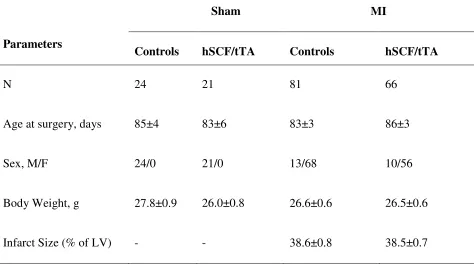
Table 2.1 General information of control and hSCF/tTA mice subjected to sham or MI surgeries ... 79
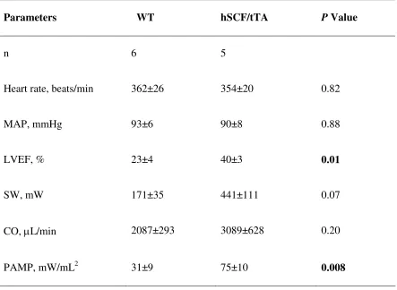
Table 2.2 Hemodynamic parameters of WT and hSCF/tTA mice 5 days post-MI ... 85
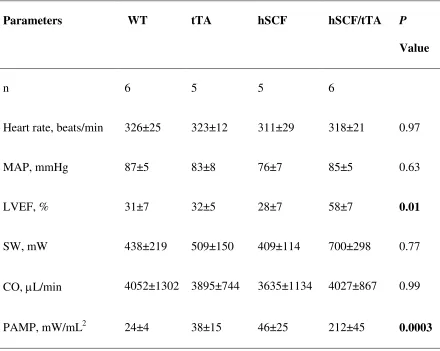
Table 2.3 Hemodynamic parameters of control and hSCF/tTA mice 30 days post-MI ... 86
xiii
List of Figures
Figure 1.1 Cardioprotective PI3K/Akt signaling pathways in ischemia/reperfusion (I/R). ... 8
Figure 1.2 Potential cell sources for cardiogenesis post-MI... 16
Figure 1.3 Illustration of soluble (S-SCF) and membrane-associated SCF (M-SCF). ... 28
Figure 1.4 Proposed cellular mechanisms by which membrane-associated SCF (M-SCF) regulates cardiac function post-MI. ... 34
Figure 2.1 Generation and characterization of the inducible hSCF/tTA transgenic mouse. .. 76
Figure 2.2 Mouse SCF expression in the heart post-MI. ... 80
Figure 2.3 Survival and LV function 30 days post-MI... 83
Figure 2.4 Cardiac function 5 days post-MI. ... 87
Figure 2.5 Myocardial apoptosis in the peri-infarct area 5 days post-MI... 90
Figure 2.6 c-kit+ and c-kit+/VEGFR2+ cells retained in the peri-infarct area 5 days post-MI.92 Figure 2.7 c-kit+ and c-kit+/VEGFR2+ cells retained in the peri-infarct area 30 days post-MI. ... 94
Figure 2.8 Mast cell staining using toluidine blue in heart tissue sections from WT and hSCF/tTA mice. ... 98
Figure 2.9 Growth factor expression in the peri-infarct area 5 days post-MI... 100
Figure 2.10 LV hypertrophy 30 days post-MI or sham operations... 102
Figure 2.11 Myocardial capillary density 5 days post-MI. ... 105
Figure 2.12 Myocardial capillary density 30 days post-MI. ... 107
xiv
Figure 3.1 Effects of hSCF overexpression on infarct size after I/R. ... 130
Figure 3.2 Myocardial apoptosis in peri-infarct area after I/R. ... 132
Figure 3.3 Myocardial Akt phosphorylation in WT and hSCF/tTA mice after I/R... 135
Figure 3.4 Myocardial eNOS phosphorylation in WT and hSCF/tTA mice after I/R. ... 137
Figure 3.5 c-kit+ cells in WT and hSCF/tTA hearts. ... 139
Figure 3.6 Cardioprotective cytokine expression in WT and hSCF/tTA mice after I/R. ... 142
Figure 3.7 Sub-population of c-kit+ cells in the myocardium after I/R. ... 145
Figure 3.8 Overexpression of hSCF in c-kit+ CSCs in vitro. ... 148
Figure 4.1 Epicardial Wt1 expression 3 days post-MI. ... 169
Figure 4.2 Arteriolar density in the peri-infarct area 5 days post-MI. ... 172
Figure 4.3 In vitro culture of EPDCs. ... 175
Figure 4.4 Outgrowth, proliferation and growth factor expression in cultured EPDCs. ... 177
Figure 4.5 Expression of hSCF protein in adenovirus treated EPDCs. ... 179
Figure 4.6 Transwell EPDC migration. ... 182
Figure 4.7 Lineage tracing of Wt1+ cell derived EPDCs 5 days post-MI... 185
Figure 4.8 Expression of hSCF protein in adenovirus treated hearts. ... 187
Figure 4.9 In vitro lineage tracing of Wt1+ cell derived EPDCs. ... 189
Figure 4.10 Fate-mapping of Wt1+ cell derived EPDCs post-MI... 192
Figure 5.1 Summarized cellular mechanisms by which M-SCF improves cardiac function and animal survival post-MI... 207
xv
Figure 5.3 Expression of soluble and membrane-associated hSCF in the serum of WT and hSCF/tTA mice. ... 214
xvi
List of Appendices
xvii
List of Abbreviations, Symbols and Nomenclature
ACE: angiotensin-converting-enzyme
Ad-GFP: adenoviral green fluorescent protein construct
Ad-hSCF: adenoviral membrane-associated human stem cell factor construct Ad-LacZ: adenoviral LacZ construct
BAD: B cell lymphoma (Bcl)-xl/Bcl-2–associated death promoter bFGF: basic fibroblast growth factor
BMP2: bone morphogenetic protein-2 BMSCs: Bone marrow-derived stem cells CDCs: cardiosphere-derived cells
CSCs: cardiac stem cells
CSPCs: cardiac side population cells DAB: diaminobenzidine tetrahydrochloride DOX: doxycycline
E: embryonic day
ELISA: enzyme-linked immunosorbent assay EMT: epithelial-mesenchymal transition eNOS: endothelial nitric oxide synthase EPCs: endothelial progenitor cells EPDCs: epicardium-derived cells EPO: erythropoietin
xviii GFP: green fluorescent protein
GSK-3β: glycogen synthase kinase 3β HF: heart failure
HGF: hepatocyte growth factor
hPGK: human phosphoglycerate kinase
hSCF: membrane-associated human stem cell factor HSCs: hematopoietic stem cells
I/R: ischemia and reperfusion IGF-1: insulin-like growth factor 1 IHD: ischemic heart disease IP: intraperitoneal
JAK/STAT: Janus kinase/signal transducers and activators of transcription LV: left ventricle
LVEF: left ventricle ejection fraction
LVW/BW: left ventricle to body weight ratio MAP: mean artery pressure
MHC: myosin heavy chain MI: myocardial infarction
MMLV: moloney murine leukemia virus MNCs: mononuclear cells
MOI: multiplicity of infection
xix mTOR: mammalian target of rapamycin
NF-κB: nuclear factor kappa B P0: postnatal day 0
PAMP: preload adjusted maximum power PDK1: phosphoinositide-dependent kinase 1 PI3K: phosphoinositide-3-kinase
PKC: protein kinase C PLC-γ: phospholipase-γ
PMA: phorbol 12-myristate 13-acetate RISK: reperfusion injury salvage kinase SCF: stem cell factor
SH2: Src homology 2
S-SCF: soluble stem cell factor SW: stroke work
TGF β: transforming growth factor β tTA: tetracycline activator
TTC: triphenyltetrazolium chloride VEGF: vascular endothelial growth factor
VEGFR2: vascular endothelial growth factor receptor 2 vWF: von Willebrand factor
WT: wild type Wt 1: Wilms tumor 1
α-SMA: α-smooth muscle actin
xx *, † and ‡are used for denoting statistical significance.
1
CHAPTER 1
Literature Review
1.1 ISCHEMIC HEART DISEASE (IHD)
IHD is a condition in which the heart muscle is damaged or works inefficiently due to an absence or relative deficiency of its blood supply. Most often caused by atherosclerosis, the clinical manifestation of IHD includes angina pectoris, acute myocardial infarction, chronic ischemic heart disease, and sudden death. IHD is the leading cause of death and a financial burden worldwide.1 For example, in the United States, IHD resulted in 445,687 deaths (about one of every five deaths) in 2005 and was reported to place a direct and indirect cost of $165.4 billion in 2009.1 Despite optimal treatment with existing drugs, the prognosis of IHD remains poor. Heart transplantation is the last but an effective option for end-stage IHD patients. The five-year survival rate of heart transplantation has achieved 70% in the United States.1 However, the limited donors for heart transplantation are not meeting the need of IHD patients. Thus, novel and effective treatments for IHD are in high demand.
1.1.1 Physiology of the Heart
capillaries that supply the heart with oxygen. The outer layer of the heart wall is the epicardium, consisting of mesothelium and connective tissues.
The heart pumps blood through two series circulations by coordinated actions of the atria and ventricles.3 Oxygenated blood from the lungs is received by the left atrium and is subsequently pumped into the aorta (systemic circulation) by the left ventricle (LV). The blood flows through arteries and capillaries to all tissues in the body to supply them with oxygen and nutrients. The deoxygenated blood returns to the right atrium of the heart from veins and is then pumped into the pulmonary circulation via the right ventricle to be re-oxygenated in the lungs. The heart generates continuous and sufficient blood circulation and can, by demand, increases its output. The coronary circulation is critical to maintain heart muscle function. Insufficient coronary perfusion caused by coronary artery narrowing or constriction results in cardiac ischemia leading to impaired cardiac function.4
1.1.2 Myocardial Infarction (MI)
and recruited to the infarcted region.6 Following acute MI, re-establishing coronary blood flow by thrombolysis or primary angioplasty is widely used to salvage viable myocardium. However, the oxidative stress and abrupt metabolic changes immediately after reperfusion have been shown to initiate further cellular injury.7,8 Healing of the infarcted myocardium leads to scar formation and ventricular remodeling, which is characterized by ventricular enlargement, fibrosis and hypertrophic growth of the peri-infarct and non-peri-infarct myocardium. The remodeled LV causes cardiac dysfunction, clinical heart failure and increased mortality.5,9
1.1.3 Heart Failure (HF)
HF is a clinical syndrome characterized by impaired cardiac function and altered
structure, leading to dyspnea and fatigue at rest or with exertion.10 Patients who survive a
MI are at an increased risk of developing HF. The extent of LV systolic and diastolic
dysfunction is associated with clinical symptoms and cardiovascular mortality. HF can be
caused by the continuous abnormal loading conditions post-MI as a result of the infarct
scar formation. The LV remodeling is a dynamic process that is driven by a number of
factors.11 During cardiac remodeling post-MI, the LV becomes dilated, hypertrophic, and
undergoes morphological and pathophysiological alterations. Pathologically, there is
extensive fibrosis as well as disarray of abnormally structured myocardial cells.
Furthermore, apoptosis becomes more active, resulting in additional loss of myocardial
cells.12 It is the combination of these pathological changes that alters the structure of the
1.1.4 Current Treatment for MI and HF
After an acute MI, patients often undergo revascularization in order to regain adequate blood supply to the myocardium. The two most widely employed treatments are angioplasty, in which the blocked artery is mechanically opened, and bypass surgery, in which vessels from elsewhere in the body are attached to bypass the blocked artery and re-establish blood flow. Also, MI patients receive pharmacological therapy to relieve angina, prevent thrombus propagation and to stabilize the “vulnerable” plaque.13 Anti-anginal drugs, which affect the balance between oxygen supply and demand, include nitroglycerin, β-adrenergic blockers, and calcium channel blockers. To reduce thrombus formation, anti-platelet and antithrombotic treatments are also recommended, unless contraindicated. Antiplatelet agents include aspirin, thienopyridines, and glycoprotein IIb/IIIa inhibitors. Commonly used antithrombotic agents include unfractionated heparin, low-molecular-weight heparin, fondaparinux, and bivalirudin. Furthermore, statin therapy promotes plaque stabilization and restores endothelial function, and has been shown to reduce the incidence of myocardial infarction, recurrent ischemia and mortality.
HF is a chronic disease that requires lifelong management including pharmacological therapy and lifestyle modification.14 Angiotensin-converting-enzyme (ACE) inhibitors are now considered a first-choice in HF drug therapy. ACE inhibitors limit the formation of angiotensin II resulting in vessel dilation as well as the prevention
of cardiac hypertrophy, fibrosis and decreased afterload and preload of the heart.
Angiotensin II receptor blockers, which have similar effects, can be used as an alternative
for patient who are unable to tolerant ACE inhibitors. Diuretics are prescribed for patients
force of cardiac contractions and relieves heart failure symptoms, especially when the
patient is not responding to ACE inhibitors and diuretics. Beta-blockers are prescribed to
reduce heart rate and lower blood pressure. They also inhibit ventricular remodeling and
reduce the risk of arrhythmias and sudden death.15 Lifestyle modifications include losing
weight, quitting smoking, avoiding alcohol and caffeine, eating a saturated-fat
low-sodium diet, exercising and reducing stress, all of which have been shown to alleviate
symptoms, slow disease progression and improve quality of life.
In spite of significant progress in clinical treatment, the prognosis of HF remains
poor. At present there is no cure for patients with end-stage HF except heart transplantation. Drug treatment can attenuate myocardial remodeling and improve
survival, but does not reduce the size of the scar. Unfortunately, angioplasty or bypass
surgery does not reverse the damage induced by MI. LV assist devices can prolong
patient lives only for a few years.16,17 On the other hand, although the survival rate after
heart transplantation is excellent, the source of transplantable donor hearts is scarce.
Therefore, there is a need for developing new and effective therapies to treat patients with
HF post-MI. Approaches to limit cardiomyocyte loss, rebuild the coronary microvascular
network and impede cardiac remodeling have been widely investigated to improve
cardiac function post-MI.
1.2 NEW APPROACHES TO TREAT IHD
Approaches to decrease myocardial cell death, enhance cardiac regeneration and impair cardiac remodeling have shown promising beneficial outcomes in animal models.
1.2.1 Inhibition of Myocardial Apoptosis
apoptosis during I/R injury have been demonstrated to decrease infarct size and improve cardiac function.28-30 Additionally, activation of pro-survival kinases including Akt has been shown to provide significant anti-apoptotic effects in MI and I/R.
1.2.2 Activation of Akt Signaling
Figure 1.1 Cardioprotective PI3K/Akt signaling pathways in ischemia/reperfusion
(I/R).
size and protects against myocardial apoptosis after I/R by phosphorylation of eNOS via the PI3K/Akt signaling pathway.34 Akt can also directly inhibit caspase-mediated cell death through the phosphorylation of BAD, releasing Bcl-2 family members to maintain mitochondrial permeability 43 and the phosphorylation of caspase-9.44 Additionally, Akt can protect against cardiomyocyte apoptosis by regulating gene expression of Glut-1, vascular endothelial growth factor (VEGF), Bcl-2 and forkhead transcription factors.45 Furthermore, Akt plays a critical role in endothelial homeostasis and angiogenesis by maintaining endothelial cell viability, regulating migration and promoting the homing of endothelial progenitor cells (EPCs) in the ischemic heart after I/R.46,47
1.2.3 Improvement of Cardiac Repair post-MI
The endogenous healing process post-MI is manifested by increased matrix synthesis, fibroblast proliferation and the release of pro-fibrotic cytokines, resulting in scar formation. The infarct size, which is critical to disease progression and prognosis, is determined by the area of coronary artery occlusion and dynamic changes during the remodeling process.48 The ideal cardiac repair would include the complete regeneration of the myocardium and neovascularization, which has been observed in Zebra fish,49 the adult newt,50 and the neonatal mouse.51 However, the ability to regenerate myocardial tissue follow injury is diminished in the adult mammals.51
1.2.3.1Neovascularization post-MI
1.2.3.2Endogenous Cardiomyocyte Regeneration post-MI
1.3 STEM CELLS IN CARDIAC REPAIR POST-MI
Stem cells are defined by their unique properties: self-renewal capacity and the potential to differentiate into one or more specialized cell types.86 Stem cells have the ability to undergo asymmetric and symmetric cell division. Asymmetric cell division gives rise to two daughter cells that have different fates, one maintaining the stem cell population and the other generating differentiated cells.87 Stem cells can be classified into 3 major categories: embryonic, induced pluripotent and adult stem cells.74,88,89 Both embryonic and induced pluripotent stem cells are pluripotent and can generate any cell type in the body. However, they present a high risk of teratoma formation, which makes them unacceptable for clinical treatment at this time. Interestingly, small populations of clonogenic, self-renewing, and multipotent cells have been found in different adult tissues including bone marrow, vascular walls, adipose tissues, skeletal muscles, heart and brain as well as the epithelium of the lung, liver, pancreas, digestive tract, skin,
limbus, retina, breast, ovaries, prostate and testis.90,91 These multipotent stem cells, referred to as adult stem cells, have the potential to differentiate into mature cell lineages in tissues from which they originate and contribute to postnatal organ homeostasis. Furthermore, bone marrow-derived stem/progenitor cells may also migrate to distant peripheral sites after intense injury and contribute to tissue repair.91,92 Adult stem cells reside in a specific cellular niche, which ensures a balance between the maintenance of the stem cell pool and the production of daughter progenitor cells engaged in lineage differentiation.93 The in vivo stimulation of adult stem cells or the use of their ex vivo
expanded and differentiated progenies is emerging as a promising approach for cell
bone marrow-derived stem cells, resident cardiac stem cells and epicardium-derived cells
have shown cardiac myogenic and vasculogenic potential, which make them favorable
candidates for cardiac repair post-MI.77,94 (Figure 1.2)
1.3.1 Bone Marrow-derived Stem Cells (BMSC)
Figure 1.2 Potential cell sources for cardiogenesis post-MI.
trophic factors released from the HSCs, which are referred to as paracrine effects.104
MSCs were originally identified as a small subpopulation of bone marrow cells which have the ability to differentiate into tissue types of mesodermal origin, such as chondrocytes, adipocytes and osteocytes.105 MSCs have been successfully isolated from various non-marrow tissues such as umbilical cord blood 106 and adipose tissue.107 DNA demethylating agents, such as 5-azacytidine, have been shown to induce MSC transdifferentiation into cardiomyocytes in vitro.108,109 Cell therapy using MSCs has been well-documented to improve cardiac performance and promote neovascularization, which are now considered mostly due to the paracrine effects instead of cardiomyocyte regeneration.110,111 There are several advantages of using MSCs in cell therapy in IHD compared to HSCs. First, they can be easily cultured and expanded in vitro to the large population needed for transplantation. Second, they can be engineered in vitro to achieve specific characteristics. Third, they have been shown to contribute to both myocardial and vascular repair post-MI. Last, they have immunological privilege by regulating immune cell activity, which makes MSCs a potential candidate for allogenetic treatment.112
be needed in order to achieve the best outcome for BMSCs stem cell therapy in IHD patients. The lack of benefit of bone marrow-derived MNCs therapy suggests that it is not an optimal stem cell type for IHD treatment.
Endothelial progenitor cells (EPCs) are a rare population of cells that circulate in the blood and have the ability to differentiate into endothelial cells and promote neovascularization. Bone marrow is the major source of adult EPCs. EPCs share many markers with HSCs, which makes it difficult to identify EPCs strictly by specific marker expression. The most widely used markers for EPCs include c-kit, CD34, CD133 and vascular endothelial growth factor receptor 2 (VEGFR2). In vitro culture of mononuclear bone marrow cells yields 2 types of EPCs: early outgrowth and late outgrowth EPCs.115 The early outgrowth EPCs have been shown to be of hematopoietic origin and potently induce angiogenesis without incorporating in blood vessels, while the late outgrowth EPCs are considered real EPCs which can be clonally expanded and give rise to endothelial cells. During neovascularization, EPCs are released from bone marrow to the peripheral circulation and mobilize to the target tissue. Mobilization of EPCs after cardiac ischemia is also observed in patients after acute MI.116 Animal studies have demonstrated that EPC transplantation or treatment with agents that enhance EPC mobilization and recruitment post-MI improves cardiac function and reduces cardiac remodeling via angiogenesis and vasculogenesis.52,117-120
1.3.2 Cardiac Stem Cells (CSCs)
low in the adult heart, they are committed to generate cardiomyocytes and their numbers can be expanded ex vivo for therapeutic purposes.66 Different approaches are employed to identify the resident CSCs, including detection of specific surface markers such as c-kit, and assessment of their unique physiological properties such as the ability to form cardiospheres in culture or to efflux Hoechst 33342 dye (side population progenitors).
Cardiosphere-derived cells (CDCs) are also CSCs identified through their ability to migrate from cultured cardiac tissues and form floating spheres. CDCs are isolated from minimally invasive percutaneous endomyocardial biopsies from the adult human heart 131 and the myocardium from other species (mice, rats and pigs).132,133 The CDCs are a mixture of c-kit+ cells and supporting endothelial and mesenchymal cells that can be amplified in culture and grown to millions in a few weeks.133 Intracoronary delivery of CDCs has been shown to improve cardiac repair and function in animal MI models.131,134 A phase I clinical trial was carried out to study the safety and efficacy of CDCs in patients with a recent MI (ClinicalTrials.gov Identifier: NCT00893360). Reduction in scar mass, increased viable heart mass and regional contractility was observed in 17 patients with MI, 6 months after CDCs treatment, while no significant changes in ejection fraction, end-diastolic and end-systolic volume were found.135 Cell therapies with autologous c-kit+ CSCs or CDCs after MI have been shown to be safe and beneficial, and warrant phase 2 studies to test their efficacy.
multipotent CSCs marked by the LIM-homeodomain transcription factor islet-1 (Isl-1) has been shown to give rise to cardiomyocytes, endothelial and smooth muscle cells both in vitro and in vivo. The Isl-1+ CSCs are mainly located in the outflow tract and right atrium of fetal or very young postnatal hearts and are negative for c-kit and Sca-1.139 The fact that Isl-1+ CSCs exist in very low numbers in the adult heart limits their therapeutic potential. Furthermore, the property of cardiac progenitor cells to expel toxic molecules through an ATP-binding cassette transporter 140 is also employed to define a pool of CSCs. They are termed cardiac side population cells (CSPs) because after exposure to the DNA-binding dye, Hoechst 33342, they can clear the fluorochrome and appear on the side of the flow cytometry profile.141 CSPs are positive for Sca-1 and negative for c-kit.142 CD31-Sca-1+ CSPs form colonies in vitro and differentiate into cardiomyocytes.142,143 When transplanted into animals with MI, CSPs can migrate to the injured myocardium and differentiate into cardiomyocyte- and endothelial-like cells.144,145
To date, whether the resident CSCs identified by different methods represent distinct populations of stem/progenitor cells or they are actually the same line of stem cells at different stage of maturation remains unknown. Exploitation of CSCs in stem cell therapy for IHD is certainly an exciting and promising strategy. However, further investigations of the endogenous regulatory mechanisms of CSCs survival, proliferation and differentiation under physiological or pathological conditions are definitely needed before stem cell therapy can be safely and effectively applied to IHD patients.
1.3.3 Epicardium-derived Cells (EPDCs)
1.4 STEM CELL FACTOR (SCF)
SCF, also known as Steel factor, mast cell growth factor, or c-kit ligand, binds to c-kit and promotes survival, proliferation, mobilization, and adhesion of all c-kit -expressing cells. c-kit is a tyrosine kinase receptor expressed in stem cells and progenitors including BMSCs, CSCs and some EPDCs 122,163,164 while SCF is ubiquitously expressed as a glycosylated transmembrane protein widely throughout the body by stromal cells,165 fibroblasts, endothelial cells and the myocardium. Alternative mRNA splicing leads to two isoforms of SCF: soluble (S-SCF) and membrane-associated (M-SCF), which differ in the absence or presence of a particular proteolytic cleavage site encoded by exon 6.166 S- and M-SCF have distinct but overlapping roles. M-SCF is the dominant isoform in vivo and induces more persistence and long-term support of target cell survival via juxtacrine, but may be less effective in stimulating growth.166-168
1.4.1 SCF Biology
melanogenesis, and works in synergy with other growth factors. In vitro, SCF treatment induces survival, proliferation and migration of HSCs, melanoblasts and primordial germ cells.173 In vivo, recombinant S-SCF elicits a multilineage response in peripheral counts and increases bone marrow cell mass, which protects against lethal irradiation by accelerating hematopoietic recovery.174-176
13-acetate (PMA) is able to increase the shedding in COS-1 cells transfected with mouse M-SCF.168 The biological activity of the S-SCF released from M-SCF is similar to that of the proteolytic product of the full length SCF.
Figure 1.3 Illustration of soluble (S-SCF) and membrane-associated SCF (M-SCF).
Thus, it is not surprising that M-SCF provides greater survival and proliferation signals compared to the soluble isoform.181,186 It also appears that the 2 isoforms of SCF have different signaling pathways for their biological activities. For example, although both S-SCF and M-S-SCF can induce proliferation of erythroid progenitors via activation of p38 and ERK pathways, M-SCF-induced signaling is more sensitive to ERK inhibition while p38 inhibition works better on S-SCF.186 Thus, through expression of different SCF isoforms, stromal cells regulate the activity, survival, proliferation, migration and adhesion of local c-kit expressing cells as well as those in remote organs and tissues.
1.4.2 SCF receptor c-kit
kinase, PI3 kinase, phospholipase-γ (PLC-γ), Src and Janus kinase/signal transducers and activators of transcription (JAK/STAT) pathways, all of which contribute to the biological effects of SCF.197 SCF/c-kit signaling can be inhibited by blockage of SCF/ c-kit binding via neutralizing antibody, ACK2,198 or inhibition of c-kit receptor activation by AG 1296.199
1.4.3 Potential Clinical Applications of SCF
Recombinant human SCF is currently used to mobilize BMSCs for collection of HSCs for hematopoietic reconstitution200 and to prevent chemotherapy-induced anemia and thrombocytopenia.201 SCF has also been indicated as a drug target for cancer treatment.202,203 Analysis of serum SCF levels is recommended to aid the diagnosis of many diseases including pancreatic cancer,184 gastrointerstitial stromal tumor,204 primary uveal melanoma,205 acute renal allograft rejection,206 delayed and slow graft function207 and Alzheimer’s disease.185
1.4.4 SCF and IHD
The role of granulocyte colony-stimulating factor (G-CSF) and S-SCF in MI was first studied by Orlic et al, showing enhanced BMSCs mobilization and recruitment to the infarcted heart and improved cardiac function in mice post-MI.68 Those beneficial effects resulted from decreased myocardial apoptosis,218 reduced arrhythmia,219 increased neovascularization 220 and impaired cardiac remodeling.221 However, Ohtsuka et al found SCF treatment alone failed to induce cardiac protective effects post-MI.220 Myocardial expression of SCF is decreased post-MI. Augmentation of SCF expression through transplanted MSCs has been shown to increase angiogenesis and cardiac function.222 Moreover, cardiac transplantation of MSCs transfected with a full length mouse SCF cDNA plasmid, which express both S- and M-SCF, enhanced EPC recruitment and myocardial angiogenesis for about 2 weeks post-MI. However, there was no change in animal survival among all treatment groups.222 This is not surprising as more than 95% of the transplanted MSCs are cleared within 7 days following transplantation into the myocardium.223 Intra-myocardial administration of SCF sustainably mobilized exogenously administered Lin-c-kit+ bone marrow-derived cells from the circulation homing to the infarcted myocardium up to 72 hours post-MI, however, cardiac function was not investigated in this study.224 The molecular and cellular mechanisms by which SCF regulates cardiac function post-MI are not fully understood.
1.5 OVERALL HYPOTHESIS AND AIMS
injections of SCF increased recruitment of BMSCs to the infarcted heart.224 Although numerous animal studies using stem cell therapy showed significant protection post-MI, only minor improvements have been observed in most clinical trials,113,114,225,226 while two recent trials showed no significant benefit at all.75,76,226 Thus, a better understanding of the pathophysiology of different types of stem cells in cardiac repair post-MI is needed.
SCF is expressed as a glycosylated trans-membrane protein by many cells and tissues, including the myocardium.227 SCF binds to its receptor c-kit and plays a critical role in cell survival, proliferation, mobilization, and adhesion. c-kit positive adult stem cells including HSCs, EPCs and CSCs122,163,164 have been demonstrated to be potential candidates for cardiac repair post-MI, which implies a possible regulatory role of SCF in MI. M-SCF is more potent than S-SCF in supporting cell survival and proliferation.19 In adult mice lacking M-SCF, cardiac morphology and function at basal conditions were normal, but significantly higher mortality and worse cardiac function were observed post-MI.228 To study the role of M-SCF in cardiac repair post-MI, we generated a novel mouse model of inducible cardiac specific over-expression of hSCF. The aim of this thesis was to study the effects of M-SCF on cardiac function post-MI and to investigate the underlying cellular mechanisms especially the role of M-SCF in regulation of c-kit+ stem/progenitor cell activity in the ischemic heart (Figure 1.4). Our overall hypothesis
was that cardiac-specific overexpression of hSCF protects the heart via inhibition of
myocardial apoptosis and enhancement of neovascularization and stem cell activity
Figure 1.4 Proposed cellular mechanisms by which membrane-associated SCF
(M-SCF) regulates cardiac function post-MI.
In order to induce cardiac-specific hSCF overexpression, a hSCF transgenic mouse was generated with the hSCF transgene expression under the control of a tetracycline-responsive element containing the α-MHC promoter. The hSCF mice were then crossed
with cardiac-specific tetracycline activator (tTA) mice to generate hSCF/tTA double transgenic mice. The hSCF/tTA transgenic mice specifically express the hSCF in the heart. These transgenic mice were healthy and fertile with no cardiac abnormalities.229 Furthermore, myocardial expression of hSCF in hSCF/tTA mice can be abrogated by treatment with doxycycline (DOX). Thus, the specific effects of hSCF can be tested in the absence and presence of DOX to turn on and off hSCF expression in the heart.
CSCs, recent studies have shown that EPDCs are another potential resource of cardiovascular progenitors.230 Thus, the third aim of my thesis was to investigate the role of hSCF in EPDC activation, migration into the sub-epicardium and contribution to cardiac repair post-MI.161 So I hypothesized in Chapter 4 that cardiac specific over-expression of hSCF promotes epicardial activation. We found that hSCF overover-expression increased EPDC activation, proliferation and mobilization which contribute to myocardial neovascularization post-MI.
1.6 REFERENCES
1. Lloyd-Jones D, Adams R, Carnethon M, De Simone G, Ferguson TB, Flegal K, Ford E, Furie K, Go A, Greenlund K, Haase N, Hailpern S, Ho M, Howard V, Kissela B, Kittner S, Lackland D, Lisabeth L, Marelli A, McDermott M, Meigs J, Mozaffarian D, Nichol G, O'Donnell C, Roger V, Rosamond W, Sacco R, Sorlie P, Stafford R, Steinberger J, Thom T, Wasserthiel-Smoller S, Wong N, Wylie-Rosett J, Hong Y. Heart disease and stroke statistics--2009 update: a report from the American Heart Association Statistics Committee and Stroke Statistics Subcommittee. Circulation. 2009;119:480-6.
2. Meessen H. Structural bases of myocardial hypertrophy. Br Heart J. 1971;33:Suppl:94-9.
3. Berne RM, Levy MN. Heart. Annu Rev Physiol. 1964;26:153-86.
4. Spaan J, Kolyva C, van den Wijngaard J, ter Wee R, van Horssen P, Piek J, Siebes M. Coronary structure and perfusion in health and disease. Philos Transact A Math Phys Eng Sci. 2008;366:3137-53.
5. Sutton MG, Sharpe N. Left ventricular remodeling after myocardial infarction: pathophysiology and therapy. Circulation. 2000;101:2981-2988.
6. Fazel SS, Chen L, Angoulvant D, Li SH, Weisel RD, Keating A, Li RK. Activation of c-kit is necessary for mobilization of reparative bone marrow progenitor cells in response to cardiac injury. FASEB J. 2008;22:930-940.
7. Anaya-Prado R, Toledo-Pereyra LH. The molecular events underlying ischemia/reperfusion injury. Transplant Proc. 2002;34:2518-9.
9. Thomas S, Rich MW. Epidemiology, pathophysiology, and prognosis of heart failure in the elderly. Heart Fail Clin. 2007;3:381-387.
10. Francis GS, Tang WH. Pathophysiology of congestive heart failure. Rev Cardiovasc Med. 2003;4 Suppl 2:S14-20.
11. Sun Y. Myocardial repair/remodelling following infarction: roles of local factors. Cardiovasc Res. 2009;81:482-90.
12. Grazette LP, Rosenzweig A. Role of apoptosis in heart failure. Heart Fail Clin. 2005;1:251-61.
13. Trost JC, Lange RA. Treatment of acute coronary syndrome: Part 1: Non-ST-segment acute coronary syndrome. Crit Care Med. 2011;39:2346-53.
14. Jessup M, Abraham WT, Casey DE, Feldman AM, Francis GS, Ganiats TG, Konstam MA, Mancini DM, Rahko PS, Silver MA, Stevenson LW, Yancy CW. 2009 focused update: ACCF/AHA Guidelines for the Diagnosis and Management of Heart Failure in Adults: a report of the American College of Cardiology
Foundation/American Heart Association Task Force on Practice Guidelines: developed in collaboration with the International Society for Heart and Lung Transplantation. Circulation. 2009;119:1977-2016.
15. Domanski MJ, Krause-Steinrauf H, Massie BM, Deedwania P, Follmann D, Kovar D, Murray D, Oren R, Rosenberg Y, Young J, Zile M, Eichhorn E,
Investigators B. A comparative analysis of the results from 4 trials of beta-blocker therapy for heart failure: BEST, CIBIS-II, MERIT-HF, and COPERNICUS. J Card Fail. 2003;9:354-63.
17. Daneshmand MA, Rajagopal K, Lima B, Khorram N, Blue LJ, Lodge AJ, Hernandez AF, Rogers JG, Milano CA. Left ventricular assist device destination therapy versus extended criteria cardiac transplant. Ann Thorac Surg.
2010;89:1205-9; discussion 1210.
18. Rodriguez M, Lucchesi BR, Schaper J. Apoptosis in myocardial infarction. Ann Med. 2002;34:470-9.
19. Foo RS, Mani K, Kitsis RN. Death begets failure in the heart. J Clin Invest. 2005;115:565-71.
20. Krijnen PA, Nijmeijer R, Meijer CJ, Visser CA, Hack CE, Niessen HW.
Apoptosis in myocardial ischaemia and infarction. J Clin Pathol. 2002;55:801-11.
21. Saraste A, Pulkki K, Kallajoki M, Henriksen K, Parvinen M, Voipio-Pulkki LM. Apoptosis in human acute myocardial infarction. Circulation. 1997;95:320-3.
22. Konstantinidis K, Whelan RS, Kitsis RN. Mechanisms of cell death in heart disease. Arterioscler Thromb Vasc Biol. 2012;32:1552-62.
23. Oyama J, Frantz S, Blais Jr C, Jr., Kelly RA, Bourcier T. Nitric oxide, cell death, and heart failure. Heart Fail Rev. 2002;7:327-34.
24. Shizukuda Y, Buttrick PM, Geenen DL, Borczuk AC, Kitsis RN, Sonnenblick EH. beta-adrenergic stimulation causes cardiocyte apoptosis: influence of tachycardia and hypertrophy. Am J Physiol. 1998;275:H961-8.
25. Sabbah HN, Sharov VG, Gupta RC, Todor A, Singh V, Goldstein S. Chronic therapy with metoprolol attenuates cardiomyocyte apoptosis in dogs with heart failure. J Am Coll Cardiol. 2000;36:1698-705.
27. Fliss H, Gattinger D. Apoptosis in ischemic and reperfused rat myocardium. Circ Res. 1996;79:949-56.
28. Lee P, Sata M, Lefer DJ, Factor SM, Walsh K, Kitsis RN. Fas pathway is a critical mediator of cardiac myocyte death and MI during ischemia-reperfusion in vivo. Am J Physiol Heart Circ Physiol. 2003;284:H456-63.
29. Brocheriou V, Hagege AA, Oubenaissa A, Lambert M, Mallet VO, Duriez M, Wassef M, Kahn A, Menasche P, Gilgenkrantz H. Cardiac functional
improvement by a human Bcl-2 transgene in a mouse model of ischemia/reperfusion injury. J Gene Med. 2000;2:326-33.
30. Chen Z, Chua CC, Ho YS, Hamdy RC, Chua BH. Overexpression of Bcl-2
attenuates apoptosis and protects against myocardial I/R injury in transgenic mice. Am J Physiol Heart Circ Physiol. 2001;280:H2313-20.
31. Jonassen AK, Sack MN, Mjos OD, Yellon DM. Myocardial protection by insulin at reperfusion requires early administration and is mediated via Akt and p70s6 kinase cell-survival signaling. Circ Res. 2001;89:1191-8.
32. Uchiyama T, Engelman RM, Maulik N, Das DK. Role of Akt signaling in mitochondrial survival pathway triggered by hypoxic preconditioning. Circulation. 2004;109:3042-9.
33. Nishihara M, Miura T, Miki T, Tanno M, Yano T, Naitoh K, Ohori K, Hotta H, Terashima Y, Shimamoto K. Modulation of the mitochondrial permeability transition pore complex in GSK-3beta-mediated myocardial protection. J Mol Cell Cardiol. 2007;43:564-70.
34. Burger D, Lei M, Geoghegan-Morphet N, Lu X, Xenocostas A, Feng Q. Erythropoietin protects cardiomyocytes from apoptosis via up-regulation of endothelial nitric oxide synthase. Cardiovasc Res. 2006;72:51-9.
36. Ping P, Zhang J, Cao X, Li RC, Kong D, Tang XL, Qiu Y, Manchikalapudi S, Auchampach JA, Black RG, Bolli R. PKC-dependent activation of p44/p42 MAPKs during myocardial ischemia-reperfusion in conscious rabbits. Am J Physiol. 1999;276:H1468-81.
37. Reid EA, Kristo G, Yoshimura Y, Ballard-Croft C, Keith BJ, Mentzer RM, Jr., Lasley RD. In vivo adenosine receptor preconditioning reduces myocardial infarct size via subcellular ERK signaling. Am J Physiol Heart Circ Physiol.
2005;288:H2253-9.
38. Nakano A, Baines CP, Kim SO, Pelech SL, Downey JM, Cohen MV, Critz SD. Ischemic preconditioning activates MAPKAPK2 in the isolated rabbit heart: evidence for involvement of p38 MAPK. Circ Res. 2000;86:144-51.
39. Weinbrenner C, Liu GS, Cohen MV, Downey JM. Phosphorylation of tyrosine 182 of p38 mitogen-activated protein kinase correlates with the protection of preconditioning in the rabbit heart. J Mol Cell Cardiol. 1997;29:2383-91.
40. Murphy E, Steenbergen C. Mechanisms underlying acute protection from cardiac ischemia-reperfusion injury. Physiol Rev. 2008;88:581-609.
41. Kim AH, Khursigara G, Sun X, Franke TF, Chao MV. Akt phosphorylates and negatively regulates apoptosis signal-regulating kinase 1. Mol Cell Biol. 2001;21:893-901.
42. Mullonkal CJ, Toledo-Pereyra LH. Akt in ischemia and reperfusion. J Invest Surg. 2007;20:195-203.
43. Datta SR, Dudek H, Tao X, Masters S, Fu H, Gotoh Y, Greenberg ME. Akt phosphorylation of BAD couples survival signals to the cell-intrinsic death machinery. Cell. 1997;91:231-41.
44. Cardone MH, Roy N, Stennicke HR, Salvesen GS, Franke TF, Stanbridge E, Frisch S, Reed JC. Regulation of cell death protease caspase-9 by
45. Cook SA, Matsui T, Li L, Rosenzweig A. Transcriptional effects of chronic Akt activation in the heart. J Biol Chem. 2002;277:22528-33.
46. O'Neill BT, Abel ED. Akt1 in the cardiovascular system: friend or foe? J Clin Invest. 2005;115:2059-64.
47. Shiojima I, Walsh K. Role of Akt signaling in vascular homeostasis and angiogenesis. Circ Res. 2002;90:1243-50.
48. McKay RG, Pfeffer MA, Pasternak RC, Markis JE, Come PC, Nakao S,
Alderman JD, Ferguson JJ, Safian RD, Grossman W. Left ventricular remodeling after myocardial infarction: a corollary to infarct expansion. Circulation.
1986;74:693-702.
49. Poss KD, Wilson LG, Keating MT. Heart regeneration in zebrafish. Science. 2002;298:2188-90.
50. Bettencourt-Dias M, Mittnacht S, Brockes JP. Heterogeneous proliferative
potential in regenerative adult newt cardiomyocytes. J Cell Sci. 2003;116:4001-9.
51. Porrello ER, Mahmoud AI, Simpson E, Hill JA, Richardson JA, Olson EN, Sadek HA. Transient regenerative potential of the neonatal mouse heart. Science.
2011;331:1078-80.
52. Kocher AA, Schuster MD, Szabolcs MJ, Takuma S, Burkhoff D, Wang J, Homma S, Edwards NM, Itescu S. Neovascularization of ischemic myocardium by human bone-marrow-derived angioblasts prevents cardiomyocyte apoptosis, reduces remodeling and improves cardiac function. Nat Med. 2001;7:430-6.
53. Freedman SB, Isner JM. Therapeutic angiogenesis for coronary artery disease. Ann Intern Med. 2002;136:54-71.
55. Tofukuji M, Metais C, Li J, Franklin A, Simons M, Sellke FW. Myocardial VEGF expression after cardiopulmonary bypass and cardioplegia. Circulation. 1998;98:II242-6; discussion II247-8.
56. Gu JW, Santiago D, Olowe Y, Weinberger J. Basic fibroblast growth factor as a biochemical marker of exercise-induced ischemia. Circulation. 1997;95:1165-8.
57. Iwabu A, Murakami T, Kusachi S, Nakamura K, Takemoto S, Komatsubara I, Sezaki S, Hayashi J, Ninomiya Y, Tsuji T. Concomitant expression of heparin-binding epidermal growth factor-like growth factor mRNA and basic fibroblast growth factor mRNA in myocardial infarction in rats. Basic Res Cardiol. 2002;97:214-22.
58. Kalka C, Masuda H, Takahashi T, Gordon R, Tepper O, Gravereaux E, Pieczek A, Iwaguro H, Hayashi SI, Isner JM, Asahara T. Vascular endothelial growth factor(165) gene transfer augments circulating endothelial progenitor cells in human subjects. Circ Res. 2000;86:1198-202.
59. Asahara T, Takahashi T, Masuda H, Kalka C, Chen D, Iwaguro H, Inai Y, Silver M, Isner JM. VEGF contributes to postnatal neovascularization by mobilizing bone marrow-derived endothelial progenitor cells. EMBO J. 1999;18:3964-72.
60. Slavin J. Fibroblast growth factors: at the heart of angiogenesis. Cell Biol Int. 1995;19:431-44.
61. Celletti FL, Waugh JM, Amabile PG, Brendolan A, Hilfiker PR, Dake MD. Vascular endothelial growth factor enhances atherosclerotic plaque progression. Nat Med. 2001;7:425-9.
62. Moulton KS, Heller E, Konerding MA, Flynn E, Palinski W, Folkman J.
63. Henry TD, Annex BH, McKendall GR, Azrin MA, Lopez JJ, Giordano FJ, Shah PK, Willerson JT, Benza RL, Berman DS, Gibson CM, Bajamonde A, Rundle AC, Fine J, McCluskey ER, Investigators V. The VIVA trial: Vascular
endothelial growth factor in Ischemia for Vascular Angiogenesis. Circulation. 2003;107:1359-65.
64. Simons M, Annex BH, Laham RJ, Kleiman N, Henry T, Dauerman H, Udelson JE, Gervino EV, Pike M, Whitehouse MJ, Moon T, Chronos NA.
Pharmacological treatment of coronary artery disease with recombinant fibroblast growth factor-2: double-blind, randomized, controlled clinical trial. Circulation. 2002;105:788-93.
65. Jones WS, Annex BH. Growth factors for therapeutic angiogenesis in peripheral arterial disease. Curr Opin Cardiol. 2007;22:458-63.
66. Stamm C, Choi YH, Nasseri B, Hetzer R. A heart full of stem cells: the spectrum of myocardial progenitor cells in the postnatal heart. Ther Adv Cardiovasc Dis. 2009;3:215-29.
67. Orlic D, Kajstura J, Chimenti S, Jakoniuk I, Anderson SM, Li B, Pickel J, McKay R, Nadal-Ginard B, Bodine DM, Leri A, Anversa P. Bone marrow cells
regenerate infarcted myocardium. Nature. 2001;410:701-5.
68. Orlic D, Kajstura J, Chimenti S, Limana F, Jakoniuk I, Quaini F, Nadal-Ginard B, Bodine DM, Leri A, Anversa P. Mobilized bone marrow cells repair the infarcted heart, improving function and survival. Proc Natl Acad Sci U S A.
2001;98:10344-10349.
70. Murry CE, Reinecke H, Pabon LM. Regeneration gaps: observations on stem cells and cardiac repair. J Am Coll Cardiol. 2006;47:1777-1785.
71. Wollert KC, Drexler H. Cell-based therapy for heart failure. Curr Opin Cardiol. 2006;21:234-9.
72. Xu M, Uemura R, Dai Y, Wang Y, Pasha Z, Ashraf M. In vitro and in vivo effects of bone marrow stem cells on cardiac structure and function. J Mol Cell Cardiol. 2007;42:441-8.
73. Martens TP, See F, Schuster MD, Sondermeijer HP, Hefti MM, Zannettino A, Gronthos S, Seki T, Itescu S. Mesenchymal lineage precursor cells induce vascular network formation in ischemic myocardium. Nat Clin Pract Cardiovasc Med. 2006;3 Suppl 1:S18-22.
74. Lee J, Terracciano CM. Cell therapy for cardiac repair. Br Med Bull. 2010.
75. Perin EC, Willerson JT, Pepine CJ, Henry TD, Ellis SG, Zhao DX, Silva GV, Lai D, Thomas JD, Kronenberg MW, Martin AD, Anderson RD, Traverse JH, Penn MS, Anwaruddin S, Hatzopoulos AK, Gee AP, Taylor DA, Cogle CR, Smith D, Westbrook L, Chen J, Handberg E, Olson RE, Geither C, Bowman S, Francescon J, Baraniuk S, Piller LB, Simpson LM, Loghin C, Aguilar D, Richman S, Zierold C, Bettencourt J, Sayre SL, Vojvodic RW, Skarlatos SI, Gordon DJ, Ebert RF, Kwak M, Moye LA, Simari RD, Cardiovascular Cell Therapy Research N. Effect of transendocardial delivery of autologous bone marrow mononuclear cells on functional capacity, left ventricular function, and perfusion in chronic heart failure: the FOCUS-CCTRN trial. JAMA. 2012;307:1717-26.
SL, Vojvodic RW, Skarlatos SI, Gordon DJ, Ebert RF, Kwak M, Moye LA, Simari RD, Cardiovascular Cell Therapy Research N. Effect of the use and timing of bone marrow mononuclear cell delivery on left ventricular function after acute myocardial infarction: the TIME randomized trial. JAMA. 2012;308:2380-9.
77. Leri A, Kajstura J, Anversa P. Role of cardiac stem cells in cardiac
pathophysiology: a paradigm shift in human myocardial biology. Circ Res. 2011;109:941-61.
78. Narula J, Haider N, Virmani R, DiSalvo TG, Kolodgie FD, Hajjar RJ, Schmidt U, Semigran MJ, Dec GW, Khaw BA. Apoptosis in myocytes in end-stage heart failure. N Engl J Med. 1996;335:1182-9.
79. Anversa P, Nadal-Ginard B. Myocyte renewal and ventricular remodelling. Nature. 2002;415:240-3.
80. Kajstura J, Cheng W, Sarangarajan R, Li P, Li B, Nitahara JA, Chapnick S, Reiss K, Olivetti G, Anversa P. Necrotic and apoptotic myocyte cell death in the aging heart of Fischer 344 rats. Am J Physiol. 1996;271:H1215-28.
81. Bergmann O, Bhardwaj RD, Bernard S, Zdunek S, Barnabe-Heider F, Walsh S, Zupicich J, Alkass K, Buchholz BA, Druid H, Jovinge S, Frisen J. Evidence for cardiomyocyte renewal in humans. Science. 2009;324:98-102.
82. Kajstura J, Urbanek K, Perl S, Hosoda T, Zheng H, Ogorek B, Ferreira-Martins J, Goichberg P, Rondon-Clavo C, Sanada F, D'Amario D, Rota M, Del Monte F, Orlic D, Tisdale J, Leri A, Anversa P. Cardiomyogenesis in the adult human heart. Circ Res. 2010;107:305-15.
83. Quaini F, Urbanek K, Beltrami AP, Finato N, Beltrami CA, Nadal-Ginard B, Kajstura J, Leri A, Anversa P. Chimerism of the transplanted heart. N Engl J Med. 2002;346:5-15.
85. Hsieh PC, Segers VF, Davis ME, MacGillivray C, Gannon J, Molkentin JD, Robbins J, Lee RT. Evidence from a genetic fate-mapping study that stem cells refresh adult mammalian cardiomyocytes after injury. Nat Med. 2007;13:970-4.
86. Zhang H, Wang ZZ. Mechanisms that mediate stem cell self-renewal and differentiation. J Cell Biochem. 2008;103:709-18.
87. Morrison SJ, Kimble J. Asymmetric and symmetric stem-cell divisions in development and cancer. Nature. 2006;441:1068-74.
88. Nesselmann C, Ma N, Bieback K, Wagner W, Ho A, Konttinen YT, Zhang H, Hinescu ME, Steinhoff G. Mesenchymal stem cells and cardiac repair. J Cell Mol Med. 2008;12:1795-1810.
89. Kearns-Jonker M, Dai W, Kloner RA. Stem cells for the treatment of heart failure. Curr Opin Mol Ther. 2010;12:432-41.
90. Hombach-Klonisch S, Panigrahi S, Rashedi I, Seifert A, Alberti E, Pocar P, Kurpisz M, Schulze-Osthoff K, Mackiewicz A, Los M. Adult stem cells and their trans-differentiation potential--perspectives and therapeutic applications. J Mol Med (Berl). 2008;86:1301-14.
91. Mimeault M, Batra SK. Recent progress on tissue-resident adult stem cell biology and their therapeutic implications. Stem Cell Rev. 2008;4:27-49.
92. Herzog EL, Chai L, Krause DS. Plasticity of marrow-derived stem cells. Blood. 2003;102:3483-93.
93. Kindler V. Postnatal stem cell survival: does the niche, a rare harbor where to resist the ebb tide of differentiation, also provide lineage-specific instructions? J Leukoc Biol. 2005;78:836-44.
95. Kubo H, Berretta RM, Jaleel N, Angert D, Houser SR. c-Kit+ bone marrow stem cells differentiate into functional cardiac myocytes. Clin Transl Sci. 2009;2:26-32.
96. Xaymardan M, Cimini M, Fazel S, Weisel RD, Lu WY, Martin U, Harvey RP, Li RK. c-Kit function is necessary for in vitro myogenic differentiation of bone marrow hematopoietic cells. Stem Cells. 2009;27:1911-20.
97. Li TS, Komota T, Ohshima M, Qin SL, Kubo M, Ueda K, Hamano K. TGF-beta induces the differentiation of bone marrow stem cells into immature
cardiomyocytes. Biochem Biophys Res Commun. 2008;366:1074-80.
98. Hruban RH, Long PP, Perlman EJ, Hutchins GM, Baumgartner WA, Baughman KL, Griffin CA. Fluorescence in situ hybridization for the Y-chromosome can be used to detect cells of recipient origin in allografted hearts following cardiac transplantation. Am J Pathol. 1993;142:975-80.
99. Balsam LB, Wagers AJ, Christensen JL, Kofidis T, Weissman IL, Robbins RC. Haematopoietic stem cells adopt mature haematopoietic fates in ischaemic myocardium. Nature. 2004;428:668-673.
100. Nygren JM, Jovinge S, Breitbach M, Sawen P, Roll W, Hescheler J, Taneera J, Fleischmann BK, Jacobsen SE. Bone marrow-derived hematopoietic cells generate cardiomyocytes at a low frequency through cell fusion, but not transdifferentiation. Nat Med. 2004;10:494-501.
101. Thompson RB, van den Bos EJ, Davis BH, Morimoto Y, Craig D, Sutton BS, Glower DD, Taylor DA. Intracardiac transplantation of a mixed population of bone marrow cells improves both regional systolic contractility and diastolic relaxation. J Heart Lung Transplant. 2005;24:205-14.
function after infarction without permanent cardiac engraftment. Eur J Heart Fail. 2005;7:722-9.
103. Templin C, Kotlarz D, Marquart F, Faulhaber J, Brendecke V, Schaefer A, Tsikas D, Bonda T, Hilfiker-Kleiner D, Ohl L, Naim HY, Foerster R, Drexler H,
Limbourg FP. Transcoronary delivery of bone marrow cells to the infarcted murine myocardium: feasibility, cellular kinetics, and improvement in cardiac function. Basic Res Cardiol. 2006;101:301-10.
104. Uemura R, Xu M, Ahmad N, Ashraf M. Bone marrow stem cells prevent left ventricular remodeling of ischemic heart through paracrine signaling. Circ Res. 2006;98:1414-21.
105. Pittenger MF, Mackay AM, Beck SC, Jaiswal RK, Douglas R, Mosca JD, Moorman MA, Simonetti DW, Craig S, Marshak DR. Multilineage potential of adult human mesenchymal stem cells. Science. 1999;284:143-7.
106. Lee OK, Kuo TK, Chen WM, Lee KD, Hsieh SL, Chen TH. Isolation of multipotent mesenchymal stem cells from umbilical cord blood. Blood. 2004;103:1669-75.
107. Zuk PA, Zhu M, Mizuno H, Huang J, Futrell JW, Katz AJ, Benhaim P, Lorenz HP, Hedrick MH. Multilineage cells from human adipose tissue: implications for cell-based therapies. Tissue Eng. 2001;7:211-28.
108. Makino S, Fukuda K, Miyoshi S, Konishi F, Kodama H, Pan J, Sano M,
Takahashi T, Hori S, Abe H, Hata J, Umezawa A, Ogawa S. Cardiomyocytes can be generated from marrow stromal cells in vitro. J Clin Invest. 1999;103:697-705.
109. Bittira B, Kuang JQ, Al-Khaldi A, Shum-Tim D, Chiu RC. In vitro
preprogramming of marrow stromal cells for myocardial regeneration. Ann Thorac Surg. 2002;74:1154-9; discussion 1159-60.
arteriogenic cytokines and promote in vitro and in vivo arteriogenesis through paracrine mechanisms. Circ Res. 2004;94:678-85.
111. Gnecchi M, He H, Noiseux N, Liang OD, Zhang L, Morello F, Mu H, Melo LG, Pratt RE, Ingwall JS, Dzau VJ. Evidence supporting paracrine hypothesis for Akt-modified mesenchymal stem cell-mediated cardiac protection and functional improvement. FASEB J. 2006;20:661-669.
112. English K, Mahon BP. Allogeneic mesenchymal stem cells: agents of immune modulation. J Cell Biochem. 2011;112:1963-8.
113. Abdel-Latif A, Bolli R, Tleyjeh IM, Montori VM, Perin EC, Hornung CA, Zuba-Surma EK, Al-Mallah M, Dawn B. Adult bone marrow-derived cells for cardiac repair: a systematic review and meta-analysis. Arch Intern Med. 2007;167:989-97.
114. Dai W, Kloner RA. Bone marrow-derived cell transplantation therapy for myocardial infarction: lessons learned and future questions. Am J Transplant. 2011;11:2297-301.
115. Yoder MC, Mead LE, Prater D, Krier TR, Mroueh KN, Li F, Krasich R, Temm CJ, Prchal JT, Ingram DA. Redefining endothelial progenitor cells via clonal analysis and hematopoietic stem/progenitor cell principals. Blood.
2007;109:1801-9.
116. Shintani S, Murohara T, Ikeda H, Ueno T, Honma T, Katoh A, Sasaki K, Shimada T, Oike Y, Imaizumi T. Mobilization of endothelial progenitor cells in patients with acute myocardial infarction. Circulation. 2001;103:2776-9.
117. Kawamoto A, Tkebuchava T, Yamaguchi J, Nishimura H, Yoon YS, Milliken C, Uchida S, Masuo O, Iwaguro H, Ma H, Hanley A, Silver M, Kearney M, Losordo DW, Isner JM, Asahara T. Intramyocardial transplantation of autologous
118. Westenbrink BD, Lipsic E, van der Meer P, van der Harst P, Oeseburg H, Du Marchie Sarvaas GJ, Koster J, Voors AA, van Veldhuisen DJ, van Gilst WH, Schoemaker RG. Erythropoietin improves cardiac function through endothelial progenitor cell and vascular endothelial growth factor mediated
neovascularization. Eur Heart J. 2007;28:2018-27.
119. Ii M, Nishimura H, Iwakura A, Wecker A, Eaton E, Asahara T, Losordo DW. Endothelial progenitor cells are rapidly recruited to myocardium and mediate protective effect of ischemic preconditioning via "imported" nitric oxide synthase activity. Circulation. 2005;111:1114-20.
120. Deindl E, Zaruba MM, Brunner S, Huber B, Mehl U, Assmann G, Hoefer IE, Mueller-Hoecker J, Franz WM. G-CSF administration after myocardial infarction in mice attenuates late ischemic cardiomyopathy by enhanced arteriogenesis. FASEB J. 2006;20:956-8.
121. Olson EN. A decade of discoveries in cardiac biology. Nat Med. 2004;10:467-74.
122. Beltrami AP, Barlucchi L, Torella D, Baker M, Limana F, Chimenti S, Kasahara H, Rota M, Musso E, Urbanek K, Leri A, Kajstura J, Nadal-Ginard B, Anversa P. Adult cardiac stem cells are multipotent and support myocardial regeneration. Cell. 2003;114:763-776.
123. Fransioli J, Bailey B, Gude NA, Cottage CT, Muraski JA, Emmanuel G, Wu W, Alvarez R, Rubio M, Ottolenghi S, Schaefer E, Sussman MA. Evolution of the c-kit-positive cell response to pathological challenge in the myocardium. Stem Cells. 2008;26:1315-24.