Scholarship@Western
Scholarship@Western
Electronic Thesis and Dissertation Repository
12-13-2012 12:00 AM
Immunohistochemical Studies of Expression and Correlation of
Immunohistochemical Studies of Expression and Correlation of
Osteopontin, CD44, and Integrin
α
V
β
3 in Selected Benign and
Osteopontin, CD44, and Integrin V 3 in Selected Benign and
Malignant Salivary Gland Tumours
Malignant Salivary Gland Tumours
Tommy Fok
The University of Western Ontario Supervisor
Dr. Mark Darling
The University of Western Ontario Graduate Program in Pathology
A thesis submitted in partial fulfillment of the requirements for the degree in Master of Science © Tommy Fok 2012
Follow this and additional works at: https://ir.lib.uwo.ca/etd Part of the Medical Pathology Commons
Recommended Citation Recommended Citation
Fok, Tommy, "Immunohistochemical Studies of Expression and Correlation of Osteopontin, CD44, and Integrin αVβ3 in Selected Benign and Malignant Salivary Gland Tumours" (2012). Electronic Thesis and Dissertation Repository. 999.
https://ir.lib.uwo.ca/etd/999
This Dissertation/Thesis is brought to you for free and open access by Scholarship@Western. It has been accepted for inclusion in Electronic Thesis and Dissertation Repository by an authorized administrator of
MALIGNANT SALIVARY GLAND TUMOURS
(Expression of OPN, CD44, integrin in salivary gland tumours)
(Thesis format: Monograph)
by
Tommy Fok
Graduate Program in Pathology
A thesis submitted in partial fulfillment of the requirements for the degree of
Master of Science
The School of Graduate and Postdoctoral Studies The University of Western Ontario
London, Ontario, Canada
ii
THE UNIVERSITY OF WESTERN ONTARIO School of Graduate and Postdoctoral Studies
CERTIFICATE OF EXAMINATION
Supervisor
______________________________ Dr. Mark Darling
Supervisory Committee
______________________________ Dr. Thomas Daley
______________________________ Dr. Henry Lapointe
Examiners
______________________________ Dr. Thomas Daley
______________________________ Dr. Keith Kwan
______________________________ Dr. Michael Shimizu
The thesis by
Tommy Chi-On Fok
entitled:
IMMUNOHISTOCHEMICAL STUDIES OF EXPRESSION AND
CORRELATION OF OSTEOPONTIN, CD44, AND INTEGRIN
α
Vβ
3IN
SELECTED BENIGN AND MALIGNANT SALIVARY GLAND
TUMOURS
is accepted in partial fulfillment of the requirements for the degree of
Master of Science
______________________ _______________________________
iii
Abstract
Osteopontin (OPN) is a phosphorylated acidic glycoprotein that has been shown to play a
vital role in tumourigenesis and metastases. The objective of this study was to determine the
expression of OPN and its receptors (CD44 and integrin αvβ3) in several salivary gland
cancers, and assess its clinical correlations.
Immunohistochemical techniques were used to semi-quantify the level of OPN expression
and its receptors in normal salivary glands and different types of salivary gland tumour.
Our results show that in tumours of salivary gland origin, higher levels of OPN were detected
in tumours which were malignant compared to those which were benign. OPN expression
levels were moderately correlated with pathological grade in MEC and ACC, and with CD44
in PLGA. Integrin αvβ3 was consistently expressed in ACA. Thus, OPN has a potential role
as a prognostic marker for salivary gland tumours.
Keywords
Osteopontin (OPN), HCAM/CD44, integrin αvβ3, salivary gland tumour, pleomorphic
adenoma (PA), acinic cell carcinoma (ACA) , adenoid cystic carcinoma (ACC),
iv
Dedication
To my parents, who nurtured me and always encouraged me to pursue my goals.
To my current residents, who have taught me so much and made these past six years enjoyable.
To my wonderful children, their energy and contagious laughter always brighten up my day.
v
Acknowledgments
I would like to express my gratitude to all those who helped make this thesis possible. I want to thank the University of Western Ontario’s Department of Pathology for giving me the opportunity to pursue my research and analyze departmental data integral to my thesis project.
I am deeply indebted to my supervisor Dr. M. Darling and advisor Dr. T. Daley whose assistance, suggestions and constant encouragement, taught me to see things in a new light and more importantly they served as role models to whom I aspire.
vi
Table of Contents
CERTIFICATE OF EXAMINATION ... ii
Abstract ... iii
Dedication ... iv
Acknowledgments... v
Table of Contents ... vi
List of Tables ... ix
List of Figures ... xi
List of Abbreviations ... xv
Chapter 1 ... 1
1 Introduction ... 1
1.1 Salivary Glands (SGs)... 1
1.2 Structure of Salivary Glands ... 2
1.3 Saliva... 3
1.4 Pathology of the Salivary Glands ... 5
1.4.2 Salivary Gland Neoplasia ... 6
1.5 Osteopontin (OPN) and its receptors ... 16
1.5.1 Osteopontin (OPN) ... 16
1.5.2 OPN gene, its structure and adhesion motifs ... 16
1.5.3 OPN physiologic functions ... 17
1.5.4 OPN and Pathologies ... 20
1.6 CD44 ... 23
1.7 Integrin ... 25
1.7.1 Integrin αvβ3 ... 26
vii
Chapter 2 ... 27
2 Hypothesis and Rationale ... 27
2.1 Hypothesis... 27
2.2 Rationale ... 27
2.3 Aims and Objectives ... 28
Chapter 3 ... 29
3 Method and Materials ... 29
3.1 Experimental Objectives ... 29
3.2 Tissue Specimens ... 29
3.2.1 Tissue Criteria ... 29
3.2.2 Normal Tissues ... 29
3.2.3 Inclusion Criteria ... 30
3.2.4 Exclusion Criteria ... 30
3.2.5 Specimens ... 30
3.3 Assay Methods ... 30
3.3.1 Antibody selection ... 30
3.3.2 Immunohistochemistry ... 31
3.4 Data collection and analysis... 32
3.4.1 Scoring Criteria ... 32
3.4.2 Statistical analysis ... 33
Chapter 4 ... 35
4 Results ... 35
4.1 Patient demographics ... 35
4.2 Antibody titers ... 35
4.3 Localization and expression of all 3 markers in Salivary Gland Tissues ... 46
viii
4.3.2 Pleomorphic Adenoma (PA) ... 49
4.3.3 Mucoepidermoid Carcinoma (MEC) ... 52
4.3.4 Adenoid Cystic Carcinoma (ACC) ... 55
4.3.5 Acinic Cell Adenocarcinoma (ACA) ... 58
4.3.6 Polymorphous Low Grade Adenocarcinoma (PLGA) ... 61
4.4 Comparison of all 3 cellular marker expressions (IHC) across all SG groups ... 64
4.5 Association studies between OPN, CD44 and integrin αvβ3 in all SG tumours ... 73
4.6 Correlation between age and expression of all 3 markers in each SG tumour type ... 77
4.7 Correlation between patients’ gender and all 3 markers in each SG tumour... 95
4.8 Correlation between type of salivary gland (major vs. minor) with all 3 markers in each SG tumour type ... 104
4.9 Correlation between all 3 markers and histological grade of the selected malignant SG tumours ... 109
4.10Expression of OPN and integrin αvβ3 in endothelial tissues ... 112
Chapter 5 ... 117
5 Discussion ... 117
5.1 OPN... 117
5.2 CD44 ... 120
5.3 Integrin αvβ3 ... 120
5.4 Correlation study with OPN and its receptor CD44 and integrin αvβ3 ... 121
Chapter 6 ... 123
6 Conclusions ... 123
References ... 125
ix
List of Tables
Table 4-1 Patients’ demographics for the mucocele (NSG) group ... 36
Table 4-2 Patients’ demographics for the PA group ... 37
Table 4-3 Patients’ demographics for the MEC group ... 39
Table 4-4 Patients’ demographics for the ACC group ... 40
Table 4-5 Patients’ demographics for the PLGA group... 41
Table 4-6 Patients’ demographics for the ACA group ... 42
Table 4-7 Association among IHC levels of OPN, CD44 and integrin αvβ3 in normal SGs as determined by Spearman’s correlation coefficient (r value) and p values in brackets. ... 75
x
Table 4-14 Association between patient’s age in PA with IHC levels of OPN, CD44 and
integrin αvβ3 as determined by Spearman’s correlation coefficient (r value) and p value
in brackets.... 82
Table 4-15 Association between patient’s age in MEC with IHC levels of OPN, CD44
and integrin αvβ3 as determined by Spearman’s correlation coefficient (r value) and p
value in brackets. ... 85
Table 4-16 Association between patient’s age in ACC with IHC levels of OPN, CD44
and integrin αvβ3 as determined by Spearman’s correlation coefficient (r value) and p
value in brackets ... 88
Table 4-17 Association between patient’s age in PLGA with IHC levels of OPN, CD44
and integrin αvβ3 as determined by Spearman’s correlation coefficient (r value) and p
value in brackets ... 91
Table 4-18 Association between patient’s age in ACA with IHC levels of OPN, CD44
and integrin αvβ3 as determined by Spearman’s correlation coefficient (r value) and p
value in brackets ... 94
Table 4-19 Association between tumour grade in MEC with IHC levels of OPN, CD44
and integrin αvβ3 as determined by Spearman’s correlation coefficient (r value) and p
value in brackets ... 110
Table 4-20 Association between tumour grade in ACC with IHC levels of OPN, CD44
and integrin αvβ3 as determined by Spearman’s correlation coefficient (r value) and p
xi
List of Figures
Figure 4-1 Serial dilutions of anti OPN antibodies for IHC staining of mucocele (NSG)
control at 200x original magnification. ... 43
Figure 4-2 Serial dilutions of IHC staining for CD44 on tonsillar tissue at 200x original
magnification. ... 44
Figure 4-3 Serial dilutions of IHC staining for integrin αvβ3 on a peripheral giant cell
granuloma (PGCG) at 200x original magnification. ... 45
Figure 4-4 IHC staining of a NSG at 400x original magnification for OPN, CD44 and
integrin αvβ3. ... 47
Figure 4-5 IHC staining score of all 3 markers in ductal and non-ductal components of
NSGs. ... 48
Figure 4-6 IHC staining of PA for OPN, CD44 and Integrin αvβ3 at 400x original
magnification. ... 50
Figure 4-7 IHC staining score of all 3 markers in ductal and non-ductal components of
Pas.... 51
Figure 4-8IHC staining of MEC for OPN, CD44 and integrin αvβ3 at 400x original
magnification. ... 53
Figure 4-9 IHC staining score of all 3 markers in various non-ductal components of
MECs. ... 54
Figure 4-10 IHC staining of ACC for OPN, CD44, and integrin αvβ3 at 400x original
magnification. ... 56
Figure 4-11 IHC staining score of all 3 markers in ductal and non-ductal components of
xii
Figure 4-12 IHC staining of ACA for OPN, CD44, and integrin αvβ3 at 400x original
magnification. ... 59
Figure 4-13IHC staining score of all 3 markers in ductal and non-ductal components of
ACAs. ... 60
Figure 4-14 IHC staining of PLGA for OPN, CD44, and integrin αvβ3 at 400x original
magnification. ... 62
Figure 4-15 IHC staining score of all 3 markers in ductal and non-ductal components of
PLGAs ... 63
Figure 4-16 Comparison of OPN expression in ductal cells in selected salivary gland
tumours. ... 65
Figure 4-17 Comparison of OPN expression in non-ductal cells in selected salivary
gland tumours.... 66
Figure 4-18 CD44 expression in ductal cells of selected salivary gland tumours. ... 68
Figure 4-19 CD44 expression in non-ductal cells of selected salivary gland tumours. ... 69
Figure 4-20 Integrin αvβ3 expression in ductal cells of salivary gland tumours. ... 71
Figure 4-21 Integrin αvβ3 expression in non-ductal cells of selected salivary gland
tumours. ... 72
Figure 4-22 IHC expression of all 3 markers in all selected salivary gland tumours. .... 74
Figure 4-23 Correlation between age of patient with expression of OPN, CD44 and
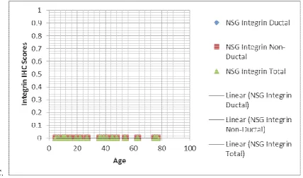
integrin αvβ3 in normal salivary gland ... 79
Figure 4-24 Correlation between age of patients with expression of OPN, CD44 and
integrin αvβ3 in PA ... 82
Figure 4-25 Correlation between ages of patients with expression of OPN, CD44 and
xiii
Figure 4-26 Correlation between age of patients with expression of OPN, CD44 and
integrin αvβ3 in ACC ... 88
Figure 4-27 Correlation between age of patients with expression of OPN, CD44 and integrin αvβ3 in PLGA ... 91
Figure 4-28 Correlation between age of patients with expression of OPN, CD44 and integrin αvβ3 in ACA ... 94
Figure 4-29 IHC expression (median) between male and female in NSG ... 95
Figure 4-30 IHC expression (median) between male and female in PA ... 96
Figure 4-31 IHC expression (median) between male and female in ACA ... 98
Figure 4-32 IHC expression (median) between male and female in ACC ... 99
Figure 4-33 IHC expression (median) between male and female in MEC ... 101
Figure 4-34 IHC expression (median) between male and female in PLGA... 103
Figure 4-35 IHC expression (median) between major and minor SGs in PA ... 104
Figure 4-36 IHC expression (median) between major and minor SGs in ACA ... 106
Figure 4-37 IHC expression between major and minor SGs in MEC ... 108
Figure 4-38 OPN IHC staining comparing low grade versus intermediate/high grade MEC ... 110
Figure 4-39 OPN IHC staining comparing low grade versus intermediate/high grade ACC ... 111
Figure 4-40 Integrin αvβ3 IHC staining in sub-epithelial endothelial cells (original magnification 200x) ... 113
xiv
Figure 4-42 Integrin αvβ3 IHC staining in endothelial cells within tumours (original
magnification 200x) ... 115
xv
List of Abbreviations
List in alphabetical order
ACA Acinic cell adenocarcinoma
ACC Adenoid cystic carcinoma
AFIP Armed Force Institute of Pathology
AJCC American Joint Committee on Cancer
bFGF Basic fibroblast growth factor
CAM Cell adhesion molecule
CT Computerized tomography
DAB 3, 3’-diaminobenzidine tetrahydrochloride
DNA Deoxyribonucleic acid
ECM Extracellular matrix
EGF Epithelial growth factor
Eta-1 Early T-lymphocyte activation gene 1
FAK Focal adhesion kinase
GRGDS Glycine-arginine-glycine-aspartate-serine
HA Hyaluronan
H&E Hematoxylin and eosin
HPF High power field
xvi
IHC Immunohistochemistry
IL Interleukin
kDa Kilodalton
LDL Low density lipoprotein
MAPK Mitogen-activated protein kinase
MEC Mucoepidermoid carcinoma
MMP Matrix metalloproteinase
MRI Magnetic resonance imaging
OPN Osteopontin
PA Pleomorphic adenoma
PAS Periodic Acid-Schiff
PDGF Platelet derived growth factor
PG Parotid gland
PGCG Peripheral giant cell granuloma
PLGA Polymorphous low grade adenocarcinoma
PTK Protein-tyrosine kinase
RFLP Restriction fragment length polymorphism
RGD Arginine-glycine-aspartate
RNA Ribonucleic acid
xvii
SCC Squamous cell carcinoma
sCD44 Soluble CD44
SG Salivary gland
SIBLING Small integrin-binding ligand N-linked glycoprotein
sIg Secretory immunoglobulin
SLG Sublingual gland
SMG Submandibular gland
TGF Transforming growth factor
uPA Urokinase-type plasminogen activator
VEG Von Ebner glands
VEGF Vascular endothelial growth factor
Chapter 1
1
Introduction
1.1 Salivary Glands (SGs)
Human saliva is produced by 3 pairs of major SGs and numerous minor mucous and
serous SGs. The 3 major SGs are: 1) parotid glands (PGs), 2) submandibular glands
(SMGs) and 3) sublingual glands (SLGs). There are approximately 600 to 1000 minor
SGs dispersed throughout the upper aerodigestive tract (1, 2, and 3).
Parotid Gland (PG)
PGs are the largest paired SGs by size but are only responsible for producing
approximately 25% of the total volume of saliva (1, and 3). Its autonomic function is
influenced by parasympathetic fibres from the inferior salivary nucleus via the lesser
petrosal nerve, a branch of the glossopharyngeal nerve (4). PGs are contained within a
dense fibrous capsule along with lymphatic tissues located in close proximity to the
ramus of the mandible. In an adult, it weighs about 15-30g. It contains 2 lobes, a
superficial lobe and a deep lobe (4). The 2 lobes are separated anatomically by the facial
nerve and its branches. PGs secrete saliva via Stenson’s ducts, which pierce through the
buccal mucosa at the level of the maxillary second permanent molar. The superficial
lobe represents 75 – 80% of a PG, while the deep lobe represents 20 – 25%. Up to 80%
of all SG neoplasms occur in the PGs, and the majority of them occur in the superficial
lobe (1, 2, and 3). PGs consist mainly of serous cells with multiple fibrous septa dividing
the individual lobules.
Submandibular Gland (SMG)
SMGs are the second largest paired SGs, and are responsible for secreting up to 70% of
the total salivary volume (3). Its autonomic function is influenced by parasympathetic
fibres from the superior salivary nucleus via the chorda tympani, a branch of the facial
nerve. It weighs approximately 7 to 10 g and is located within the submandibular triangle
individual, the inferior aspect of the SMG is often visible below the angle of the
mandible. Saliva produced from SMGs is secreted into the oral cavity via Wharton’s duct
which opens just lateral to the frenulum of the tongue in the anterior floor of the mouth.
SMGs account for up to 80% of sialolithiasis due to the torturous and up-sloping course
of Wharton’s duct (1). SMGs contain mostly mucous glands that are capped with
crescent-shaped serous demilunes.
Sublingual Gland (SLG)
SLGs are the smallest of the major SGs. They are located within the oral cavity just
above the mylohyoid muscle and immediately below the mucosa of the floor of the
mouth (2, and 3). They have the same innervation as the SMGs, and each weighs only 3
g (2, and 3). SLGs often secrete saliva via multiple small ducts, called the ducts of
Rivinus, which can drain either directly into the floor of the mouth or into Wharton’s duct
or through the sublingual duct of Bartholin. Neoplasms are rarely found in SLGs, but,
the neoplasms of the SLG are often malignant. Their lymphatic drainage is into both the
submental and submandibular lymph nodes (4). SLGs contain mixed cell types,
composed of both mucous cells and serous demilunes.
Minor Salivary Glands
There are more than 600 minor SGs scattered throughout the oral cavity and upper
aerodigestive tract (1, 2, and 3). They are composed of aggregates of mucous glands and
ducts which are not encapsulated. They secrete saliva directly into the oral cavity via
individual ducts. There is a set of unique minor salivary glands, called Von Ebner glands
(VEG), located within the crypts of circumvallate papilla in the dorsum of the tongue.
The saliva from VEG contains lipase as well as normal salivary proteins (4).
1.2 Structure of Salivary Glands
SGs empty into the oral cavity via a main excretory duct, which progressively divides
into smaller interlobar and intralobular ducts that drain the lobes of SGs (4). The striated
duct, which is part of intralobular duct, plays a significant role in the modification of
the striated duct. Salivary gland units contain secretory and non-secretory acinar cells
and ductal cells (4).
Cell Types
There are 3 main cell types found within the secretory units: mucous cells, myoepithelial
cells and serous cells (4). Mucous and serous cells are the main secretory cells of the
SGs, which differ in their structure and secreted end products (4).
Serous cells are pyramidal in shape with spherical nuclei. They produce proteins and
glycoproteins that contain antimicrobial, enzymatic and calcium-binding functions. Their
secretory granules are deeply basophilic with hematoxylin and eosin (H&E) stains (4).
Mucous cells are tubular in shape and are arranged adjacent to each other around a large
central lumen. The end product of mucous cells is mainly mucins which function
primarily as lubricants. The mucous appears unstained with routine H&E, however,
mucous can be stained using special stains such as Periodic Acid-Schiff (PAS) or Alcian
blue (1, and 4).
Myoepithelial cells are epithelial in origin but contain contractile elements similar to
smooth muscle cells. They surround the intercalated ducts and secretory units of SGs.
These cells appear flattened and lie between the basal cytoplasmic membrane of secretory
or ductal cells and the basement membrane. Contractions of myoepithelial cells aid the
secretion of saliva by acinar cells into the lumen, and the propulsion of saliva from the
intercalated duct towards the striated duct and eventually to the excretory duct (4).
1.3 Saliva
The production and secretion of saliva occurs in 2 stages under the control of the
autonomic nervous system, gustatory system and olfactory system. The first stage occurs
in the acinar cells, under the influence of neurotransmitters, and results in the activation
of ion channels causing fluid secretion, which is an isotonic secretion. As this fluid
travels down the ductal system, the second stage of salivation occurs. This involves fluid
trace amounts of serum protein. However, in disease states, especially inflammatory
conditions, the level of secreted proteins can greatly increase (5).
The main functions of saliva are: 1) to lubricate the oral mucosa to prevent drying and
trauma, 2) to aid in the digestion of food, 3) to mediate taste, 4) to control microbial
overgrowth, and 5) to maintain oral pH to aid remineralization of teeth (5).
(1) Saliva acts as a barrier against irritants, carcinogens and desiccation from mouth
breathing. It also protects the oral mucosa from physical trauma during mastication and
speech production (5).
(2) Saliva contains the hydrolytic enzymes amylase and lipase. Their role in
digestion is somewhat redundant in a healthy adult because (1) the transit time in the oral
cavity and the esophagus is insufficient for the enzyme to work effectively, and these
enzymes are degraded by acid in the stomach, (2) bile salts produced from the liver and
excreted from the gall bladder emulsify lipids and allow lipase to work more effectively
in the small intestine, and (3) the pancreas secretes amylase and numerous
disaccharidases to breakdown polysaccharides into absorbable forms. However, in young
infants, especially those born pre-term, the production of pancreatic enzymes is very low,
and infants are dependent on oral amylase to digest breast milk or infant formula (6).
(3) Saliva acts as a solvent for chemicals in food. It also acts as a vehicle by which
these chemicals are delivered to the taste buds. It should be noted that patients with
xerostomia complain of a decreased sensation of taste (7).
(4) Proteins related to the innate and humoral immune systems are secreted in saliva.
These proteins include lysozymes, salivary peroxidases, lactoferrin, histatins and
secretory IgA (sIgA) (5). These secretory proteins have antimicrobial properties and
assist in viral neutralization. Histatins are low molecular weight proteins rich in histidine
and have potent antifungal activity. It has been shown that people whose saliva is low in
histatins are more susceptible to oral thrush (8).
(5) Acidic environments can cause the demineralization of teeth. Saliva is
surface. As well, the bicarbonate in the saliva helps buffer the pH of the oral cavity to
maintain a neutral pH which favours remineralization of dentition (5).
Over the past decade, saliva has been a useful adjunct in the diagnosis of clinical
diseases, for example, measuring the cortisol levels in saliva for Cushing syndrome (9).
Recently, other cellular proteins have been identified in whole saliva such as IL-1β, IL-6,
IL-8, and OPN (10, and 11).
1.4 Pathology of the Salivary Glands
There are many different abnormalities and diseases of the salivary glands, including
developmental conditions, infections, immune-related disorders, neoplasms and
inflammatory conditions. Inflammatory conditions are the most common, especially the
mucocele, cases of which are readily available to use as salivary gland control tissue in
the study of salivary gland neoplasia.
1.4.1.1
Mucocele
Mucoceles are often found in the minor salivary glands, especially on the lower lip in a
paramedian fashion. Mucoceles are often painless. However, larger lesions can cause
mild discomfort. They are best described as soft, fluid-filled vesicles that are fluctuant
and often measure less than 1 cm in diameter. They appear bluish in colour because of
the mucin. However, deeper mucoceles are frequently grayish in colour, and found on
the ventral surface of the tongue and the floor of the mouth (1, 2, and 3).
Clinical Behaviour
Mucoceles are benign and occur commonly on the lower lip in all age groups. They are
associated with a history of trauma. However, many patients do not recall a traumatic
insult. Other lesions that may appear blue must also be considered in the differential
diagnosis; especially low grade mucoepidermoid carcinomas. Cavernous hemangiomas,
lymphangiomas and venous varices may also give a similar appearance. Although rare, a
vascular leiomyoma appears blue and has a predilection for the lower lip as well (1, 2,
Histological Features
Microscopically, mucoceles appear as granulation tissue surrounding mucin without a
true epithelial lining. This is a distinguishing feature from mucous retention cysts. In
addition, neutrophils and foamy histiocytes are usually present in mucoceles (1, and 3).
Treatment and Prognosis
Minor salivary gland mucoceles are managed by local excision of the associated salivary
gland as well as the overlying epithelium. Excision can help establish the diagnosis and
cure the condition with little chance of recurrence. However, care must be taken during
the excision as to not transect another minor salivary gland, as this can produce a new
mucocele in the same location (2).
1.4.2
Salivary Gland Neoplasia
SG carcinomas represent approximately 3% of all head and neck malignancies excluding
lymphoma. In general SG tumours comprise less than 0.5% of all malignancies. In
Canada, the annual incidence of SG tumours is between 2 – 3 cases per 100,000
population. The trend of SG tumours in Canada over the past 15 years had been
relatively stable. Unlike the most common head and neck neoplasm, squamous cell
carcinoma (SCC), smoking and heavy alcohol consumption do not appear to be risk
factors for SG tumour development. However, there are suggestions from the literature
that occupational exposures to volatile solvents and early exposure to ionizing radiation
may lead to the development of certain SG tumours (1, and 3).
1.4.2.1
Benign Salivary Gland Tumours
There are a number of different benign salivary gland tumours including basal cell
adenoma, canalicular adenoma, ductal papillomas, Warthin’s tumour, oncocytoma and
myoepithelioma. By far the most common is the pleomorphic adenoma (PA).
1.4.2.1.1
Pleomorphic Adenoma (PA)
Pleomorphic adenomas (PAs) are the most common form of salivary gland tumours. Up
superficial lobe. However, they can arise from submandibular, sublingual and minor
salivary glands as well. The peak incidence of PAs occurs between the 4th and 7th
decade of life, and there is a slight female predilection with a 1.4 to 1 female to male ratio
(1, 2, and 12).
Clinical Behaviour
Patients with PAs often present with a slow-growing, mobile, painless mass of unknown
duration. The usual presentation is commonly noted as an incidental finding by a
physician or a dentist during a clinical examination for an unrelated issue. Most PAs are
found in the superficial lobe of parotid gland, and they rarely cause facial paralysis due to
compression. However, when a PA is found in the deep lobe of the parotid gland, it can
extend into the parapharyngeal space causing obstructive symptoms. When present in
minor salivary glands, PAs present as a submucosal mobile mass usually without
ulceration, but when noted in the palate, it is often fixed because palatal mucosa is tightly
bounded to underlying bone. Although, benign and slow growing in nature, if left
untreated, up to 3 – 4% of PAs may become carcinoma ex-pleomorphic adenomas, which
are aggressive malignancies that may metastasize and result in death (13).
In general, the etiology of PAs is unknown. However, in a subset of PAs, there appears
to be a decreased expression of p53, a tumour suppressor gene, and Ki-67, a cellular
marker for proliferation (13, and 14). As well, the MUC1 gene appears to play a role in
the recurrence of PAs after surgical intervention (15).
PAs are composed of neoplastic myoepithelial cells mixed with neoplastic ducts and
stroma. Certain cellular markers had been implicated in the malignant transformation of
PAs. The down-regulation of neural cell adhesion molecule (N-CAM) and a cytoplasmic
accumulation of β-catenin in PA are suggestive of malignant transformation (16).
A thorough history and physical examination is vital to aid in the diagnosis and treatment
of PA. However, imaging such as CT scans and MRIs can determine the location and
indeterminate, an incisional biopsy of a representative tissue sample can help yield a
definitive diagnosis (2).
Histological Features
PAs are characterized by heterogeneity of cytology and architecture. Heterogeneous in
nature, PAs can be broadly sub-classified into cellular and myxoid types (1, and 12).
The cellular type of PAs is mainly derived from scattered mucoid material separating the
epithelial elements into small groups of ductal structures, cellular nests, anastomosing
cords or foci of keratinizing squamous cells. In comparison, the myxoid type of PA is
composed of mesenchymal-like elements such as myxoid, chondroid, hyaline and
osseous regions with fewer epithelial components (12). Cellular PA is often surrounded
by a complete fibrous capsule of varying thickness with occasional small extensions of
neoplastic cells (12). However, myxoid types often have incomplete capsules, yet they
are often well demarcated from normal salivary gland tissues which allows for complete
surgical excision. Recurrence of PA is rare, and it is mainly due to incomplete excision
of the primary tumour or seeding of tumour in the surrounding tissue due to the rupture of
the tumour during excision. When it recurs, it is often multi-focal (1, and 2).
Treatment
The treatment of choice for PA is excision with a negative margin. As the majority of
PAs arise from the superficial parotid gland, the preferred treatment is a superficial or
total parotidectomy with facial nerve preservation (2). However, if the tumour is located
close to the main trunk of the facial nerve, capsular dissection has been shown to have a
greater than 95% success rate in achieving local control of the tumour. Because of the
high success rate of local control with surgical intervention alone, post-operative
radiation therapy (RT) is no longer advised, however, RT maybe used when PAs are
located in minor salivary glands that are not amenable for surgical resection (1, and 2).
The clinical outcome for surgical resection with negative margins for primary PAs has
been reported to have a 99% success rate without recurrence for greater than 10 years
(17). However, the success rate is only 70% at 10 years if repeated surgical intervention
is required (18). The risk of major complications is low; however, the risk of
complications is increased with salvage surgery for recurrent PA. The likelihood of
malignant transformation increases with the age of the patient and the duration of the
neoplasm (1, and 2).
1.4.2.2
Malignant Salivary Gland Tumours
As with benign salivary gland tumours, there is a variety of different malignant salivary
gland neoplasms, some of which are rare. To ensure sufficient available tissue for
research, this thesis investigates the 4 most common types.
1.4.2.2.1
Acinic Cell Adenocarcinoma (ACA)
Acinic cell adenocarcinomas (ACAs) are rare salivary gland tumours which account for
approximately 3% of all salivary gland malignancies. ACAs are almost exclusively
found in the parotid gland, in up to 80 - 99% of cases (1, 2, 3, and 12), but only accounts
for 2 – 4% of all parotid gland tumours.
Clinical Behaviour
ACA can appear in all age groups with a median age of occurrence at 52 years, which is
slightly earlier than most other salivary gland malignancies. Like other malignant
salivary gland neoplasms, ACA affects slightly more females than males with a ratio of
3:2. ACA often presents as a painful mass within the parotid gland. As it enlarges it can
affect peripheral nerves and nodal metastases can occur in 1 out of 5 cases. Another
unique aspect of ACAs is that 3% are found bilaterally, which ranks second only to
Warthin’s tumour (up to 15%) in bilateral occurrence (1, 2, and 3).
Histological Features
ACAs are often well circumscribed and may show some encapsulation, especially in the
eccentric nuclei and PAS positive cytoplasmic granules. Ductal cells tend to form a
syncytium and lack features of the other cells. Atypia may be present, but mitotic
figures are infrequent and are usually found in ductal cells. The stroma is usually
fibrovascular but may be hyalinized. A unique feature is the frequent presence of a
lymphoid infiltrate which contains germinal centres. Although encapsulated, evidence of
infiltration is often seen, yet, this does not correlate with clinical behaviour.
Multinodularity, hyalinization and infiltration are often seen in recurrent and
metastasizing tumours (1, and 12).
Treatment and Prognosis
ACAs tend to have a favourable outlook with a 10-year survival rate of 70-85%. Some
long term data suggests 20-year survival rates of over 65%. However, the poorest
prognosis of ACA has been associated with high grade lesions or the presence of
metastatic disease at the time of diagnosis (1, and 3).
It is widely believed that surgical excision is the primary mode of therapy for ACA.
Although, there is debate over which form is preferable: local excision versus radical
excision. Recent literature has suggested a less aggressive surgical approach for a
localized lesion with negative node status (2). Post-operative radiation therapy is often
employed to improve the clinical outcome. However, emerging data suggests that
neutron beam radiation is more effective than conventional radiation. Unfortunately, like
many salivary gland malignancies, ACA is highly resistant to chemotherapy (2).
1.4.2.2.2
Adenoid Cystic Carcinoma (ACC)
Adenoid cystic carcinomas (ACCs) are the second most common type of malignant
salivary gland neoplasms and account for 25% of all malignant salivary gland tumours.
It is most commonly found in the submandibular gland. In parotid glands, it constitutes
up to 15% of all malignancies. It is rarely found in the sublingual gland. In minor
salivary glands, more than half of ACCs occur in the palate. It has a wide age
distribution; however, it is often diagnosed between the 4th and 6th decades of life, with
salivary gland malignancies, ACC has no known etiology. However, there is evidence
that some forms of aggressive ACC are involved with p53 tumour suppressor gene
inactivation (19, and 20).
Clinical Behaviour
ACCs often grow very slowly and have a relatively indolent but relentless course. Often
it can present as a firm painless mass that progressively enlarges. Advanced tumours
may present with pain and nerve paralysis. ACC have a propensity to invade peripheral
nerves. Although, ACC have a good 5 year survival rate of approximately 90%, because
of the associated perineural invasion at the time of definitive treatment, it often recurs
and recurrences have a poor 15 year survival rate of less than 40% (1, 2, and 3).
Unlike most carcinomas, ACC seldom metastasizes to regional lymph nodes. However,
distant metastases are common in late diagnoses. The lung is the most common site of
metastases, while the liver is the second most common location. Bone metastases
indicate a fulminant course of ACC and are associated with a guarded prognosis (1, 3).
Histological Features
The diagnosis of ACC is made by histological assessment of a biopsy or surgical
resection specimen. There are three major histological patterns of ACC: (1) cribriform,
(2) tubular and (3) solid. The solid pattern is often associated with a clinically aggressive
course. The tumours are classified based on the predominant histological type, as ‘pure’
patterns rarely occur. Rather, there tends to be a combination of two or three patterns
within a single tumour (1, and 12).
The classic cribriform type is composed of small, basophilic, uniform cells that are
arranged into islands or anastomosing cords, with a pathognomonic “Swiss-cheese”
pattern. The tubular type is composed of either single or multiple layers of basaloid cells
that are arranged in small ducts with a single lumen. The solid type, unlike the cribriform
or tubular types, is composed of sheets of basaloid cells with scanty cytoplasm and often
Treatment
Primary treatment of ACC is complete surgical resection of the tumour if possible.
However, some studies had advocated post-operative radiotherapy, which can limit local
failure from microscopically positive margins. Unfortunately, ACCs are resistant to
chemotherapy, thus radiation therapy is used for metastatic disease (2).
Prognosis
The solid type of ACC shows the worst clinical outcome of all 3 histological patterns (1).
The long term survival of ACC remains low. This is due to its high rate of perineural
invasion and its infiltrative characteristics which pose a challenge for the surgeon to be
able to achieve a clean surgical margin (2).
1.4.2.2.3
Mucoepidermoid Carcinoma (MEC)
Mucoepidermoid carcinomas (MECs) are the most common form of all malignant
salivary gland tumours. However, it only accounts for 10 - 15% of all salivary gland
tumours. It is most commonly found in the parotid gland. The peak incidence of MEC
occurs between the 3rd and 5th decades of life, with a slight female predilection. It is
also the most common salivary gland malignancy in children (1, 3, 4, and 12).
Clinical Behaviour
Patients with MEC often complain of a painless, solitary, firm mass. The majority of
patients are asymptomatic during the early course of the disease, though, over time,
patients with high grade or advanced lesions will report symptoms such as tenderness,
pain, drainage, and dysphagia. When lesions are located in the parotid gland, trismus and
facial nerve paralysis can occur (2). In rare instances, MECs show an accelerated growth
pattern without a period of slow or absent growth (2).
In minor salivary glands, MEC are often discovered incidentally during a dental
examination. They are often found in the palate, retromolar pad, floor of the mouth,
buccal mucosa, lips or tongue. These MECs present as a bluish smooth submucosal
through the mucosal surface may be interpreted as a draining dental abscess. Over time,
these swellings become symptomatic with paresthesia or numbness of the dentition,
ulceration or hemorrhage (1, 2, and 3).
Histological Features
The etiology of MEC is unknown. Diagnosis of MEC is based on clinical and
microscopic examination of the lesion. Histologically, it is usually divided into 3 main
types of cells: (1) mucin producing mucous cells, (2) intermediate cells, and (3)
epidermoid cells with squamous differentiation. The term ‘mucoepidermoid’ is derived
from the combination of mucous and epidermoid cells found within the lesion. Aside
from the 3 main cell types, columnar and clear cells are occasionally found, which can
proliferate in a focal area or combine with the main cell types in a cystic or solid pattern
(1, and 12).
Mucous cells are the neoplastic cells containing mucin that are important for the
diagnosis and subsequently for determining the prognosis of the MEC. Mucous cells are
relatively large with polymorphism and have foamy cytoplasm resembling the mucous
acini of normal salivary glands. In most MECs, mucous cells often constitute less than
10% of the lesion. They are more prevalent in low grade rather than in high grade
lesions (1, and 12).
Intermediate cells are highly proliferative, and are found in small clusters or in solid
sheets combined with mucous and epidermoid cells. There is a gradual transition of
intermediate cells from the smaller basaloid cells to larger cells that are 2 to 3 times the
size of the original basaloid cells. Further increases in size and content lead to the
identification of epidermoid cells. Thus, at the late stage of differentiation of
intermediate cells, it is often difficult to distinguish large intermediate cells from
epidermoid cells (1, and 12).
Epidermoid cells tend to form small nests or they line the cystic spaces that contain
mucous and intermediate cells. Occasionally, they form keratin pearls or they are
Grading
MEC is divided into low, intermediate and high grades. In low grade lesions, prominent
cystic structures of varying sizes are filled with mucin. Cysts are mainly lined by a
single layer of mucus-secreting columnar cells with few mitotic figures and little cellular
atypia (12). With intermediate lesions, there are solid areas of squamous cells and
intermediate basaloid cells with increasing cellular atypia, mitotic figures and an
increased nuclear to cytoplasmic ratio (1, and 12). In high grade lesions, nests of
intermediate basaloid cells and epidermoid cells are present with few cystic components.
Ductal structures are rarely found. There are high numbers of mitotic figures, greater
than 4/10 high power fields (hpf), sometimes with necrosis and perineural invasion (9).
Tumour makers (p27 and Ki-67) have been identified which correlate with aggressive
behavior and a poor prognosis. (21)
Treatment and Prognosis
The main treatment of MEC is surgical intervention. The overall 5-year survival rate of
MEC is over 85%. More specifically, the 5-year survival rate of low grade lesions is
greater than 90%, while for high grade lesions, it is below 50% (1, and 2). Yet, overall
outcome mainly depends on the clinical stage, tumour grade and location of the MEC.
Although, tumour grade may predict the aggressiveness of the lesion, it is now known
that the TNM clinical staging system is a better indicator of prognosis in patients with
MEC. MEC often metastasizes to bone, the lungs and the brain, which implies poor
prognosis (1, and 2). Patients usually succumb to the disease due to local recurrences or
distant metastases.
1.4.2.2.4
Polymorphous Low Grade Adenocarcinoma (PLGA)
Polymorphous low grade adenocarcinoma (PLGA) is almost exclusively found on minor
salivary glands, with the palate being the most common site. Its peak incidence is
between the 3rd to 7th decades of life, with a median at the mid-6th decade. PLGA has a
slight female predilection (1, and 12).
PLGA is a slowly growing, often painless mass. It is considered to be a low grade
neoplasm with a good long term prognosis and almost no metastatic potential. Early
lesions can often be mistaken for a benign lesion such as a pleomorphic adenoma
microscopically if the surgical biopsy specimen is small and inadequate. Because it
presents as a palatal mass with an intact mucosal surface, it can be clinically mistaken for
pleomorphic adenoma as well. Adenoid cystic carcinomas and low-grade
mucoepidermoid carcinomas should be considered in the clinical differential diagnosis
(1, 3, and 12).
Histological Features
Microscopically, it appears as a non-encapsulated lesion with infiltrative borders and a
variable growth pattern. Several patterns occur: tubular, solid, papillary, microcystic,
cribriform, fascicular, pseudo-adenoid cystic, or strand-like. Occasionally, the nuclei are
hyperchromatic but are usually open faced. Mitotic figures are rare. The stroma may be
fibrous, myxoid, or hyalinized. PLGA may demonstrate significant infiltration with
invasion of small nerves, vessel walls, and bone, which belies its low-grade behaviour.
Central necrosis is not seen. The peculiar histologic pattern of the tumour does not
appear to relate to its clinical behaviour. Special cellular markers such as alpha-smooth
muscle actin, and c-kit have been used to distinguish equivocal cases of ACC and PLGA
(22).
Treatment and Prognosis
Primary treatment for PLGA is surgical excision with 1.5 cm peripheral margins. This is
despite the tumour’s tendency for perineural invasion and its lack of a capsule (2). The
decision to pursue a palatectomy depends on radiographic or clinical evidence
demonstrating erosion of the palate. Radiotherapy is not as effective as surgery and is
often not required (2).
PLGA generally has a good prognosis with a 5 year survival rate of 85%. Recurrence
and distant metastases are rare if the surgical margins are negative. Like many
lungs and bones. Primary treatment remains surgical excision with wide peripheral
margins and often involves partial maxillectomies or mandibulectomies (2). Radiation
therapy may be used post-operatively to improve clinical outcomes (2).
1.5 Osteopontin (OPN) and its receptors
1.5.1
Osteopontin (OPN)
Osteopontin (OPN) is an acidic phosphoglycoprotein and is a member of the SIBLING
(Small integrin-binding ligand N-linked glycoprotein) family, which also includes bone
sialoprotein, dentin matrix protein-1, dentin sialophosphoprotein, and matrix extracellular
phosphoglycoprotein, which can modulate cell behaviour by autocrine and paracrine
mechanisms by interacting with cell surface receptors such as integrin (23). This
glycine-arginine-glycine-aspartate-serine (GRGDS) integrin binding motif is also presented in
fibronectin, vitronectin and a variety of extracellular proteins that bind to members of the
integrin family of cell surface receptors.
OPN is a monomer protein which undergoes extensive post-translational modification
including phosphorylation, glycosylation, and cleavage at variable sites. This results in a
variable molecular mass that ranges from 25 to 75 kDa. Originally, OPN expression was
generally considered to be limited to the bones and dentition of adults, because OPN is
highly expressed in bone matrix, and is regulated by osteotropic hormones (24).
However, studies since the mid-1990s have found OPN expression in numerous soft
tissues such as kidneys, mammary glands, and salivary glands (25, 26, 27, 28, 29, and
30).
1.5.2
OPN gene, its structure and adhesion motifs
In 1992, Crivello isolated the cDNA and gene encoding for OPN and concluded that
OPN is poorly to moderately conserved between several species of animals including
humans. Yet, its functional domains are conserved. Its amino and carboxylic terminal
regions and the approximately 50 amino acids surrounding the RGD signaling sequence
RGD integrin binding domains, similar phosphorylation and glycosylation motifs and
sites for controlled proteolysis (23).
In humans, the OPN gene is mapped to chromosome 4q13, and Bg/II restriction fragment
length polymorphism (RFLP) has been described. A particular RLFPs mouse OPN allele
has been described to enhance resistance to infection by bacteria. The mechanism of
resistance may involve the interference of Ca2+ uptake or the interaction of the causative
agent with the host cell surface receptor (32).
Numerous growth factors, hormones, tumour promoters and oncogenes are involved in
the regulation of OPN expression. Denhardt sequenced 1kb upstream of the mouse OPN
promoter region and searched for transcription factor recognition sites. Aside from the
characteristic promoter regions such as TATA box, CCAAT box, and GC box, other
recognition sites such as Sp1, CTF/NF-1 and AP-1, were found. These regions and sites
may have contributed to the ubiquitous nature of OPN gene expression in various tissues
(26). OPN expression is also augmented at higher levels in different transformed cell
lines compared to the non-tumourigenic counterparts (33).
The RGD integrin binding domain mediates interactions via αvβ1, αvβ3, αvβ5, αvβ6, α5β1,
and α8β1 (34, 35, 36, and 37). Adjacent to the RGD domain at the C-terminus is a cryptic
SVVYGLR sequence that is exposed upon cleavage with thrombin. This cryptic domain
interacts with more integrins such as α4β1, α4β7, and α9β1 (38, 39, and 40). Aside from
binding with integrin, OPN also interacts with CD44, a hyaluronic acid receptor (41). A
number of different CD44 isoforms exist, as the mRNA of CD44 has a variable splicing
region where 10 variant exons exist via differential splicing. OPN does not bind to the
standard isoform (42), but, OPN binds to some of the CD44 splice variants such as v6
and v7 (43). However, the exact mechanism of domain binding has not yet been
elucidated (44).
1.5.3
OPN physiologic functions
An increase in OPN transcription has the ability to activate T-lymphocytes, thus the
alternative name for OPN is ‘the early T-lymphocyte activation gene 1’ (Eta-1) (32, and
33). Early research has shown that Eta-1/OPN has the ability to enhance resistance to
flaviviruses and Rickettsia infection. In mouse models, rotavirus infections induce OPN
expression, which decreases the severity of the disease. OPN deficient mice infected
with rotavirus demonstrated more severe diarrhea when compared to wild type mice.
OPN acts as a potent chemoattractant that aids the maturation of macrophages, which
upregulate their own OPN expression via autocrine routes. It has been demonstrated that
upregulation of OPN occurs with ischemic injury such as stroke, acute tubular necrosis
and myocardial necrosis (45, 46, and 47). However, with activation of T-lymphocytes
and the recruitment of activated macrophages, increased OPN expression may actually
increase the amount of tissue damage.
Although, there are low levels of OPN expression in neutrophils, OPN is important for
the recruitment and migration of neutrophils. In two separate experiments using
OPN-null mice, a reduced chemotaxis of neutrophils was demonstrated in sodium periodate
induced peritonitis and concanavalin-A induced hepatitis in liver (48, and 49).
Interaction of OPN with neutrophils has been identified via α9β1 integrin binding (50).
Osseous function
OPN, a secreted acidic glycosylated phosphoprotein, contains a high content of serine
and aspartate residues, which are associated with osteocalcin (51). It has the ability to
bind to the hydroxyapatite matrix of bone via the aspartic acid-rich region. OPN is
secreted by osteoblasts, osteocytes and osteoclasts, and highly expressed in chondrocytes
days before the onset of mineralization (52). It is found in abundance in cell-matrix and
matrix-matrix interfaces in mineralized tissues. It is hypothesized that OPN may protect
the surface of exposed bone or prime it for cell-matrix interaction (53). It acts as an
opsonin to facilitate macrophage binding to mineralized tissue debris. It can bind to type
I collagen, fibronectin, and osteocalcin to improve the physical strength of the matrix
in OPN knock out mice, yet it appears that OPN is involved in bone remodeling, but it is
not essential for it to occur (55, and 56).
Osteopetrosis, a condition of impaired bone resorption caused by a failure of normal
osteoclastic activity, is associated with higher levels of OPN expression in rats (57).
High levels of calcitriol (Vitamin D3) are also associated with osteopetrosis. This is
likely because OPN expression is stimulated by calcitriol, which normally promotes
osteoclast differentiation and bone resorption (57). It is now known that phosphorylation
of the serine residues of OPN is required for proper cell-matrix interaction with β3
integrin and the GDS sequence to stimulate osteoclast attachment (58). Osteoblast
attachment is independent of β3, yet it still requires the GDS sequence.
In vivo experiments found that mechanical stresses on osteoblast-like cells lead to
elevated OPN and Type 1 collagen mRNA (59). It is postulated that the stimulation of
mechanosensitive channels lead to the influx of intracellular calcium ions and modify
gene expression.
Wound Healing
OPN is an important excreted molecule that recruits inflammatory cells to the site of
injury. Whether OPN is pro-inflammatory or pro-healing has not yet been established.
An incisional wound on OPN-null mice showed alterations in the architecture of the
extracellular matrix and collagen fiber diameter, and resulted in more residual debris
(60). Research of localized OPN knockdown using antisense oligodeoxynucleotides,
further supported earlier findings of smaller diameter collagen fibrils when compared to
controls (61).
Macrophages at the wound site of the OPN-null mice expressed increased levels of
mannose receptors, which is associated with reduced pro-inflammatory cytokine
secretion and increased pro-healing cytokines (62). As well, decreased migration of
inflammatory cells at the wound site resulted in a reduction of granulation tissue
formation and scarring with accelerated healing (61). OPN directly binds to type I
organization, ECM formation and myofibroblast differentiation via TGF-β1 (63, 64, and
65).
1.5.4
OPN and Pathologies
Atherosclerosis and Vascular Diseases
Atherosclerosis is considered to be a result of chronic inflammation of the vasculature.
Given the role of OPN in inflammatory processes such as T-cell activation and the
recruitment of macrophages, a number of studies have investigated the role of OPN in the
progression of atherosclerosis. A hyperlipidemic mouse model, comprised of mice with
ApoE-deficiency showed an accelerated formation of atherosclerotic lesions as compared
to wild type mice. However, OPN and ApoE deficient mice demonstrated a significant
reduction of atherosclerotic lesions when compared to ApoE only deficient mice after 36
weeks of a normal diet (66). A similar study with a triple knockout of ApoE, LDL
receptor and OPN showed that the OPN deficiency decreased the lesion size of
atherosclerotic plaques (67). In humans, OPN is expressed in smooth muscle cells,
endothelial cells and macrophages in atherosclerotic lesions (68, and 69). Animal models
showed that OPN promotes smooth muscle cell migration and proliferation (70). Thus,
OPN enhances the proliferation, migration and accumulation of smooth muscle cells and
endothelial cells in repair and remodeling processes of the vasculature.
Renal stone and glomerulonephritis
In mice, OPN is abundant in the nephron, particularly at the loop of Henle and distal
convoluted tubules (71). In pathology, OPN can be found in the epithelial lining of
Bowman’s capsule. In tubulointerstitial nephritis, focal increases of OPN can be found at
sites where macrophages accumulate (68).
OPN appears to play a dual role in both the formation of and protection against calcium
oxalate stone formation. When OPN is secreted into the tubular fluid, often in a
hypo-phosphorylated form, it acts to suppress the growth of calcium oxalate crystals by
disrupting the crystallization process (72). In contrast when calcium oxalate crystals are
epithelial cells. OPN upregulation appears in response to hypoxic and reperfusion
injury. Young kidneys can readily increase OPN production in response to hypoxic
injury as compared to old kidneys, and the OPN production decreases intra-cellular Ca2+
and nitric oxide production thus protecting the cells from injury (26).
Tumourigenesis
Tumourigenesis is a multi-step process (Knudson hypothesis) that transforms normal
cells into neoplastic cells (73, and 74). There are numerous mechanisms within the body
to defend against malignant transformations such as the host immune system and cellular
repair system. However, when tumourigenesis steps escape or evade the cellular and/or
immunological check points, a gradual build-up of non-lethal transformations can
eventually lead to a neoplasm. In order for cells to evade and escape cellular and
immunological mechanisms that can lead to the cells’ destruction, these cells have to
have acquired the following cellular changes and abilities: 1.Evading apoptosis, 2.
Sustaining growth signaling, 3. Inducing angiogenesis, 4. Evading growth suppression, 5.
Replication immortality, and 6. Ability to invade and metastasize.
OPN has been implicated in several of the 6 key characteristics of metastatic neoplasms.
Studies using OPN deficient mice models injected with melanoma cells (75) and breast
cancer cells (76) have implicated OPN in sustaining a more rapid tumour growth.
Recently, He et al. found that the presence of 2 other variant splicings of OPN was a
prognostic factor for tumour aggressiveness and metastasis, especially OPN-c (77, and
78).
Evasion of Apoptosis
Transformed cells are distinct from normal cells, in that transformed cells express
different cell surface antigens that can be recognized by the host immune system and be
removed from the environment via signal induced apoptosis. However, some
transformed cells acquire the ability to alter the apoptotic signal cascade to resist
apoptotic cell death. OPN promotes the migration of lung cancer cells through integrin
pathways (79). OPN may enhance cancer cell survival by activating the Akt pathway,
upregulating GAS6 (80), and its interaction with CD44 and integrins via inside-out
signaling activation of src (81).
Induction of Angiogenesis
In order to maintain limitless growth potential of tumour cells, tumour cells must acquire
and maintain an independent vascular supply to maintain adequate nutrition. It has been
demonstrated that OPN binding to certain integrin receptors induces angiogenesis (34).
Binding of OPN to integrin αvβ3 increased the expression of vascular endothelial growth
factor (VEGF) in endothelial cells. This overexpression allowed recruitment of
neovascularization. Aside from upregulating VEGF, OPN interacts with integrin αvβ3
and activates connective tissue growth factor and cysteine-rich angiogenic inducer 61,
which enhances neovascularization and tumour growth in vivo (80). OPN signaling via
NF-κB also prevents endothelial cell apoptosis (82).
Invasion and Metastasis
For a malignant tumour to establish a secondary growth site within the host, numerous
events must take place: invasion of the basement membrane and extracellular matrix;
evasion of host immunity via either lymphatic or hematological spread, attachment to a
distant site, and invasion of the basement membrane at the distant site.
The first step of metastasis is the invasion of the tumour cell into the extracellular matrix.
To penetrate through the basement membrane, the tumour cells need to secrete
appropriate proteases. One group of proteases that can enable the degradation of the
basement membrane is the matrix metalloproteinases (MMP). OPN had been shown to
increase expression of MMP-1 and MMP-2 (83). MMP-2 levels correlate with tumour
invasiveness and metastasis in murine melanoma cells (84). Breast cancer cell lines,
when treated with OPN, develop increased levels of urokinase-type plasminogen
activator (uPA). This uPA can indirectly activate MMP-1, 2, 3, 9 and 14 (85, 86, and
87). Each of these MMPs can digest different ECM components thereby playing an
Once the tumour cells invade through the ECM and into the lymphatic or vascular
channels, these cells need to survive and avoid detection from the host immune system in
the vascular or lymphatic channels in order to establish a secondary growth centre.
Tumour derived OPN has been shown to inhibit NO-mediated tumour cell apoptosis in
colon adenocarcinoma via inhibition of inducible nitric oxide synthase in lymphocytes
and macrophages and blocking their functions (88).
OPN also plays an important role in establishing bone metastasis. It can activate TGF-β
and bFGF via the upregulation of uPA. Nemoto et al. and Chellaiah et al. were able to
demonstrate a lower incidence of bone metastasis in OPN knock-out mice models
injected with breast cancer cell lines (75, and 89). In addition, they were able to elicit
bone metastasis with the introduction of OPN.
Therapeutic Targets
There are currently several strategies and therapeutic approaches to modulate OPN
activity in tumour cells, especially those of breast cancer. OPN binding to CD44v has
been shown to promote migration and invasion of tumour cells; however, this interaction
requires thrombin cleavage of OPN to expose the SVVYGLR binding site. With this in
mind, a novel treatment using thrombin-inhibitor Argratroban has demonstrated
decreased cell growth, adhesion, and migration in breast cancer cells (90). Another
method is to directly inhibit OPN expression. Agelastatin A inhibits OPN and its use
causes reduced growth, adhesion and invasion of breast cancer cell lines (91). Thus,
targeting OPN appears to be a good approach in cancer management.
1.6 CD44
CD44 is a transmembrane glycoprotein with variable N and O-linked glycosylation sites
belonging to the family of cell adhesion molecules (CAMs) (92). They are ubiquitously
expressed in human tissues especially in the epithelium (93). They control cellular
behaviour by interacting with soluble proteins and the extracellular matrix (ECM) to
maintain tissue integrity. CD44 has 11 isoforms (1 standard and 10 variants) which are
total of 20 exons (94, and 95). All isoforms contain a large ectodomain, a transmembrane
domain, and a cytoplasmic domain. Variants are generated by alternate splicing of exons
6 to 15 mRNA inserting into the variable region in the large extracellular ectodomain
(96). The molecular weight for CD44 ranges from 80 to over 200 kDa (97), however, the
smallest standard isoform of CD44 (CD44s) is approximately 85 kDa (98).
CD44 is involved in many cell-cell and cell-matrix interactions which include adhesion,
migration and lymphocytic functions (99). Thus, by variant splicing, CD44 interacts with
many soluble proteins and ECM, however, the main ligand is hyaluronan (HA) (100). As
well, CD44 interacts with TGF-β, osteopontin, laminin and others (41).
The main physiological functions of the CD44 – HA interaction are: T cell proliferation,
IL-2 production, NK cell cytotoxicity and macrophage production of cytokines and
chemokines. Recent studies have shown that CD44 is involved in various mechanisms of
metastasis. The CD44 isoforms that have been implicated with malignancy include:
CD44s, CD44v3, CD44v5, CD44v6 and CD44v10. Although, HA is the principal ligand
of CD44, it is by no means the only one, osteopontin is one of several non-ECM ligands
which binds to CD44. Binding between OPN and CD44 and its variants occurs via the
RGD independent domain. It has been shown that OPN-CD44s and its variant
interactions facilitate adhesion, migration and stimulation of both normal and
transformed cells via activation of mitogen-activated protein kinase (MAPK) and
PI3K/Akt pathways. As well, CD44 variants co-operate with β1 containing integrins to
bind with OPN to stimulate cell motility and chemotaxis (41). Altered levels of
expression of OPN and CD44v6 were found in odontogenic tumours but not in
inflammatory or developmental odontogenic cysts (101).
The presence of CD44s and CD44v in salivary gland tissue has been reported (102, 103,
104, 105, and 106). Fonseca et al. further characterizes the expression pattern of CD44v
within normal salivary gland tissue (107). They found that 3 main variants exist: 1)
CD44v3 is positive in acinar and myoepithelial cells, but negative in mucus-producing
cells, 2) CD44v5 was consistently negative in all salivary gland tissues, and 3) CD44v6
further studies with CD44 in salivary gland pathology, a down regulation of v3 and v6
was shown to be associated with malignancy and an up-regulation was only associated
with PA (108). Recently, Wang et al. demonstrated CD44v3, v6 and v10 were associated
with head and neck squamous cell carcinoma (HNSCC) (101).
Soluble CD44 (sCD44) had been shown to modulate the function of cell surface CD44
and has been demonstrated to be up-regulated in many cancers. Chang et al. showed that
sCD44v6 was detected in oral squamous cell carcinomas, but was not a clinical
prognosticator for disease progression (109).
1.7 Integrin
Integrins are a family of transmembranous cell adhesion receptors, which are
heterodimeric glycoproteins that are composed of α and β subunits (110). The molecular
weight of an integrin is approximately 280 kDa, the larger α subunit ranges from 150 –
180 kDa, and a smaller β subunit is about 90 kDa (111). There are at least 18 α subunits
and 8 β subunits forming at least 24 combinations known to date (112). They bind to
collagens, laminins, or RGD-containing proteins such as OPN. As well, several integrins
can bind to cellular receptors and viruses such as adenovirus and human
immunodeficiency virus (113). Its net effect is cell-type specific and context dependent,
as integrin mediated adhesion via “outside-in” and “inside-out” activation can regulate
apoptosis, mitogenesis, cell differentiation, cell migration, as well as cell to cell adhesion
(111). Integrin is also involved in the progression of many common diseases such as
neoplasms, tumour metastasis, immune dysfunction, infections, osteoporosis and
coagulopathies (114, and 115).
There are several signal transduction pathways via integrin interactions, one of which is
focal adhesion kinase (FAK), which is a non-receptor protein-tyrosine kinase (PTK).
Immunofluorescence and in vitro and in vivo assays show that FAK is co-localized at the
C-terminus of integrin receptors and the phosphorylation of FAK at various tyrosine
residues transduces signals downstream and causes activation of the MAPK cascade.
Seig et al. showed that platelet derived growth factor (PDGF) and epithelial growth factor