pISSN 2320-1770 | eISSN 2320-1789
Research Article
Maternal history and second trimester uterine artery Doppler in the
assessment of risk for development of early and late onset pre-eclampsia
and intra uterine growth restriction
Humera Noor
1*, Mahapara Manzoor
1, Saima Sadiq
1, Naveed Shahzad
2INTRODUCTION
Pre-eclampsia is a multi-factorial, multi-system disorder
of human pregnancy defined by hypertension
(BP>140/90mm Hg) and proteinuria after 20 weeks of gestation in a previously normotensive women.1 It is more common in women who are pregnant for the first time, who have a family or past history of hypertensive pregnancy.2 It is thought to be consequence of impaired trophoblastic invasion of the maternal spiral arteries and their conversion from narrow to wide non-muscular channels.3,4 The classic concept that pregnancy induced
hypertension primarily originates from defective
placentation has recently been challenged. The concept of early and late onset pre-eclampsia is more modern and
widely accepted that two entities have different etiologies and should be regarded as different from the disease.5,6 Ness and Roberts suggested that it may be the common clinical end result of abnormal placentation as well as maternal factors.7
Early onset pre-eclampsia (<32 weeks>)8,9
Comprises small subgroup 5-20%
Impaired trophoblastic invasion of the spinal arteries.
Uterine artery Doppler waveform abnormalities
Signs of intra-uterine growth restriction
1Department of Obstetrics and Gynaecology, Government Medical College, Srinagar, India
2Department of Health and Medical Education, Kashmir, India
Received: 11 March 2016
Accepted: 07 April 2016
*Correspondence:
Dr.Humera Noor,
E-mail: [email protected]
Copyright: © the author(s), publisher and licensee Medip Academy. This is an open-access article distributed under the terms of the Creative Commons Attribution Non-Commercial License, which permits unrestricted non-commercial use, distribution, and reproduction in any medium, provided the original work is properly cited.
ABSTRACT
Background: To evaluate the value of one step uterine artery Doppler and maternal history in the prediction of early onset pre-eclampsia and intra uterine growth restriction in a random population
Methods: This study was conducted from 2012 to 2014 Lalla Ded hospital which is a tertiary care hospital associated with GMC Srinagar. This was a prospective study conducted on 200 pregnant women in the second trimester between 19-22 weeks of gestation. Singleton pregnant women were recruited from those attending antenatal care clinics at this hospital over a period of two years. Obstetric and medical history was taken. Transabdominal ultrasound was done and then patients were subjected to Doppler examination of uterine arteries.
Results: Mean age of study population was 26.3 years with maximum number of patients in 25-29 year age group. Total numbers of primigravida were 59.5%. Maternal history revealed that 9.5% had previous history of hypertensive pregnancy, 3.5% had history of IUGR. Early onset pre-eclampsia (<32 weeks) was present in 42.9% and late onset pre-eclampsia (>32 weeks) was present in 57.1% cases. Doppler abnormalities were present in 7.5%. In the patients who developed pre-eclampsia 57.1% had uterine artery Doppler abnormalities.
Conclusions: Maternal history and uterine artery Doppler at 19-22 weeks gestation is a single step test for the prediction of early onset(<32 weeks) and late onset pre-eclampsia and intra uterine growth restriction.
Keywords: Pregnancy, Primigravida, Pre-eclampsia, Doppler, IUGR
Late onset pre-eclampsia (>32 weeks)9
>80% cases worldwide.
Normal or slightly altered behaviour of the uterine artery vascular resistance expressed by an increased
pulsatilityindex or a diastolic notch in pregnancy having
pre-eclampsia or fetal growth restriction.10
The addition of maternal history to the uterine artery Doppler velocimetric studies in second trimester of pregnancy should be a single step for accurate prediction of pre-eclampsia and fetal growth restriction so as to allow judicious allocation of resources for monitoring and preventive measures to improve maternal and perinatal outcome.
Aims and objectives
1. To evaluate the value of one step uterine artery
Doppler in the prediction of early onset pre-eclampsia and intrauterine growth restriction in a random population
2. To ascertain whether uterine artery Doppler and
maternal history is effective in high risk pregnancies
3. To develop a predictive model for pre-eclampsia
using maternal history and uterine artery Doppler.
METHODS
This study was conducted in Lalla Ded Hospital which is a referral/tertiary care Obstetric and Gynaecology Hospital in Srinagar city catering to whole Kashmir province. This was a prospective study conducted on 200 pregnant women in the second trimester between 19-22 weeks of gestation. Singleton pregnant women were recruited from those attending antenatal care clinics at this hospital over a period of two years. Both primigravida and multigravida were included. The examination was performed after explaining the procedure and an informed consent was taken from all the women. Women who agreed to participate in the study, a detailed Obstetrics and medical history was taken with reference to the history of PIH in the previous pregnancy, history of fetal size and weight in context of fetal growth restriction in the previous pregnancy was recorded. Transabdominal ultrasound was done for measurement of fetal growth and examination of fetal anomalies.
This study was conducted in Lalla Ded Hospital which is a referral/tertiary care Obstetrics and Gynaecology Hospital in Srinagar city catering to whole Kashmir province. This was a prospective study conducted on 200 pregnant women in the second trimester between 19-22
recruited from those attending antenatal care clinic at this hospital over a period of two years. Both primigravida and multigravida were included. The examination was performed after explaining the procedure and an informed consent was taken from all the women. Women who agreed to participate in the study, a detailed Obstetrics and medical history was taken with reference to the history of PIH in the previous pregnancy, history of fetal size and weight in context of fetal growth restriction in the previous pregnancy was recorded. Transabdominal ultrasound was done for measurement of fetal growth and examination of fetal anomalies.
Inclusion criteria
All singleton pregnancies (primigravida and
multigravida)
Gestational age of 19-22 weeks
Exclusion criteria
Twin pregnancy
Fetal congenital or structural abnormalities
Chronic hypertension (pre-gravid BP>140/90
mmHg)
Renal disease
Diabetes
Gestational age was calculated according to the date of last menstrual period and confirmed by a second trimester USG performed between 16 and 18 weeks of pregnancy, if there was a discrepancy (>10), USG was used to determine gestational age. In all cases baseline investigations like haemogram, blood group, blood group screening, LFT, KFT were evaluated. Ultrasound examination: for the USG examination the women were placed in supine position with300 left lateral tilt and real time sonography was done to determine the gestational age, presentation, placental localization , baby weight and amount of liquor.
Doppler examination
All the women were examined by the same observer, in dorsal decubitus position, table tilted at 450. Equipment used to conduct the Doppler velocimetric study was GE, LOGIC 500 MD, MR3 Doppler machine, with the frequency of 3.5-5 MHz the wall filter was kept at a minimum and the images were recorded on an online
computer. The Doppler study was performed
insonation was maintained as low as possible and always
less than 500.The pulsatility index was measured and the
presence or absence of early diastolic notch, either unilateral or bilateral was noted. The same was then repeated on the contralateral uterine artery and the mean pulsality index (mPI) was calculated.
Uterine artery Doppler results were considered to be abnormal if any of the subjects had:
1. Mean pulsatility index (mPI) >95th percentile.
2. Presence of diastolic notch.
An early diastolic notch was defined as V-shaped deflection towards the baseline in early diastole, just after systolic flow but prior to maximum diastolic flow. The criteria used to determine presence of a notch were those described by Bower et al: a notch was considered to be present ‘no matter how small the notch was seen to be, from either uterine artery.11
Mean Pulsatility Index (mPI)
Pulsatility index of right uterine artery + Pulsatility index of left uterine artery/2.
Pre-eclampsia was defined according to the guidelines of the International Society for the study of hypertension in pregnancy. This required two recordings of diastolic blood pressure of >90 mmHg at least 4 hours apart in previously normotensive women, and proteinuria of 300 mg or more in 24 hours, or two recordings of at least ++ on dipstick analysis of midstream or catheter urine specimens, if no 24-hour collection was available. Fetal growth restriction was defined as a birth weight < 10th percentile for gestation17.
Every subject was followed up at 24, 28, 32 and 36
weeks with ultrasound scans, blood pressure
measurements and urinalysis (24-hour) for proteins. Blood pressure was recorded at each antenatal visit in immediately if abnormal symptoms, such as generalized edema, headache, blurred vision or epigastric pain existed. All subjects were then followed through the whole pregnancy period to see whether pre-eclampsia and fetal growth restriction developed or not. If pre-eclampsia developed, gestational age (in weeks) was noted and birth weight of the neonates was recorded and was correlated with the data pertaining to the maternal history and uterine artery Doppler findings.
For the purpose of analysis the study population was divided into:
Uncomplicated pregnancies (without pre-eclampsia, fetal growth restriction)
Pre-eclampsia before 32 weeks (early onset).
Pre-eclampsia after 32 weeks (late onset).
Fetal growth restriction (birth weight less than 10th percentile for gestation).
The statistical analysis was done by using Mann-Whitney U test. The data was expressed as mean +_ standard deviation (SD) and percentages. Any p-valueless than 0.05 i.e. (p<0.05) was considered to be significant. All
Table 1: Age distribution of subjects.
Age (year) n %
Table 2: Pre-eclampsia across the age (years) of studied subjects.
Table 4: Distribution of cases with respect to onset of
Table 5: Parity wise PE and IUGR distribution of the studied population.
Parity Pre-eclampsia IUGR
Present Absent Present Absent
Primi 9(64.3%) 110(59.6) 7(46.7%) 112(60.5%)
Multi 5(35.75%) 76(40.9%) 8(53.3%) 73(39.5%)
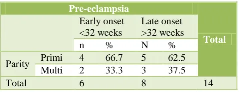
Table 6: Parity wise distribution of cases in relation to gestation age (weeks) of onset of PE.
Pre-eclampsia
Table 7: Blood pressure of the studied population.
BP mean±sd
Systolic BP(mmHg) 118.6±14.2(110,156)
Diastolic BP(mmHg) 76.4±10.3(70,106)
Table 8: Maternal and family history in the studied subjects.
Table 9: Maternal history and family history wise distribution of PE.
For the purpose of analysis study population was divided into those who had a normal course of the pregnancy, those who developed pre-eclampsia before 32 weeks, those who developed pre-eclampsia after 32 weeks and those who had fetal growth restriction.
Table 10: Doppler examination of the studied population.
Doppler n %
Normal 185 92.5
Diastolic notch 4 2
Pulsatility index 11 5.5
Table 11: Doppler abnormalities detected among the pre-eclampsia.
The analysis of age distribution showed that the mean age was 26.3 years. It was observed that there was statistically significant increased risk of pre-eclampsia among women who were in age group of 30-34 years with OR(Odds Ratio) of 3.6, this was comparable with study of FANG et al.12 The parity distribution revealed that majority of the women who developed pre-eclampsia were primigravida and this was consistent with study
done by Odegard et al.13 The distribution of cases
according to BMI even though non-significant
women who developed pre-eclampsia uterine artery Doppler abnormalities were present in 57.1% and in women who had IUGR uterine artery Doppler abnormalities were present in 40%. Doppler study of the uterine arteries had a sensitivity of 57.1% and specificity of 96.2% for pre-eclampsia and for IUGR it was 40% and 95.1% respectively.
In the study subjects who had pre-eclampsia of early onset (<32 weeks), mean pulsatility index (Mpi) >95th percentile was present in 50% cases whereas in late onset group (>32 weeks) mPI >95th percentile was present in only 25% cases. In cases who had IUGR mean pulsatility index >95th percentile was present in 33.33% cases. This was comparable with studies done by Yu et al(2008)16 where mPI >95 percentile was present in 43.8% cases. In women who developed pre-eclampsia before 32 weeks diastolic notch was present in 33.33% cases compared to the women who developed pre-eclampsia after 32 weeks where notch was present in only 12.5% cases. In cases that had IUGR, notch was present in 11.1%. The finding from the present study show that the uterine artery Doppler in the second trimester of pregnancy can identify 83.33% of pregnancies that will subsequently develop early onset pre-eclampsia (<32 weeks) and 40% of pregnancies who will have IUGR.
In early onset (<32 weeks) pre-eclampsia, IUGR was present in 66.7% and in late onset (>32 weeks) IUGR was present in 33.3% cases. In present study uterine artery Doppler had a positive predictive value (PPV) of 53.5% and negative predictive value (NPV) of 96.8%. In the present study the test result had a low sensitivity (57.1%) and positive predictive value of 53.3% for pre-eclampsia. This test has a high negative predictive value 96.8% which implies that only 3.2% of women with normal Doppler results will develop pre-eclampsia.
CONCLUSION
From present study, we concluded that,
1. Single step uterine artery Doppler at 19-22 weeks
gestation is a feasible test for the prediction of early onset (<32 weeks) pre-eclampsia associated with IUGR.
2. Early onset (<32 weeks) pre-eclampsia associated
with IUGR, defective placentation might have a causative role to play.
3. The combined model of second trimester uterine
artery Doppler and maternal history helps us to categorize pre-eclampsia cases into early onset and late onset.
4. The combined model of second trimester uterine
artery Doppler and maternal history can be used as a screening test for assessing the development of
pre-eclampsia and IUGR and initiating appropriate preventive modalities.
Funding: Not required
Conflict of interest: None declared
Ethical approval: The study was approved by the Institutional Ethics Committee
REFERENCES
1. Brown MA, Lindheimer MD, De Swiet M, Van
Assche A, Moutquin JM. The classification and diagnosis of the hypertensive disorders of pregnancy: statement from the international Society for the study of hypertension in pregnancy (ISSHP). Hypertens Pregnancy. 2001;20:ix-xiv.
2. Hjartardoltir S, Leifsson BG, Geirsson RT,
Stein-throsdottir V. Paternity change and the recurrence risk in familial hypertensive disorder in pregnancy. Hypertension in pregnancy. 2004;23(2):219-25.
3. Pijnenborg R, Bland JM, Robertson WB, Brosens I.
Uteroplacental arterial changes related to interstitial trophoblast migration in early humans pregnancy placenta. 1983:4397-413.
4. Lyall F. Priming and remodeling of human placental
bed spiral arteries during pregnancy- a review. Placenta. 2005:31-6.
5. Von Dadelszen P, Magee LA, Roberts JM. Sub
classification of pre-eclampsia. Hypertens
pregnancy. 2003;22:143-8.
6. Huppertz B. Placental origins of preeclampsia
challenging the current hypothesis. Hypertension. 2008;51:970-5.
7. Ness RB, Roberts JM. Heterogenous causes
constituting single syndrome of preeclampsia: a hypothesis and its implication. Am J Obstet Gynecol. 1998;179:1317-23.
8. Murphy DJ, Striate GM. Mortality and morbidity
associated with early onset pre-eclampsia. Hypertns Pregnancy. 2009;9:221-31.
9. Ness RB, Sibai BM. Shared and disparate
components of the pathophysiology of fetal growth restriction and preeclampsia. Am J Obstet Gynecol. 2008;201:121-6.
10. Irion O, Masse J, Forest JC. Prediction of
preeclampsia, low birthweight for gestation and prematurity by uterine artery blood flow velocity waveforms analysis in low risk nulliparous women. Br J Obstet Gynaecol. 1998;105:422-9.
11. Bower S, Bewley S, Campbell S. Improved
prediction of pre-eclampsia by screening of uterine arteries using the early diastolic notch and color Doppler imaging. Obstet Gynecol. 1993;82:78-83.
12. Fang R, Dawson A, Vitool L. Williams asian
biomedicine. 2009;5:477-86
13. Odegard RA, Vatten LJ, Nilsen ST. Risk factors and
clinical manifestations of pre-eclampsia. BJOG. 2000;107:1410-6.
14. Oosterhof H, Aaanoudse JG. Ultrasound pulsed
influence of sampling site and placental implantation. Gyn Obstet Invest. 1992;33;75-79.
15. Walker JJ. Pre-eclampsia. Lancet 2000;356:1260-5.
16. Yu CK, Khouri O, Onwudine N. Prediction of
pre-eclampsia by uterine artery Doppler imaging;
relationship to gestational age at delivery.
2008;31:310-3.
Cite this article as: Noor H, Manzoor M, Sadiq S,
Shahzad N.Maternal history and second trimester