4(9): 1873-1882, 2014 SCIENCEDOMAINinternational
www.sciencedomain.org
Morphometry of Human Fetal Ear Ossicles:
A Human Cadaveric Study
M. Pramila Padmini
1*and B. Narasinga Rao
11Department of Anatomy, MIMS Medical college, Vizianagaram, Nellimarla, A.P, India.
Authors’ contributions
This work was carried out in collaboration between both authors. Author MPP designed the study, wrote the first draft of the manuscript and managed the literature searches. Author BNR managed the analyses of the study. Both authors read and approved the final manuscript.
Received 23rdJuly 2013 Accepted 10thOctober 2013 Published 9thJanuary 2014
ABSTRACT
Aim: Most of the studies were on adult ossicles. In this present work, the aim is to study
the morphometry of the ear ossicles in the human fetuses and use of the study in medical applications.
Materials and Methods:This study is performed on 100 sets of middle ear ossicles, each set consisting of Malleus, Incus and Stapes, which were taken from 50 fetal cadavers left and right sides of both.
Result: The morphometric data of malleus and incus in their length are 5.21mm and 4.85mm, the height of the stapes is 2.52mm. The indices of malleus, incus and stapes are 51.28, 67.54 and 88.12mm.
Conclusion: The study of morphometry in the ear ossicles of the human fetal cadavers can be useful for prosthetic surgical reconstruction.
1. INTRODUCTION
Researchers have long known that the middle ear bones of a mammal evolved from the jawbones of their reptilian fore bearers, and that the “repurposing” of the bones, for the sake of improved hearing occurred in parallel with the refinement and elaboration of mammalian dentition [1]. The knowledge about these ossicles goes back to the 15th century [2]. Since that time, extensive studies have been carried out on their morphometry [3,4,5] embryology [6] function and structure [7,8,9] as well as the surgical reconstruction [10,11]. Most of these previous studies were on adult ossicles. It has been reported that these ossicles arrive at maximal size in fetal life [12]. In this present work, the aim is to study the morphometric of the ear ossicles in the human fetuses and use of the study in medical applications.
2. MATERIALS AND METHODS
This study is performed on 100 sets of middle ear ossicles(50 left and 50 right ), each set consisting of malleus, incus and stapes, which were taken from 50 fetal cadavers left and right sides of both sexes, obtained from local government, and private hospitals from vizianagaram, andhra pradesh, india. All the brains are severed at the level of medulla oblongata and have been used for project study. The fetuses in which the brains are dissected out have been used for the present study of ear ossicles. The ossicles have been obtained from tympanic cavity after opening tegmen tympani, which is the roof of middle ear [13]. This is an innovative method used to remove the ossicles of the ear since 2004 in the anatomy department. The measurements were estimated with a digital vernier callipers which has an accuracy of 0.01 mm which was purchased from precision instruments, model no.1112 made in korea. Measurements were taken three times of the same ear ossicle and are done for all the ossicles and the average is tabulated.
3. RESULTS
All the photographs were taken using a dissecting microscope (Focus microscopes, Basis for laboratories since 1967) of 10x magnification. The following parameters have been recorded:
Measurements of malleus:
1. Total length (maximal distance between the top of the head and the end of the umbo-Fig.1a),
2. Length of head and neck (maximal distance between the top of the head and the end of the lateral process-Fig.1b),
3. Length of manubrium mallei (distance from the end of the lateral process to the end of manubrium-Fig.1c),
Measurements of (Incus):
5. Total length (maximal distance between the superior edge of the body and the end of the long process-Fig.2a),
6. Total width (maximal distance between the superior edge of the body and the end of the short process-Fig.2b)
Measurements of (stapes):
9. Total height (maximal distance between the top of the head and the basis stapedis-Fig.3a),
10. Length of the base of stapes (maximal length of the long axis of basis stapedis-Fig.3b),
11. Width of the stapes (maximal width between the two crus-Fig.3c), 12. Index: Length of the base of stapes X 100 / total height of stapes.
3.1 Morphometry of Fetal Ear Ossicles
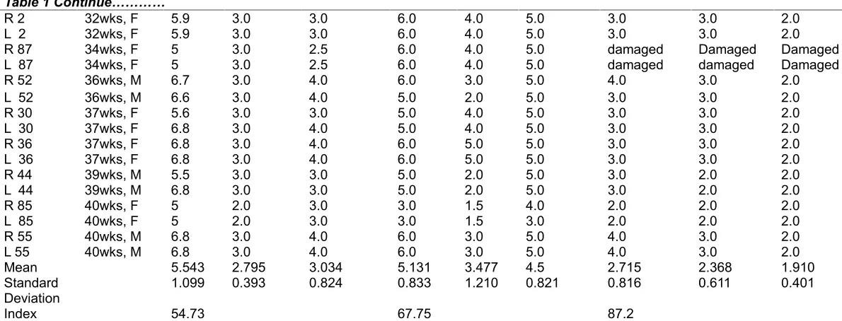
Table 1. Showing the measurements of malleus, incus and stapes
Side, s.no of fetus
Age in cms and
sex
Malleus in mm incus in mm stapes in mm
Total
length Length ofhead and neck
Length of
manubrium Totallength Width Distancebetween processes
Height Length of
Base Width
R 24 20wks, F 4.8 2.5 2.5 4.0 3.5 3.0 2.0 2.0 2.0
L 24 20wks, F 4.8 2.5 2.5 4.0 3.5 3.0 2.0 2.0 2.0
R 28w 22wks, M 4.5 3.0 2.0 5.0 4.0 5.0 3.0 2.5 2.8
L 28w 22wks, M 4.8 3.0 2.0 5.0 4.0 5.0 3.0 2.5 2.8
R 40 22wks, M 4.9 3.0 2.5 4.0 3.5 3.0 2.0 2.0 2.0
L 40 22wks, M 4.8 3.0 2.5 4.0 3.5 3.0 2.0 2.0 2.0
R 13 24 wks, M 5.0 2.5 3.0 5.5 4.5 5.0 3.5 2.5 1.5
L 13 24wks, M 5.0 2.5 3.0 5.5 4.5 5.0 3.0 2.0 1.5
R 84 24wks, M 6.6 3.0 4.0 6.0 5.0 5.0 3.0 3.0 2.0
L 84 24wks, M 6.6 3.0 4.0 6.0 5.0 5.0 3.0 3.0 2.0
R 88 24wks, F 4.8 3.0 2.0 5.0 2.0 3.0 2.0 2.0 0.5
L 88 24wks, F 4.8 3.0 2.0 5.0 2.0 3.0 2.0 2.0 1.5
R 39 25wks, M 6.6 3.0 4.0 6.0 5.0 5.0 3.0 3.0 2.0
L 39 25wks, M 6.6 3.0 4.0 6.0 5.0 5.0 3.0 3.0 2.0
R 28 26wks, F 5 3.0 2.5 4.0 3.0 4.0 absent absent absent
L 28 26wks, F 5 3.0 2.5 4.0 3.0 4.0 absent absent absent
R 11 28wks, M 5.8 2.5 3.5 5.2 5.0 5.0 1.0 1.0 1.5
L 11 28wks, M 5.8 2.5 3.5 5.2 5.0 5.0 1.0 1.0 1.5
R 78 28wks, M 2 1.5 1.0 5.0 2.0 3.0 1.2 2.5 1.0
Table 1 Continue…………
R 2 32wks, F 5.9 3.0 3.0 6.0 4.0 5.0 3.0 3.0 2.0
L 2 32wks, F 5.9 3.0 3.0 6.0 4.0 5.0 3.0 3.0 2.0
R 87 34wks, F 5 3.0 2.5 6.0 4.0 5.0 damaged Damaged Damaged
L 87 34wks, F 5 3.0 2.5 6.0 4.0 5.0 damaged damaged Damaged
R 52 36wks, M 6.7 3.0 4.0 6.0 3.0 5.0 4.0 3.0 2.0
L 52 36wks, M 6.6 3.0 4.0 5.0 2.0 5.0 3.0 3.0 2.0
R 30 37wks, F 5.6 3.0 3.0 5.0 4.0 5.0 3.0 3.0 2.0
L 30 37wks, F 6.8 3.0 4.0 5.0 4.0 5.0 3.0 3.0 2.0
R 36 37wks, F 6.8 3.0 4.0 6.0 5.0 5.0 3.0 3.0 2.0
L 36 37wks, F 6.8 3.0 4.0 6.0 5.0 5.0 3.0 3.0 2.0
R 44 39wks, M 5.5 3.0 3.0 5.0 2.0 5.0 3.0 2.0 2.0
L 44 39wks, M 6.8 3.0 3.0 5.0 2.0 5.0 3.0 2.0 2.0
R 85 40wks, F 5 2.0 3.0 3.0 1.5 4.0 2.0 2.0 2.0
L 85 40wks, F 5 2.0 3.0 3.0 1.5 3.0 2.0 2.0 2.0
R 55 40wks, M 6.8 3.0 4.0 6.0 3.0 5.0 4.0 3.0 2.0
L 55 40wks, M 6.8 3.0 4.0 6.0 3.0 5.0 4.0 3.0 2.0
Mean 5.543 2.795 3.034 5.131 3.477 4.5 2.715 2.368 1.910
Standard 1.099 0.393 0.824 0.833 1.210 0.821 0.816 0.611 0.401
Deviation
Index 54.73 67.75 87.2
Note: 50 sets of other 25 fetuses belonging to 12wks-2 fetuses, 16 wks-3 fetuses,18wks-3 foetuses,25wks-6 fetuses, 36wks-4 fetuses, 39wks-7 fetuses showed similar measurements, and are excluded from the Table.1.(R-RIGHT SIDE, L-LEFT SIDE). Mean and standard deviation has been taken from
4. DISCUSSION
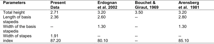
There are few studies in the literature on individual differences in these ossicles and these studies were on either adult or different species. The present morphometric results of the malleus, incus and stapes were compared with previous results (Table 2 [14,15,16,3,17,2,18], Table 3 [14,15,16,,3,17,2,18], Table 4 [14,15,2]). The present values of malleus and incus are less when compared to Erdoğan Unur et al. [14]. The index of malleus is approximately closer to the values of Arensberg et al. [2]. The morphometric index values of stapes are more when compared to the previous studies. This may be due racial variation.
Table 2. Comparative morphometric data of middle ear ossicles of present study with previous studies. Metric values of malleus given in mm
Parameters Present
data Erdognanet al. 2002 Bouchet& Giraut, 1969
Masali
1968 Arensberg &nathan,1972 Harada1972 ArensbergEt al, 1981 Aycanet al. 1990
total
length 5.54 7.7 7.9 7.6 7.3 8 7.8 8.1
length of
manubrium 3.03 4.7 4.7 4.6 3.5 4.2 4.4 4.9
length of head & neck
2.79 4.9 -- -- -- 5 -- 5.1
Index 54.73 61 -- -- -- -- 56.6
--Table 3. Comparative morphometric data of middle ear ossicles of present study with previous studies. Metric values of incus given in mm
Parameters Present
data Erdognanet al. 2002
Bouchet& Giraut, 1969
Masali
1968 Arensberg& nathan,1972
Harada
1972 ArensbergEt al,1981 Aycanet al. 1990
Total
length 5.13 6.5 6.5 6.4 6.8 6.8 6.4 6.7
Total
Width 3.47 4.9 5.1 4.8 5.1 4.8 5.1 5.1
Distance between the processes
4.5 6.1 -- -- -- 4.2 -- 6.1
Index 67.75 80 -- -- -- -- --
--Table 4. Comparative morphometric data of middle ear ossicles of present study with previous studies. Metric values of stapes given in mm
Parameters Present
Data Erdognanet al. 2002 Bouchet &Giraut, 1969 Arensberget al. 1981
Total height 2.71 3.20 3.50 3.20
Length of basis
the same size as adults, might be observed in ossicle banks for future use in ossiculoplasty. In addition, these ossicles can be used as homografts to replace eroded adult middle ear ossicles [14].
There are two types of middle-ear prosthesis, partial ossicular replacement prosthesis (PORPs) and total ossicular replacement prosthesis (TORPs). A PORP is used in cases when either the incus or the incus and malleus have degenerated. It connects the head of the stapes to either the manubruim of the malleus or directly to the tympanic membrane. A TORP is used in cases when either both the incus and stapes have degraded or when all three ossicles have degraded. It connects the manubrium of the malleus or the tympanic membrane directly to the stapedial footplate [19,20].
There are many conditions like cholesteatoma and otitis media an inflammation of the middle ear leading to ossicular degradation [20] that require surgical reconstruction of the ossicular chain. The goal of ossicular reconstruction is the restoration of conductive hearing. Ossicular chain reconstructions have been performed since 1875, using various forms of natural materials and, since 1952, by using synthetic implants [21]. Kenji Homma et al. [22] in his study on Ossicular resonance modes of the human middle ear for bone and air conduction used 5 human adult cadaveric temporal bones. Homografts evolved and are now available in a variety of sizes that can easily be sculpted to fit a patient's unique middle ear anatomy. The homograft bone becomes living tissue over time as it is incorporated by the host ear and provides superior audiologic results compared with autografts [23]. Other prostheses that may be used include a homograft prosthesis made of prefashioned labyrinthine bone or cadaveric ossicle, a stainless steel piston prosthesis, a wire prosthesis, or a polymeric silicone prosthesis [24]. Homografts from cadavers gained acceptance and are available in many presculpted designs, thereby decreasing the time needed for surgical reconstruction [25]. Precise measurements of stapes and incus are essential in the design of the middle ear implants and electromagnetic implants. The knowledge of variations of these ossicles and its morphometric data will help the otologist during reconstructive surgery and provide necessary information for the prosthesis designer [26]. The ossicles attain adult size in foetal life at around six months. Ossicles obtained from new born cadaver can be preserved in ossicle bank for future use in ossiculoplasty [27]. Congenital bilateral absence of stapes and oval window were confirmed by history, audiologic examination, high-resolution computed tomography scanning, and/or surgery in the study of Yi Z et al. [28]. The modified Lempert's fenestration operation of the horizontal semicircular canal is a safe and good choice for the patients and a better choice than a hearing aid throughout life [29]. The present study shows that the ossicles do not grow after birth, because of this only their morphometric measurements are useful in prosthetic designs.By using the foetal cadaveric temporal bones and ear ossicles, an ossicular replacement prosthesis can be made which would be very useful in improving number of prosthetic designs.
5. CONCLUSION
The present study shows that the ossicles do not grow after birth, because of this only their morphometric measurements are useful in prosthetic designs. By using the foetal cadaveric temporal bones and ear ossicles, an ossicular replacement prosthesis can be made which would be very useful in improving number of prosthetic designs.
CONSENT
Not applicable.
ETHICAL APPROVAL
Not applicable as the study is on still born foetuses obtained from local hospitals.
ACKNOWLEDGEMENTS
I am thankful to all the authors mentioned in the manucsript.
COMPETING INTERESTS
Authors have declared that no competing interests exist.
REFERENCES
1. Natalie Angier In Mammals, a Complex Journey to the Middle Ear Published: October
12, 2009.
2. Arensburg B, Harell M, Nathan H. The human middle ear ossicles, morphometry and
taxonomic implications. Journal of Human Evolution. 1981;10:199-205.
3. Arensburg B, Nathan H. Observations on a notch in the short (Superior or Posterior)
process of the incus. Acta Anat. 1971;78: 84-90.
4. Sarrat R, Guzman G, Tores A. Morphological variations of Human ossiculatmpani.
Acta Anat. 1988;131:146-149
5. Unur E, Aycan K, Ekinci N, et al. The study of incus from morphometric view. Erciyes
Medical Journal. 1993;15:16-19.
6. Louryan S. Develompment of auditory ossicles in the human embryo: correlations with
data obtained in mice. Bull Assoc Anat (Nancy). 1993;77:29-32.
7. Sarrat R, Torres A, Guzman A.G, et al. Functional structure of human auditory
ossicles. Acta Anat. 1992;144:189-195.
8. Huttenbrik K.B. The mechanics and function of middle ear. Part I: The ossicular chain
and middle ear muscles. Laryngorhinootologie. 1992;71:545-51.
9. Beer HJ, Bornitz M, Hardtke H.J, et al. Modelling of components of the middle ear and
13. Pramila Padmini M, Narasinga Rao B. Morphological variations in human fetal ear ossicles-A study. Int J Anat Res. 2013;02:40-42.
14. Erdoğan Unur, Harun Ülger, Nihat Ekinci, Erciyes Tip Dergisi Morphometrical And
Morphological Variations Of Middle Ear Ossicles In The Newborn (Erciyes Medical Journal). 2002;24(2):57-63.
15. Bouchet A, Giraud M. Contrubituon a l’etude morphologique et radiologique des osselets de l’ouie. Compte rendu de l’ Association des Anatomists 53 Congrés. 1968;141:588-600.
16. Masali M. The ear bones and the vertebral column as indications of taxonomic and postural distinctions among old world primates with reference to the origin of man. Rosenberg and Sellier, Torino; 1968.
17. Harada O, Ishii H. The Condition of the auditory ossicles in microtia. Plast Reconst Surg. 1972;50:48-53
18. Aycan K, Unur E, Bozkır MG, et al. Anatomical study of malleus. Journal of Health Sciences. 1990;1:152-158.
19. Koike T, Wada H, Kobayashi T, Takasaka T. Finite-element method (FEM) analysis of human middle-ear. Proceedings of the ARO Meeting. 1996;778.
20. Prendergast PJ, Ferris P, Rice HJ, Blayney AW. Vibroacoustic modelling of the outer and middle ear using the "nite element method. Audiology and Neuro-Otology. 1999a.;4:185-191.
21. Lobel K. Ossicular replacement prosthesis. Taken from ‘Clinical Performance of Skeletal Prosthesis’, Chapman & Hall; 1999.
22. Kenji Homma, Yu Du. Ossicular resonance modes of the human middle ear for bone and air conduction Journal of Acoustical Society of America February. 2009;125(2):968–979. doi:10.11.21/130565 64, PMCID: PMC2852437.
23. Wehrs RE. Homograft ossicles in tympanoplasty. Laryngoscope 1982; 92:540-546. 24. Schuknecht HF. Otosclerosis surgery. In: Nadol JB, Jr, Schuknecht HF, eds. Surgery of
the ear and temporal bone. New York, NY: Raven. 1993;223-244.
25. Kartush JM. Ossicular chain reconstruction: capitulum to malleus. Otolaryngol Clin North Am. 1994;27:689-715.
26. Farahani RM, Nooranipour M. Anatomy and Anthropometry of human stapes. American Journal of Otolaryngology. 2008;29:42-47.
27. Rodriguez K, Shah R K, Kenna M. Anomalies of the middle and inner ear. Otolaryngologic clinics of North America. 2007;40:81-96.
28. Yi Z, Yang J, Li Z, Zhou A, Lin Y. Bilateral congenital absence of stapes and oval window in 2 members of a family. Etiology and Management Otolaryngology head Neck Surgery. 2003;128(6):777-82.
29. Schmidt JL, Cole TM 3rd, Silcox MT. Technical note: a landmark-based approach to the study of the ear ossicles using ultra-high-resolution X-ray computed tomography data. Am J Phys Anthropol. 2011;145(4):665-71.
© 2014 Padmini and Rao; This is an Open Access article distributed under the terms of the Creative Commons Attribution License (http://creativecommons.org/licenses/by/3.0), which permits unrestricted use, distribution, and reproduction in any medium, provided the original work is properly cited.
Peer-review history: