Proteomes and Identification of LipL32 as a Biomarker for Potency
P. C. Humphryes,aM. E. Weeks,bA. Gielbert,aG. Thomson,aand N. G. Coldhama
Animal Health and Veterinary Laboratories Agency, Addlestone, New Haw, Surrey,aand UCL Institute of Child Health, London,bUnited Kingdom
The current batch potency test for
Leptospira interrogans
serovar Canicola vaccines requires the use of a large number of
ham-sters and has severe effects (i.e., hepatic and renal failure resulting in death); while this vaccine is effective, a safer, cheaper, more
ethical replacement is desired. The aim of this study was to analyze vaccine proteomes and identify target molecules common to
all
L. interrogans
serovar Canicola vaccines which could be used to design an
in vitro
potency test. Initial analysis of
L.
interro-gans
serovar Canicola vaccines (A to E) from different manufacturers, using the
Limulus
amebocyte lysate assay and
silver-stained sodium dodecyl sulfate polyacrylamide gels, indicated that lipopolysaccharide was not present in all vaccines, preventing
it from being a suitable target molecule. The protein contents of vaccines A to E were therefore determined by two-dimensional
liquid chromatography mass spectrometry ([2D-LC/MS] 221
ⴞ
31, 9
ⴞ
8, 34
ⴞ
4, 21
ⴞ
5, and 34
ⴞ
17 proteins [mean
ⴞ
1
stan-dard deviation] found, respectively). The outer membrane protein LipL32 was established to be common to all and to be present
at a significantly higher (
P
<
0.05) relative spectral abundance in a batch of vaccine which passed the
in vivo
potency test than in
one which had failed. Further analysis using multiple reaction monitoring revealed that the concentration of the N terminus of
LipL32 was significantly lower (
P
<
0.01) in failed batches (
n
ⴝ
2) of vaccine than in passed batches (
n
ⴝ
2); the concentration of
the C terminus between the two batches was approximately the same. An
in vitro Leptospira
vaccine potency test, based on
N-terminal amino acid quantification of LipL32, was subsequently developed.
P
revalent worldwide,
Leptospira interrogans
is a waterborne
zoonotic pathogen capable of infecting virtually any mammal
it comes into contact with (1). Primarily contracted through
ex-posure to infected urine, infection causes symptoms that can be
diverse, ranging from fever to hepatic failure, and that often result
in death if left untreated (22). Other symptoms, such as cessation
of milk production and miscarriage, are frequently observed in
cattle (8), making leptospirosis particularly economically
damag-ing to the farmdamag-ing community. Vaccination is widely used to
pro-tect against infection; however the vaccines currently available are
serovar specific (21), and regular boosters are required to
main-tain immunity (20). Large volumes of vaccine are therefore
pro-duced every year to protect the domestic animal population. The
majority of
Leptospira
vaccines available on the market are either
bi- or multivalent and are derived from heat-inactivated cultures
of the bacteria which have been grown (19) in medium containing
a high level of Tween 80 and albumin (typically 1%, wt/vol).
Potency testing of
Leptospira
vaccines is required prior to new
batches being released onto the market. The current test, as laid
out by the European monograph status review (25), requires that
for each serovar the vaccine provides protection against, 10
ham-sters be challenged with a virulent strain of that serovar. Half of
these receive the vaccine prior to challenge while the control group
does not; the endpoint of this test is death, and euthanasia is not
typically employed. Additionally,
Leptospira
has a predisposition
to lose virulence during
in vitro
culture (39) and liquid nitrogen
storage (28), due in part to alterations in the expression of
lipo-polysaccharide (LPS) and outer membrane proteins (15, 17, 26); it
is therefore routine practice to maintain the virulence of strains by
passage through hamsters (34).
An
in vitro
replacement for the hamster potency test has been
sought for some time as the use of such a large number of animals,
accrued through regular usage of the test and
in vivo
challenge
strain maintenance, is ethically and financially undesirable.
En-zyme-linked immunosorbent assay (ELISA)-based vaccine batch
potency tests for some
Leptospira
serovars, such as
Leptospira
borg-petersenii
serovar Hardjo, have been successfully developed and
implemented (12); however similar attempts at the development
of an
in vitro
potency test (10) for
Leptospira interrogans
serovar
Canicola have had mixed success. In the United States ELISA
po-tency tests for certain specific
L. interrogans
serovar Canicola
vac-cines are in usage (30); however, they require that adjuvants be
removed prior to testing, which is impractical for widespread
im-plementation due to the wide variety of adjuvants used by
differ-ent manufacturers. It has been clearly established (13, 29),
how-ever, that both protein and LPS derived from
Leptospira
are
capable of eliciting an immune response.
Prior studies (24, 31) have successfully described the proteome
of
L
.
interrogans
. The proteins LipL32 (Hap-1), LipL41, LipL45,
and OmpL1 have been identified as possible vaccine candidates (7,
9, 38) and, in some cases, used in
in vivo
trials (16, 33). A LipL32
DNA vaccine has been shown to confer protective immunity
against
L. interrogans
serovar Canicola in the gerbil model (3);
however, the protective effect of LipL32 against
L. interrogans
se-rovar Canicola in the hamster model has yet to be determined. The
exact mechanism by which
Leptospira
vaccines provide protective
immunity remains poorly understood, and to date no reliable
in
vitro
method exists for their analysis or quantitation. The aim of
Received16 November 2011Returned for modification17 January 2012
Accepted1 February 2012
Published ahead of print8 February 2012
Address correspondence to N. G. Coldham, [email protected].
Supplemental material for this article may be found athttp://cvi.asm.org/.
Copyright © 2012, American Society for Microbiology. All Rights Reserved.
doi:10.1128/CVI.05622-11
on August 17, 2020 by guest
http://cvi.asm.org/
the present study was to identify biomarkers present in
L.
interro-gans
serovar Canicola vaccines that could be used as the basis for
the development of an
in vitro
vaccine batch potency test. The
protein and LPS content of
L. interrogans
serovar Canicola
vac-cines were chosen for analysis due to their known immunogenic
effects.
MATERIALS AND METHODS
Vaccine selection and preparation.Bivalent vaccines (giving protection againstL.interrogansserovars Canicola and Icterohaemorragiae) which had passed thein vivovaccine batch potency test and been released for commercial sale were purchased from five different manufacturers for analysis. Vaccine C was of subunit manufacture comprising outer mem-brane proteins, whereas vaccines A, B, D, and E were derived from heat-inactivated bacteria. Test batches of vaccine C (n⫽4) which had not been subjected to the vaccine potency test were also obtained from the manu-facturer; test batches were available only for vaccine C.
The vaccines (1 ml) were concentrated using 5-kDa molecular-weight-cutoff concentrators (Sartorius Stedim, France), washed once with 0.5 ml of 2.5 mM ammonium bicarbonate (pH 8.0), and concen-trated again to a final volume of 0.5 ml (the protein content of vaccine B was too low to process in this manner, so it was therefore analyzed without washing). The protein concentration in the washed vaccines was then determined using the Bradford method (Sigma-Aldrich, United King-dom), with bovine serum albumin as the calibration standard. Estimation of LPS in the vaccines was determined using an Endochrome K kit (Charles River, United Kingdom) which is based on theLimulus amebo-cyte lysate (LAL) assay.
Vaccine batch potency test.The test batches (n⫽4) of vaccine C were assessed for potency according to the guidelines laid out in the most recent European monograph (25). For each batch analyzed, five female hamsters (weight,ⱕ120 g) (Charles River, Germany) were inoculated subcutane-ously with 0.5 ml of vaccine (diluted 1/40 with 0.9% [wt/vol] physiological saline); five control hamsters were kept unvaccinated. Fifteen days follow-ing vaccination all the hamsters were challenged by intraperitoneal inoc-ulation with 1 ml of virulentL. interrogansserovar Canicola (⬃1⫻108 cells/ml). For the test to be valid at least four of the five unvaccinated controls had to die within 14 days of infection. Vaccine batches were deemed potent if at least four of the five vaccinated hamsters survived for 14 days longer than the unvaccinated controls. All animal procedures used in this study were covered under the Animals (Scientific Procedures) Act 1986 by Home Office Project License number PPL 70/7249 and were approved by the Animal Ethics Committee at the Animal Health and Veterinary Laboratories Agency (AHVLA) where this work was per-formed.
Proteome analysis by two-dimensional liquid chromatography mass spectrometry (2D-LC/MS).Three replicates of each vaccine, nor-malized by dilution in 2.5 mM ammonium bicarbonate (pH 8.0) to 100
g of protein, were heat denatured at 95°C for 5 min and then digested overnight with 2g of sequencing grade trypsin (Promega) (5). Digestion was terminated by the addition of 1l of 25.2 M formic acid (Fluka). Due to the low concentration of protein found in vaccine B (see Fig. 2), only 6
g was used for each replicate of that vaccine.
Tryptic digests were centrifuged (5,000⫻gfor 1 min) to remove
particulates, and 50l of supernatant was taken for strong cation ex-change (SCX) fractionation by high-pressure liquid chromatography (HPLC). Tryptic peptides were chromatographed using a Hewlett-Pack-ard 1100 system on a Biobasic SCX HPLC (2.1 by 100 mm) column (Thermo Scientific). Analytes were chromatographed at a flow rate of 0.25 ml/min with mobile phases comprising 75:25 2.5 mM ammonium ace-tate-acetonitrile, pH 4.5 (A), and 75:25 250 mM ammonium acetate-acetonitrile, pH 4.5 (B), and a binary gradient (time [t]⫽0 min, 100% A; t⫽5 min, 100% A;t⫽18 min, 65% A;t⫽20 min, 100% B;t⫽22 min, 100% A;t⫽32 min, 100% A). The effluent was monitored at 280 nm, and 15 fractions of 1 ml were collected between 8 and 23 min. The SCX frac-tions were dried at 60°C under vacuum using an Eppendorf 5301 centrif-ugal concentrator (Eppendorf, United Kingdom).
Dried SCX fractions (containing 50g of protein) were resuspended in 0.1% (vol/vol) formic acid (20l) and analyzed on an Agilent 6520 quadrupole time-of-flight (Q-TOF) mass spectrometer (Agilent Technol-ogies) with an HPLC chip cube source. The chromatography chip con-sisted of a 40-nl enrichment column (Zorbax 300 SB C18; 5-m particle size) and a 75-m by 150-mm analytical column (Zorbax 300 SB C18; 5-m particle size) driven by the Agilent Technologies 1200 series nano/ capillary liquid chromatography system. Both systems were controlled by MassHunter Workstation Data Acquisition for Q-TOF (version B.02.00, patches 1 and 2; Agilent Technologies). Tryptic peptides (1l) were loaded onto the enrichment column of the chip and washed with eight column volumes of 0.1% trifluoroacetic acid (TFA). Tryptic peptides were separated on the analytical column using an acetonitrile gradient (4.5% to 72% [vol/vol]) over 47 min and eluted directly into the mass spectrometer. The mass spectrometer was run in positive ion mode, and MS survey scans were run over a range ofm/z250 to 3,000 and at five spectra per second. Precursor ions were selected for automatic tandem MS (MS/MS) at an absolute threshold of 2,000 and a relative threshold of 0.01, with a maximum of five precursors per cycle, and active exclusion set at 1 spectra and released after 3 min. Precursor charge-state selection and preference were set to 2⫹and then 3⫹. A synthetic peptide mix (10 ng/ml; MRFA, AVDQLNEQSSEPNIYR, VTALYEGFTVQNEANK, ARPQELPF LASIQNQGR, ISVNNVLPVDFNLMQQK, and NYINQYSEVAIQMVM HMQPK) was analyzed (see Fig. S3 in the supplemental material) prior to each run (defined as 15 SCX fractions), and two blank samples (com-prised of 0.1% formic acid and 95:5 acetonitrile-H2O) were used at the end. The mass spectrometer was also calibrated prior to each run using electrospray-time of flight (ES-TOF) Tuning Mix (Agilent, United King-dom) to within a residual error of 2 ppm.
Protein identification.The search engine Spectrum Mill (Agilent, United Kingdom) was used to extract MS/MS data from MassHunter acquisition files, and proteins were subsequently identified by comparison of tryptic peptide product ion mass spectra against those generatedin silicofrom a protein database. Search parameters included selection of trypsin as the proteolytic enzyme with up to two missed cleavage sites and a variable modification for oxidation of methionine residues; precursor and product mass tolerances were set to 20 and 50 ppm, respectively. Identified protein lists (and associated information) with a Spectrum Mill protein score higher than 11 were exported as tab-separated files for bioin-formatics analysis; proteins identified had at least two distinct tryptic pep-tides and were present in all three technical replicates. To date the
com-TABLE 1Tryptic peptides and product ions selected for detection and quantification of LipL32 using LC-MRMa
Peptide sequence Terminus Molecular mass (Da) Precursor ion for MRM (m/z) Product ion for MRM (m/z) Retention time (min)
SSFVLSEDTIPGTNETVK N 1924.07 962.50 845.40 13.16
ISFTTYKPGEVK C 1369.56 457.30 628.80 10.80
aThe full amino acid sequence of LipL32 and the locations of the tryptic peptides used for quantitation (underlined) are as follows: MKKLSILAISVALFASITACGAFGGLPSLK SSFVLSEDTIPGTNETVKTLLPYGSVINYYGYVKPGQAPDGLVDGNKKAYYLYVWIPAVIAEMGVRMISPTGEIGEPGDGDLVSDAFKAATPEEKSMPHWFDTWIRVER MSAIMPDQIAKAAKAKPVQKLDDDDDGDDTYKEERHNKYNSLTRIKIPNPPKSFDDLKNIDTKKLLVRGLYRISFTTYKPGEVKGSFVASVGLLFPPGIPGVSPLIHSNPEEL QKQAIAAEESLKKAASDATK.
on August 17, 2020 by guest
http://cvi.asm.org/
plete genome ofL.interrogansserovar Canicola has not been sequenced; therefore, a custom-made database derived from chromosomes I and II of L. interrogansserovar Copenhageni (ftp://ftp.ncbi.nlm.nih.gov/genomes /Bacteria/Leptospira_interrogans_serovar_Copenhageni; accessed on 6 December 2010) was used for database interrogation.
The relative abundance of proteins present in all three technical re-peats was determined through spectral counting (40). The normalized spectrum abundance factor (NSAF), calculated as described by Zybailov et al. (41), was used to account for differences in peptide length, allowing for comparison of protein abundance between individual vaccines.
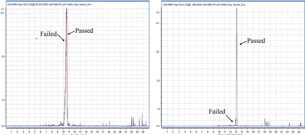
Quantitation of LipL32 by LC-MS/MS.Synthetic peptide analogues, corresponding to N- and C-terminal tryptic peptides found in LipL32, were obtained at a purity of 98% (Peptides Synthetics, United Kingdom) for detection and quantification using multiple reaction monitoring (MRM) (Table 1 and Fig. 1). Passed (n⫽2) and failed (n⫽2) batches of vaccine C (PH1 to PH4) (Table 2) were prepared, quantitated, and di-gested as described above. SCX separation was also performed as de-scribed above; however, the 15 fractions of each replicate (containing 50
g of total protein equivalents) were recombined prior to being taken to dryness, dissolved in 0.1% (vol/vol) formic acid (50l), and analyzed on an Agilent 6410 Triple Quadrupole mass spectrometer (Agilent Technol-ogies) with an HPLC chip cube source. The chromatography chip con-sisted of a 160-nl enrichment column (Zorbax 300 SB C18; 5-m particle size) and a 75-m by 150-mm analytical column (Zorbax 300 SB C18; 5-m particle size) driven by the Agilent Technologies 1200 series nano/ capillary liquid chromatography system. Both systems were controlled by MassHunter Workstation Data Acquisition for Triple Quadrupole
(ver-sion B.02.01; Agilent Technologies). Tryptic peptides (1l) were loaded onto the enrichment column of the chip and washed with eight column volumes of 0.1% TFA. Peptides were then separated on the analytical column using an acetonitrile gradient (4.5% to 90% [vol/vol]) over 25 min and eluted directly into the mass spectrometer. The mass spectrom-eter was run in positive ion mode, with the electrospray voltage set to 1900 V and gas temperature and pressure set at 300°C and 4 liters/min, respec-tively. Optimal transitions and conditions for the peptides of interest (Ta-ble 1 and Fig. 1) were obtained using the MS and MS/MS data from previous Q-TOF analysis of vaccine C. The acquired data were quantified using the Agilent MassHunter Quantitative Analysis software (version B.03.01; Agilent Technologies).
Analysis of total LPS content in vaccines by PAGE.The vaccine(s) LPS content was visualized by sodium dodecyl sulfate-polyacrylamide gel electrophoresis (SDS-PAGE) on the Xcell Surelock system (Invitrogen) using a 4 to 12% gradient NuPAGE gel run at 150 W for 80 min. Gels contained a 3.5-kDa protein ladder (Invitrogen), two concentrations of LPS (10g and 500 ng,Escherichia coliderived; Sigma, United Kingdom) and aliquots (20g) of untreated vaccine (A, C, D, and E); vaccine B was loaded in a smaller amount (2.5g) due to its lower concentration of protein (Fig. 2). LPS was detected in the gel using the silver staining method described by Tsai et al. (37).
Statistical analysis.All results are presented as means of the standard deviations of the mean (except proteomics data). Comparison of the con-centration and relative abundance of proteins between vaccines was per-formed using a Student’sttest; aPvalue of⬍0.05 was taken to be statis-tically significant.
RESULTS
Evaluation of vaccine LPS content.
The LPS content of the five
vaccines (A to E) was evaluated by PAGE using an LPS-specific
silver stain (Fig. 3). This staining method works through
interac-tion with the polysaccharide part of the molecule (11) and showed
that vaccine C did not contain any detectable LPS. For
confirma-tion, the LAL assay (Fig. 2), which functions by interaction with
the lipid A (36) and core region (6) of LPS, was also performed on
vaccines A to E. The LAL assay did not detect LPS in vaccine C,
FIG 1LC-MRM chromatograms of passed (shown in blue) and failed (shown in red) batches of vaccine C. The chromatograms presented on the left are for the
C-terminal tryptic peptide (ISFTTYKPGEVK; transitionm/z457.3 to 628.8), and those on the right are for the N-terminal tryptic peptide (SSFVLSEDTIPGT-NETVK; transitionm/z962.5 to 845.4).
TABLE 2Hamster potency test performed on four batches of vaccine C
Vaccine C batch name
No. of surviving hamsters/total no. of hamsters
Test outcomea
PH1 5/5 Pass
PH2 5/5 Pass
PH3 3/5 Fail
PH4 3/5 Fail
aAt least four hamsters (of five) had to survive the test for a batch to pass.
on August 17, 2020 by guest
http://cvi.asm.org/
suggesting that it is either absent or present at levels below the
detection limits of these assays.
Vaccine proteome characterization.
Analysis of vaccines A
to E revealed that the total number of proteins in each varied
considerably (see Table S1 in the supplemental material).
However, two proteins, LipL32 and flagellin, were found to be
common to the five vaccines analyzed (Table 3); due to the low
number of proteins identified in vaccine B and their
corre-spondingly low coverage, a separate comparison of vaccines A
and C to E was also performed, which identified two additional
conserved proteins (Table 3, shaded area). Two batches of
vac-cine C were available to establish whether LipL32 or flagellin
protein could be used as a biomarker for vaccine potency; one
had passed the
in vivo
potency test, batch PH1 (Table 2), while
the other, batch PH3, had failed (Table 2). Both were
subse-quently analyzed by 2D-LC/MS and quantitated using the
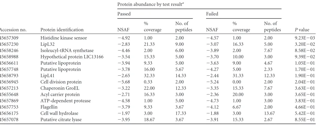
NSAF (Table 4; see also Table S2 in the supplemental material).
The average number of proteins detected in the three replicates
of the failed batch was lower than that found in the passed
batch (Table 4); however, the total numbers of conserved
pro-teins (i.e., propro-teins detected in all three replicates) found in the
passed and failed batches (see Table S2) were approximately
the same (22 and 18 proteins, respectively). Based on the NSAF,
LipL32 and histidine sensor kinase protein were shown to be
FIG 3Vaccines A to E (lanes 2, 4, 5, and 6 contain 20g; lane 3 contains 2.5
g) were run on a 4 to 12% NuPAGE gel stained with silver to detect LPS. Lanes 1 and 9 contain a 3.5-kDa protein ladder, and lanes 7 and 8 contain 10g and 500 ng, respectively, ofEscherichia coliLPS as a positive control.
FIG 2Concentrations of protein and LPS in vaccines A to E (as sold) as
determined by the Bradford and LAL assays, respectively. Two replicates of each vaccine were analyzed for each assay; mean and standard deviation of the
mean are shown. TABLE
3 Conserved proteins present in commercially available vaccines from five different manufacturers Accession no. Protein identification Protein abundance by vaccine a AB C D E NSAF % Coverage No. of peptides NSAF % Coverage No. of peptides NSAF % Coverage No. of peptides NSAF % Coverage No. of peptides NSAF % Coverage No. of peptides 45657230 LipL32 ⫺ 4.11 27.00 12.00 ⫺ 2.64 6.00 2.00 ⫺ 2.48 26.67 6.67 ⫺ 2.22 24.33 7.33 ⫺ 2.88 19.00 5.00 45657753 Flagellin ⫺ 3.41 31.00 25.00 ⫺ 2.44 5.00 2.33 ⫺ 3.01 17.00 4.00 ⫺ 2.35 26.00 6.67 ⫺ 2.59 21.00 6.33 45658793 LipL41 ⫺ 3.35 32.67 33.33 ND ND ND ⫺ 2.09 30.67 13.00 ⫺ 2.08 30.67 11.00 ⫺ 2.39 26.67 10.00 45656311 LIC10411 b ⫺ 2.45 38.67 36.00 ND ND ND ⫺ 3.13 15.00 2.00 ⫺ 2.96 15.00 2.00 ⫺ 3.03 15.33 2.33 aValues are means. NSAF, ln relative abundance (higher values indicate greater abundance); ND, protein not detected. The total numbers of proteins pe r vaccine group (mean ⫾ 1 standard deviation) are as follows: A, 221 ⫾ 31; B, 9 ⫾ 8; C, 34 ⫾ 4; D, 21 ⫾ 5; E, 34 ⫾ 17. Shaded areas represent additional conserved proteins identified when vaccine B is excluded from comparison. bHypothetical protein.
on August 17, 2020 by guest
http://cvi.asm.org/
present at a higher abundance (
P
ⱕ
0.05) in the batch of
vac-cine which had passed the
in vivo
potency test than in the failed
batch; since Lipl32 was also known to be highly conserved in
vaccines A to E, it was chosen for further analysis.
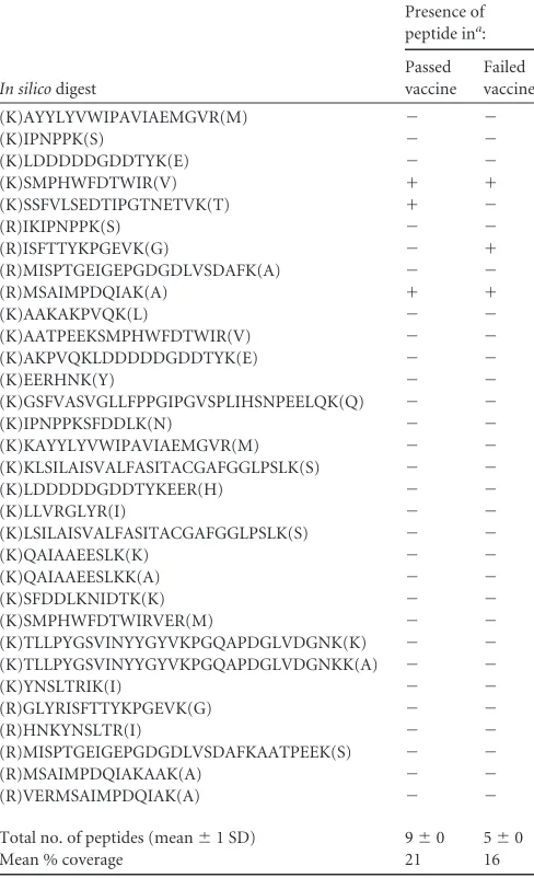
Spectrum Mill was subsequently used to identify LipL32
tryptic peptides which were present in each batch (Table 5).
The number of LipL32 vaccine peptides identified was
signifi-cantly lower than that predicted by
in silico
digestion; this can
be attributed to a variety of factors including alterations to the
protein during vaccine manufacture, peptide loss during
chro-matographic fractionation (which is consistent with
2D-LC/MS experiments), and the fact that not all tryptic peptides
can be detected using a mass spectrometer (32). However, both
batches were analyzed in parallel, and so any losses would be
consistent between the two.
Quantitation of LipL32 in passed and failed batches of
vac-cine.
To enable accurate determination of the concentration of
LipL32 in the vaccines, two tryptic peptides from Table 5,
cor-responding to the N and C termini of the protein, were selected
for quantitation using multiple reaction monitoring (MRM).
The limits of detection (LOD) of the N and C terminus peptides
were determined (2) as 0.025 and 0.018 fmol/
g, respectively,
using the empirical method. Whereas the limits of
quantifica-tion (LOQ; defined as the lowest concentraquantifica-tion of the standard
with a coefficient of variation lower than 20% [2]), were 0.02
and 0.05 fmol/
g for the N and C terminus peptides.
Calibra-tion curves for both peptides were constructed using
Mass-Hunter software (Agilent, United Kingdom) over the tested
concentration range (0.01 to 100 fmol/
g) allowing
determi-nation of sample concentration (Table 6) by interpolation. The
concentration of the N terminus region was significantly lower
(
P
ⱕ
0.01) in failed batches (Table 2, PH3 and PH4) of vaccine
(Table 6) than in passed batches (Table 2, PH1 and PH2); no
statistical difference was observed in the concentration of the C
terminus regions. The C-terminal peptide ISFTTYKPGEVK was
not detected in the passed batch (PH1) of vaccine C during
Q-TOF analysis (Table 5); however, subsequent targeted MRM
anal-ysis revealed it to be present (Table 6) as this method is more
sensitive than Q-TOF analysis.
DISCUSSION
Leptospira
-derived LPS has been shown to be highly immunogenic
(29), and it is has been suggested that mutation of LPS can elicit
cross-protection against multiple serovars (35). However, it was
not detected in vaccine C, suggesting that at least in this vaccine it
may not be necessary to induce protective immunity against
Lep-tospira
; further work is required to elucidate its function in the
other vaccines analyzed. An alternative biomarker candidate,
which is conserved across all the vaccines, was sought by
compar-ison of the proteomes of the five vaccines under investigation.
Protein content was assessed initially by the Bradford assay, which
revealed that the concentration of protein in vaccine B was much
lower than that found in the other vaccines. A smaller quantity of
vaccine B was therefore analyzed using 2D-LC/MS which could
account for the lower number of proteins identified (Table 3).
Subsequent proteomic analysis of vaccines A and C to E has
re-vealed four conserved proteins. Of these, LipL32, LipL41, and the
flagellin protein are of interest as they are known to be potential
immunogens (16, 18); when vaccine B was included in the
com-parison, the only proteins identified as being conserved were
flagellin and LipL32.
LipL32, also known as hemolysis associated protein-1
(Hap-1), is a major surface-expressed outer membrane protein (7)
found in pathogenic
Leptospira
species (14). It is known to
provide cross-protection against
L
.
interrogans
(4) in the gerbil
model, and various different methods for presenting it as a
vaccine have been used in trials (3, 33). A recent study (23) has
reported that recombinant LipL32 does not confer protective
immunity against leptospirosis in hamsters; however, this
study was limited to three serovars and did not include the
L
.
interrogans
serovar Canicola.
Initial analysis of passed and failed batches of vaccine (Table 4,
PH1 and PH3) using spectral counting (NSAF) estimated that
LipL32 was present at a statistically (
P
ⱕ
0.05) higher abundance
TABLE 4Abundance of conserved proteins present in a passed and failed batch of vaccine C
Accession no. Protein identification
Protein abundance by test resulta
Pvalue
Passed Failed
NSAF % coverage
No. of
peptides NSAF
% coverage
No. of peptides
45657309 Histidine kinase sensor ⫺4.92 1.00 2.00 ⫺4.57 1.00 2.00 9.23E⫺03
45657230 LipL32 ⫺2.83 21.33 9.00 ⫺3.07 16.33 5.00 3.20E⫺02
45658246 Isoleucyl-tRNA synthetase ⫺4.46 2.00 6.00 ⫺3.89 2.00 7.67 8.58E⫺02
45658988 Hypothetical protein LIC13166 ⫺3.54 15.33 5.00 ⫺3.70 10.00 3.00 9.39E⫺02
45656611 Putative lipoprotein ⫺3.94 9.33 5.00 ⫺3.63 9.00 4.67 1.05E⫺01
45657748 Putative lipoprotein ⫺3.78 16.00 5.67 ⫺4.27 5.00 2.33 1.70E⫺01
45658793 LipL41 ⫺2.65 32.33 14.33 ⫺2.44 31.33 12.33 1.90E⫺01
45656945 Cell division protein ⫺5.68 0.33 2.00 ⫺5.24 0.00 2.00 2.04E⫺01
45657213 Chaperonin GroEL ⫺3.22 22.00 12.33 ⫺3.35 15.33 7.67 3.63E⫺01
45655648 Acyl carrier protein ⫺2.71 16.33 3.00 ⫺2.36 20.00 3.00 3.65E⫺01
45657869 ATP-dependent protease ⫺4.58 1.00 5.00 ⫺4.73 1.00 3.00 3.83E⫺01
45657753 Flagellin ⫺3.79 9.33 3.67 ⫺4.12 6.67 2.00 4.66E⫺01
45656175 Cell wall hydrolase ⫺1.97 3.00 17.33 ⫺1.88 3.00 13.67 5.42E⫺01
45657078 Putative citrate lyase ⫺3.95 18.67 3.67 ⫺3.91 15.33 2.67 8.55E⫺01
aValues are means. NSAF, ln relative abundance (higher values indicate greater abundance). The mean total numbers of proteins (⫾1 standard deviation) were 54⫾8 in passed batches and 42⫾4 in failed batches.
on August 17, 2020 by guest
http://cvi.asm.org/
in passed vaccines than in failed vaccines, indicating that it could
make a good target molecule candidate for an
in vitro
potency test.
To investigate this further, multiple reaction monitoring (MRM)
was used to accurately quantitate (27) the concentration of LipL32
in passed (PH1 and PH2) and failed (PH3 and PH4) batches
(Ta-ble 6) of vaccine C. MRM quantitates proteins based on specific
peptide sequences; to ensure good coverage of LipL32, two
pep-tide sequences, corresponding to the start (N terminus) and end
(C terminus) of the protein, were selected for quantitation. The
concentration of the C terminus peptides was approximately the
same across both conditions (0.88 and 0.79 fmol/
g in the passed
and failed batches, respectively) (Table 6); however the
concen-tration of the N terminus peptide was substantially (
P
ⱕ
0.01)
lower in failed batches. This suggests that LipL32 is somehow
al-tered in failed batches of vaccine, which would result in fewer
spectra being detected during Q-TOF analysis, accounting for the
lower abundance observed through spectral counting. It is
pro-posed that this N-terminal region of LipL32 may be used as an
effective biomarker for quantifying
Leptospira
vaccine potency
in
vitro
. Its effectiveness as an epitope for protective efficacy has not
been determined; however, it is plausible that this truncation to
the primary full-length structure of LipL32 in failed batches could
alter its conformational structure, which could conceivably be the
cause of its inability to convey protective immunity; to determine
this, further work using X-ray crystallography of the LipL32 found
in passed and failed batches will be required.
In conclusion, this study demonstrates 2D-LC/MS to be an
effective tool for vaccine analysis which could be utilized in the
development of
in vitro
potency tests for a number of different
bacterial vaccines (tests for other serovars of
Leptospira
in
partic-ular); 2D-LC/MS-based techniques could also be developed for
the quality assurance stage of vaccine manufacture. This study has
also shown that MRM-based N-terminal amino acid quantitation
of LipL32 has the potential to act as a novel assay target for the
determination of potency
in vitro
. Further validation will be
re-quired, with access to a larger sample cohort of passed and failed
batches from multiple manufacturers, to determine if it can
re-place the hamster challenge batch potency test.
ACKNOWLEDGMENTS
This work was supported by a grant from The National Centre for the Replacement, Refinement and Reduction of Animals in Research.
We thank William Newell for his assistance with the NSAF calcula-tions.
REFERENCES
1.Adler B, de la Pena Moctezuma A.2010.Leptospiraand leptospirosis.
Vet. Microbiol.140:287–296.
2.Armbruster DA, Pry T.2008. Limit of blank, limit of detection and limit
of quantitation. Clin. Biochem. Rev.29(Suppl 1):S49 –S52.
3.Branger C, et al.2005. Protection againstLeptospira interroganssensu lato
challenge by DNA immunization with the gene encoding hemolysin-associated protein 1. Infect. Immun.73:4062– 4069.
4.Branger C, et al.2001. Identification of the hemolysis-associated protein
1 as a cross-protective immunogen ofLeptospira interrogansby adenovi-rus-mediated vaccination. Infect. Immun.69:6831– 6838.
5.Coldham NG, Woodward MJ.2004. Characterization of theSalmonella
typhimuriumproteome by semi-automated two dimensional HPLC-mass spectrometry: detection of proteins implicated in multiple antibiotic re-sistance. J. Proteome Res.3:595– 603.
6.Conrad ML, Pardy RL, Armstrong PB.2001. Response of the blood cell
of the American horseshoe crab,Limulus polyphemus, to a lipopolysaccha-ride-like molecule from the green algaChlorella. Biol. Bull.201:246 –247.
7.Cullen PA, et al.2005. Surfaceome ofLeptospiraspp. Infect. Immun.
73:4853– 4863.
8.Department for Environment, Food, and Rural Affairs.2008. Zoonoses
report: United Kingdom 2008. Department for Environment, Food, and Rural Affairs, London, United Kingdom.http://archive.defra.gov.uk.
9.Dong H, et al.2008. Characterization of the ompL1 gene of pathogenic
Leptospira species in China and cross-immunogenicity of the OmpL1 protein. BMC Microbiol.8:223.
TABLE 5Comparison of tryptic peptides detected in two batches of
vaccine C against anin silicodigest of LipL32
In silicodigest
Presence of peptide ina:
Passed vaccine
Failed vaccine
(K)AYYLYVWIPAVIAEMGVR(M) ⫺ ⫺
(K)IPNPPK(S) ⫺ ⫺
(K)LDDDDDGDDTYK(E) ⫺ ⫺
(K)SMPHWFDTWIR(V) ⫹ ⫹
(K)SSFVLSEDTIPGTNETVK(T) ⫹ ⫺
(R)IKIPNPPK(S) ⫺ ⫺
(R)ISFTTYKPGEVK(G) ⫺ ⫹
(R)MISPTGEIGEPGDGDLVSDAFK(A) ⫺ ⫺
(R)MSAIMPDQIAK(A) ⫹ ⫹
(K)AAKAKPVQK(L) ⫺ ⫺
(K)AATPEEKSMPHWFDTWIR(V) ⫺ ⫺
(K)AKPVQKLDDDDDGDDTYK(E) ⫺ ⫺
(K)EERHNK(Y) ⫺ ⫺
(K)GSFVASVGLLFPPGIPGVSPLIHSNPEELQK(Q) ⫺ ⫺
(K)IPNPPKSFDDLK(N) ⫺ ⫺
(K)KAYYLYVWIPAVIAEMGVR(M) ⫺ ⫺
(K)KLSILAISVALFASITACGAFGGLPSLK(S) ⫺ ⫺
(K)LDDDDDGDDTYKEER(H) ⫺ ⫺
(K)LLVRGLYR(I) ⫺ ⫺
(K)LSILAISVALFASITACGAFGGLPSLK(S) ⫺ ⫺
(K)QAIAAEESLK(K) ⫺ ⫺
(K)QAIAAEESLKK(A) ⫺ ⫺
(K)SFDDLKNIDTK(K) ⫺ ⫺
(K)SMPHWFDTWIRVER(M) ⫺ ⫺
(K)TLLPYGSVINYYGYVKPGQAPDGLVDGNK(K) ⫺ ⫺
(K)TLLPYGSVINYYGYVKPGQAPDGLVDGNKK(A) ⫺ ⫺
(K)YNSLTRIK(I) ⫺ ⫺
(R)GLYRISFTTYKPGEVK(G) ⫺ ⫺
(R)HNKYNSLTR(I) ⫺ ⫺
(R)MISPTGEIGEPGDGDLVSDAFKAATPEEK(S) ⫺ ⫺
(R)MSAIMPDQIAKAAK(A) ⫺ ⫺
(R)VERMSAIMPDQIAK(A) ⫺ ⫺
Total no. of peptides (mean⫾1 SD) 9⫾0 5⫾0
Mean % coverage 21 16
a
The presence (⫹) or absence (⫺) of the tryptic peptide is indicated.
TABLE 6Concentrations of the N- and C-terminal tryptic peptides of
LipL32 in passed and failed vaccines
Region of protein
Concn in vaccine (fmol/g [mean⫾1 SD])a
Fold difference
P value Passed
(n⫽2)
Failed (n⫽2)
N terminus 1.28⫾0.68 0.08⫾0.03 15.87 0.007
C terminus 0.88⫾0.29 0.79⫾0.23 1.11 0.586
aThree replicates of each vaccine were analyzed.
on August 17, 2020 by guest
http://cvi.asm.org/
10. Ebert E.1999. Guinea pig serology as an alternative to the hamster chal-lenge test for potency testing ofLeptospirahardjo vaccines, p 102–109.In Proceedings of the 1999 International Conference on Alternatives to Animal Challenge Tests in the Batch Control of Leptospira Vaccines for
Veterinary Use.European Directorate for the Quality of Medicines and
Healthcare, Strasbourg, France.
11. Fomsgaard A, Freudenberg MA, Galanos C.1990. Modification of the
silver staining technique to detect lipopolysaccharide in polyacrylamide gels. J. Clin. Microbiol.28:2627–2631.
12. Goddard RD, Hopkins IG, Thornton DH.1986. The development of a
potency test for Leptospira hardjo vaccines: a comparison of protection in calves and serology in guinea-pigs. J. Biol. Stand.14:337–344.
13. Guerreiro H, et al. 2001. Leptospiral proteins recognized during the
humoral immune response to leptospirosis in humans. Infect. Immun. 69:4958 – 4968.
14. Haake DA, et al.2000. The leptospiral major outer membrane protein
LipL32 is a lipoprotein expressed during mammalian infection. Infect. Immun.68:2276 –2285.
15. Haake DA, et al.1998. Characterization of leptospiral outer membrane
lipoprotein LipL36: downregulation associated with late-log-phase growth and mammalian infection. Infect. Immun.66:1579 –1587.
16. Haake DA, et al.1999. Leptospiral outer membrane proteins OmpL1 and
LipL41 exhibit synergistic immunoprotection. Infect. Immun.67:6572– 6582.
17. Haake DA, et al.1991. Changes in the surface ofLeptospira interrogans
serovar grippotyphosa during in vitro cultivation. Infect. Immun.59: 1131–1140.
18. Hauk P, et al.2008. In LipL32, the major leptospiral lipoprotein, the C
terminus is the primary immunogenic domain and mediates interaction with collagen IV and plasma fibronectin. Infect. Immun.76:2642–2650.
19. Johnson RC, Harris VG.1967. Differentiation of pathogenic and
sapro-phytic leptospires. I. Growth at low temperatures. J. Bacteriol.94:27–31.
20. Klaasen HL, Molkenboer MJ, Vrijenhoek MP, Kaashoek MJ. 2003.
Duration of immunity in dogs vaccinated against leptospirosis with a bi-valent inactivated vaccine. Vet. Microbiol.95:121–132.
21. Koizumi N, Watanabe H.2005. Leptospirosis vaccines: past, present, and
future. J. Postgrad. Med.51:210 –214.
22. Levett PN.2001. Leptospirosis. Clin. Microbiol. Rev.14:296 –326.
23. Lucas DS, et al.2011. Recombinant LipL32 and LigA from Leptospira are
unable to stimulate protective immunity against leptospirosis in the ham-ster model. Vaccine29:3413–3418.
24. Malmstrom J, et al.2009. Proteome-wide cellular protein concentrations
of the human pathogenLeptospira interrogans. Nature460:762–765.
25. Marbehant P.1999. Leptospiral vaccines batch potency testing. European
monograph status review, p 11–16.InProceedings of the 1999 Interna-tional Conference on Alternatives to Animal Challenge Tests in the Batch Control ofLeptospiraVaccines for Veterinary Use. European Directorate for the Quality of Medicines and Healthcare, Strasbourg, France.
26. Matsunaga J, et al.2002. Novel 45-kilodalton leptospiral protein that is
processed to a 31-kilodalton growth-phase-regulated peripheral mem-brane protein. Infect. Immun.70:323–334.
27. Ong SE, Mann M. 2005. Mass spectrometry-based proteomics turns
quantitative. Nat. Chem. Biol.1:252–262.
28. Reed NE, Varney WC, Goddard RD, Wyeth PJ.2000. The maintenance
of challenge strains used in the potency test for canine leptospira vaccines. Biologicals28:25–28.
29. Ruby KW.1999. Development of in vitro assays for measuring relative
potencies of leptospiral bacterins containing serovars Pomona, Canicola, Grippothyphosa and Icterohaemorrhagiae, p 35– 45.InProceedings of the 1999 International Conference on Alternatives to Animal Challenge Tests in the Batch Control of Leptospira Vaccines for Veterinary Use. European Directorate for the Quality of Medicines and Healthcare, Stras-bourg, France.
30. Ruby KW, Walden DM, Wannemuehler MJ.1996. Development of an in
vitro assay for measuring the relative potency of leptospiral bacterins con-taining serovar Canicola and its correlation to the hamster potency assay. Dev. Biol. Stand.86:341.
31. Sakolvaree Y, et al.2007. Proteome and immunome of pathogenic
Lep-tospiraspp. revealed by 2DE and 2DE-immunoblotting with immune se-rum. Asian Pac. J. Allergy Immunol.25:53–73.
32. Sanders WS, Bridges SM, McCarthy FM, Nanduri B, Burgess SC.2007.
Prediction of peptides observable by mass spectrometry applied at the experimental set level. BMC Bioinformatics8(Suppl 7):S23.
33. Seixas FK, et al.2007. Evaluation of different ways of presenting LipL32
to the immune system with the aim of developing a recombinant vaccine against leptospirosis. Can. J. Microbiol.53:472– 479.
34. Silva EF, et al.2008. Characterization of virulence of Leptospira isolates
in a hamster model. Vaccine26:3892–3896.
35. Srikram A, et al.2011. Cross-protective immunity against leptospirosis
elicited by a live, attenuated lipopolysaccharide mutant. J. Infect. Dis. 203:870 – 879.
36. Takayama K, et al.1984. Influence of fine structure of lipid A onLimulus
amebocyte lysate clotting and toxic activities. Infect. Immun.45:350 –355.
37. Tsai CM, Frasch CE.1982. A sensitive silver stain for detecting
lipopoly-saccharides in polyacrylamide gels. Anal. Biochem.119:115–119.
38. Wang Z, Jin L, Wegrzyn A.2007. Leptospirosis vaccines. Microb. Cell
Fact.6:39.
39. Woodward MJ.2001. Leptospira, p 2137–2158.InSussman M (ed),
Mo-lecular medical microbiology. Academic Press, New York, NY.
40. Zybailov B, Coleman MK, Florens L, Washburn MP.2005. Correlation
of relative abundance ratios derived from peptide ion chromatograms and spectrum counting for quantitative proteomic analysis using stable iso-tope labeling. Anal. Chem.77:6218 – 6224.
41. Zybailov B, et al.2006. Statistical analysis of membrane proteome
ex-pression changes inSaccharomyces cerevisiae. J. Proteome Res.5:2339 – 2347.