1
The Role Of Apoptotic Signaling In Axon Guidance
Riley Kellermeyer*, Leah M. Heydman*, Grant S. Mastick and Thomas Kidd.
Dept. of Biology/MS 314, University of Nevada, 1664 N. Virginia St., Reno, NV 89557,
USA.
*Equal contribution
Abstract
Navigating growth cones are exposed to multiple signals simultaneously and have to
integrate competing cues into a coherent navigational response. Integration of guidance
cues is traditionally thought to occur at the level of cytoskeletal dynamics. Drosophila
studies indicate that cells exhibit a low level of continuous caspase protease activation,
and that axon guidance cues can activate or suppress caspase activity. We base a model
for axon guidance on these observations. By analogy with other systems in which caspase
signaling has non-apoptotic functions, we propose that caspase signaling can either
reinforce repulsion or negate attraction in response to external guidance cues by cleaving
cytoskeletal proteins. Over the course of an entire trajectory, incorrectly navigating axons
may pass the threshold for apoptosis and be eliminated, whereas axons making correct
decisions will survive. These observations would also explain why neurotrophic factors
can act as axon guidance cues and why axon guidance systems such as Slit/Robo
signaling may act as tumor suppressors in cancer.
Keywords:
Axon guidance, growth cone, cytoskeleton, caspases, apoptosis, signal integration, basal
level of caspase activity, death associated inhibitor of apoptosis, axon branching, Netrin,
Introduction
The navigational center of growing axons is the growth cone, a highly dynamic
expansion of the axon shaft that samples the environment and integrates multiple cues to
generate directed extension, retraction, and turning [1,2]. Traditionally, axon attractants
such as Netrins are thought to increase cytoskeletal outgrowth towards a cue, whereas
axon repellents such as Slits inhibit cytoskeletal growth [3,4]. The net effect of attractive
and repulsive cues on the cytoskeleton results in growth towards attractive cues and away
from repulsive cues. Integration has been demonstrated to also occur through interactions
between cell surface receptors, and through intracellular kinases [5,6]. More recent
evidence suggests that the traditional view of growth cones navigating up or down
gradients of guidance cues such as Netrin may not be accurate or even valid in vivo [7-11].
Our own work led us to identify a role for the apoptotic machinery in the growth cone
that is likely functioning to integrate opposing guidance cues. This review examines
models for how the cell death machinery could be involved in axon guidance with an
emphasis on results from Drosophila.
Drosophila Netrin-B is a neurotrophic factor that blocks cell death
Netrins are diffusible axon guidance cues most famous for attracting axons to the
CNS midline [12]. The fly has two Netrin genes, NetA and NetB, that are required for
midline and motor neuron axon guidance [13,14]. The genes appear to be the product of
tandem duplication and display a high degree of functional overlap. Netrins are expressed
by the CNS midline and embryos lacking both Netrins (NetAB) have axon guidance
of either gene. Localized sources of Netrins therefore appear to provide a navigational
cue. In contrast to midline expression, pan-neuronal expression of either Netrin in wild
type embryos leads to axon phenotypes, either due to a lack of positional information
specific to the midline or through attraction to non-midline areas [13,14]. It was therefore
surprising to find that pan-neuronal expression of NetB alone can rescue axon guidance
defects in NetAB embryos [15]. In contrast, pan-neural expression of NetA increases the
severity of the NetAB mutant phenotype, establishing a clear difference between the
proteins. NetB was subsequently identified as a neurotrophic factor when over-expressed,
because blocking cell death using the baculovirus p35 caspase inhibitor in discrete
subsets of neurons can rescue NetAB midline guidance defects [15]. These findings
substantiate a model for a non-apoptotic role for caspases in the growth cone, as
Figure 1 Apoptotic machinery and axons
(A)Schematic of axon guidance signaling pathways that potentially interact with the
apoptotic machinery. Candidate downstream effectors are shown based on
proteins that can promote survival and are known to interact with the receptors
shown in Drosophila [19-24]. Cytoplasmic signaling components may act on
caspase regulators, such as inactivating RHG proteins, promoting Diap1 function
or could act directly on the caspases (uncertainty indicated by dotted lines).
(B) A Drosophila embryo lacking the Netrin-A and Netrin-B axon guidance genes
(NetAB) stained to reveal eagle positive axons (brown), whose axon guidance
Anterior is to the top and growth cones of the EW neuron cluster are indicated by
arrows. The growth cones/axons are growing at different rates from segment to
segment, with some having crossed the midline and fasciculated with the
contralateral homologue and other axons having not yet crossed the midline. In
older embryos 90% of the EW axons cross the midline, as opposed to about 50%
in NetAB embryos alone. Image courtesy of G. Newquist.
The Apoptotic Machinery and Guidance Receptors
Programmed cell death or apoptosis is a key part in the development of
multicellular organisms and in the maintenance of the correct number of cells in mature
animals [27]. The classic role for cell death in neural development is to eliminate
unneeded connections that fail to compete for a limiting survival factor, the neurotrophic
hypothesis [28]. For example, axons that are misguided and fail to reach their target tissue
die [29]. Further research has shown that neurotrophic factors can also induce death in
certain contexts, and this activity is functionally conserved in the fly [30-32]. Apoptosis
operates through molecular cascades, offering several potential signaling nodes that could
intersect with axon guidance signaling pathways. A crucial event in apoptosis is the
activation of specialized cysteine-aspartic acid proteases called caspases (Figure 1A).
Signals derived from the mitochondria, in response to external pro-apoptotic signals or
the withdrawal of trophic support molecules, trigger activation of initiator caspases
[33-35]. Activation relies on multimerization and/or conformational changes of initiator
caspases, like Dronc, leading to proteolytic activation of effector caspases such as Drice
proteolytic cleavage that occurs during apoptosis. Caspase activity is buffered by the
death-associated inhibitor of apoptosis protein family (Diap1/thread), the viral p35
protein and other specialized inhibitors which oppose caspase activation. Many caspase
inhibitors, like Diap1, are continuously active to prevent apoptosis by marking initiator
caspases for degradation [36,37]. To initiate apoptosis, Diap1 is cleaved by inhibitor of
apoptosis antagonists, notably Hid, Grim and Reaper (RHG proteins), ultimately allowing
caspase activation [34]. In addition to apoptosis, caspases have been implicated in actin
dynamics of Drosophila spermatid individualization [38,39], sense organ precursor
selection [36,40], and dendrite retraction [41,42] (reviewed in [35]). Together, this cascade
of apoptotic regulators is present in all cells at low levels, but normally kept in check by
specific and tightly regulated modulators.
The apoptotic machinery is present in the growth cone of extending axons and can
be activated by external signals such as Netrin [16]. The principal vertebrate Netrin
receptor is DCC (Deleted in Colorectal Cancer; Frazzled in the fly), which dimerizes
upon ligand binding to stimulate signaling pathways that alter cytoskeletal dynamics [12].
Netrins are also capable of repelling axons through the Unc-5 receptor [43]. In
vertebrates, failure of DCC to homodimerize triggers cell death via the initiator caspase-9
and the effector caspase-3 [44]. The dual function of DCC to transduce both migratory
and apoptotic signals is known as the dependence receptor hypothesis, wherein cell
survival is dependent on ligand occupancy of receptors. Dependence receptors such as
DCC are characterized by having caspase cleavage sites in their cytoplasmic domains and
that the absence of the ligand triggers caspase cleavage of the receptor. The DCC caspase
tumor suppression [45,46]. However, the ability of Netrin to act as a survival factor,
particularly in spinal cord development is controversial as conflicting survival
phenotypes have been observed by different groups [47,48]. Additional recent evidence
strongly suggests that the dependence receptor mechanism is not operating in the mouse
spinal cord [49].
In flies, the DCC homologue, frazzled (fra; also called Unc-40), lacks the caspase
cleavage site in the cytoplasmic domain, suggesting Fra is not a dependence receptor,
though alternative sites could exist [48,50]. Additionally, loss of fra activity triggers apoptosis in some tissues, rather than being protective from cell death, as would be
expected from the loss of a dependence receptor [51]. The ability to rescue the axon
guidance defects of NetAB mutants by blocking apoptotic signaling therefore requires an
alternative explanation to the dependence receptor hypothesis.
How could caspase signaling operate in growth cone guidance?
Apoptosis requires major changes in the structure of cells via rearrangements of
the cytoskeleton, so it is not surprising that caspases cleave a large number of
cytoskeletal and structural proteins such as actin, -tubulin, and Spectrin [52-55].
Caspase-3 cleaves Spectrin in growth cones in culture [56]. Changes in levels and
localization of cytoskeletal proteins are observed during apoptosis of larval salivary
glands, and these changes are prevented by inhibiting caspases [57]. Many proteins that
modulate the cytoskeleton are also cleaved during apoptosis such as cofilin, GAP43, and
rho kinase (Rock) as well as many cell adhesion molecules [58-60]. These molecules could
protrusion (actin driven extension of filopodia and lamellipodia) and inhibit axon
outgrowth. Inhibiting caspase activity would protect cytoskeletal components from
proteolysis and potentially increase protrusion and forward movement. Migrating border
cells in the Drosophila ovary provide an apoptosis-independent example for this model.
Normal border cell migration requires the activity of the Diap1 caspase inhibitor [61].
Diap1 forms a complex with the actin cytoskeletal modulators Rac and Profilin, and
Diap1 protects these modulators from degradation by Dronc. In ovary border cell
migration, Diap1 alters cytoskeletal dynamics independent of apoptosis inhibition, as loss
of Diap1 in these cells does not result in cell death and migration phenotypes are not
rescued with p35. Similarly, Diap1 promotes F-actin assembly in polarized elongation of
sensory organ progenitors by blocking Dronc activation, in a caspase-dependent,
apoptosis-independent manner [62,63]. F-actin turnover at the cell margin therefore
requires inhibition of Diap1 without any effect on cell survival.
There are several important implications from these and other studies for
non-apoptotic functions of caspase protease activity. The first is that activation of the
apoptotic machinery does not necessarily lead to cell death. In vertebrates, molecular
mechanisms have been identified that prevent complete activation of the apoptotic
cascade [64]. Studies of the synapse suggest that transient and local activation of caspases
can remodel the synapse [65], and observations of activated caspase-3 restricted to the
growth cone and sites of axon branching are consistent with this model [16,66]. Second,
Drosophila studies have demonstrated that there is a continuous low level of initiator
caspase activation through auto-processing and the role of Diap1 is to counteract this
wing discs using the FRET-based SCAT3 caspase activity probe [36]. A vertebrate Diap1
homologue, X-linked inhibitor of apoptosis (XIAP) plays a role in limiting caspase
activation [71,72], and appears central to restricting caspase activation to subcellular
compartments of neurons [73].
Returning to the fly CNS and the observation that Netrin mutant phenotypes can
be rescued by anti-apoptotic factors, the simplest model to explain the effects of caspase
inhibition in Netrin mutants is that there is a basal level of caspase activation in growth
cones which has to be overcome for maximal forward growth (Figure 2A). This model
was first proposed by Gilman and Mattson, after demonstrating that addition of caspase
inhibitors to neuronal cultures increases axon outgrowth [65]. The implication is that a
basal level of caspase proteolysis keeps normal axon growth below maximum levels,
although the molecular targets of caspases in growth cones remain undefined. To support
any of the models described, cleavage of specific substrates, such as cytoskeletal
components, will have to be demonstrated in the growth cone itself, which may require
the study of larger growth cones from other invertebrate or vertebrate species, given the
small size of Drosophila growth cones.
It is worth noting that caspases can also modulate the actin cytoskeleton
independently of their protease roles, such as promoting Aip1/cofilin mediated actin
polymerization in migrating lymphocytes, though it is not known how widespread this
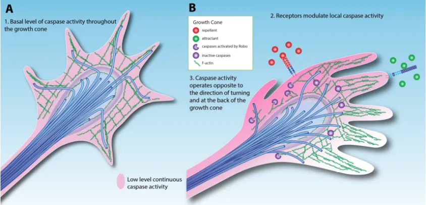
Figure 2 Models for caspase activity in the growth cone
(A)Simple low-level activation of caspases throughout the growth cone.
(B) Attractant and repellent cues modulate caspase activity via cell surface receptors,
altering the growth cone trajectory. In this model, repellents increase caspase
activity while attractants decrease caspase activity.
Extracellular modulation of caspase activity in the growth cone
An exciting possibility arising from this simple model of caspase-mediated
guidance is that caspase activity could be actively modulated in response to external cues
(Figure 2B). Mehlen has proposed that axon guidance signaling pathways actively
modulate tumor cell survival, explaining why axon guidance molecules are implicated in
preventing cancer [74]. Despite a large number of studies implicating Slit/Robo signaling
as tumor suppressors in cancer, a direct link of Slit/Robo signals to apoptosis has not yet
been shown [75]. DCC has been strongly linked to caspase activity [44], and DCC acts as a
frazzled appears to act as a tumor suppressor, because while fra mutant clones are usually
not viable, they can be rescued with p35 expression blocking cell death [51]. Additionally,
loss of fra activity leads to invasive cell phenotypes reminiscent of metastasis [51,76]. In
the embryo, NetB likely promotes axon growth by inhibiting caspase activity in the
growth cone. Double mutants for the fra and Dscam1 Netrin receptors display an increase
in cell death, whereas there is no change in either mutant alone [15], suggesting multiple
receptors may mediate this activity.
Emerging evidence reveals that repellent signaling pathways are able to activate
caspases, in some cases through direct binding. The Slit/Robo, Eph/Ephrin and
Sema/Plexin pathways all recruit and/or activate caspases [17,66,77-81]. Slit/Robo
signaling in zebrafish axons has been shown to genetically interact with caspases, in a
manner that suggests localized activation [66]. Consistent with Slit/Robo regulation of
caspases, we have observed low levels of activated caspase in a pattern that matches
Robo localization in the ventral nerve cord (Figure 3). This suggests that caspase activity
in the growth cone is increased by axon repellents and decreased by attractants, allowing
axon outgrowth in the direction of attractant cues (Figure 2B). Drosophila motor neurons
similarly integrate information concerning the levels of attractants and repellents
emanating from their target muscle [82]. Thus caspase activation and inhibition could sum
the input signals to determine lowered caspase activity domains where forward growth
occurs, while a widespread basal level of inhibitors like Diap1 restrict the spread of local
caspase activation by repellents. In this respect, the model resembles the synapse where
the duration and intensity of caspase activation determines the difference between
If axon repellents activate caspases at low levels, they could mediate contact
inhibition or increase sensitivity to survival factors in tumors. Alternatively, it could be
that cytoskeletal rearrangements in response to both positive and negative cues require
caspase activation, or that caspase activation controls growth cone protein levels [16].
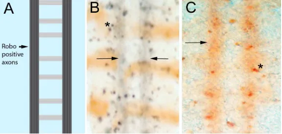
Figure 3 Low level caspase activity in longitudinal axons
A. Schematic of the CNS axon scaffold in the fly ventral nerve cord. The axons form a ladder-like pattern. The Robo repulsive receptor is only found in the longitudinal portions
of CNS axons (arrow). B, C. Drosophila embryonic nerve cords stained with an antibody raised against activated vertebrate Caspase-3 that appears to detect Dronc activation in
flies [85]. The panel B is enhanced with nickel staining to give a black precipitate and is counterstained with wingless-lacZ (brown horizontal stripes). Panel C is only stained for activated caspase (brown). Dying cells are visible as densely stained regions, usually oval
in shape (asterisks). A continuous low level of staining can be seen in the region occupied
by the longitudinal axons (arrows). This pattern matches the pattern of Robo localization,
with the Robo protein excluded from axons segments crossing the midline while
upregulated in axons using active Slit/Robo signaling to avoid the midline [86],
Caspase signaling at the fly CNS midline
In fly NetAB mutants, most growth cones orient towards the CNS midline but
many fail to cross it [87]. The original interpretation of this observation is that Netrins are
required for axons to extend across the midline, rather than to attract axons to the
midline. The ability of caspase inhibition to rescue axon crossing in NetAB mutants
suggests that caspase activity inhibits midline crossing or axon outgrowth in general. To
explain this observation, we propose that there is a low level of caspase activity in the
growth cone that needs to be overcome for forward growth. Alternatively, or in parallel,
strict temporal control of caspase activation may allow for the necessary cytoskeletal
rearrangements [35]. While many axons don’t, a significant number of axons do cross the
midline in NetAB mutants, revealing that there are undiscovered attractants expressed at
the midline. This suggests that caspase inhibition may increase axon outgrowth enough to
allow growth cones to use these other cues to locate and grow towards the midline.
Interestingly, the fly CNS midline is a source of other neurotrophic factors like the
Drosophila neurotrophins DNTs [88], acting through neuronal Toll receptors [31].
Classically, the axonal target tissue produces these neurotrophic factors. An increasing
number of examples are now known where intermediate targets supply neurotrophic
factors a phenomenon termed en passant or pre-target neurotrophic action [48,89-91]. As
NetB and DNTs are expressed by the CNS midline intermediate targets, and required for
motor axon targeting [82,92], both gene families may function as en passant neurotrophic
factors and as guidance cues. Interestingly, midline glia require the axon derived
epidermal growth factor Spitz to survive, and when the axons fail to contact the midline
Artificially promoting survival by increasing MAPK signaling, which inhibits Hid,
removes the necessity for migration suggesting that Spitz is acting as a caspase-mediated
attractive signal. Searching for survival factors could be a general mechanism that
influences cell and growth cone migration. MAPK signaling in vertebrate growth cones
and commissural axons has been implicated in the response to Netrin supporting this
model [16,44]. Together, Drosophila axon navigation across the ventral midline suggests
that there remains much to be discovered about the functional links between the classical
axon guidance problem of midline crossing and caspase signaling.
Apoptotic signaling in axon branching
As axons extend, particularly as they enter their target tissues, they also branch,
with each branch forming its own growth cone. The process of axon branching is also
likely regulated by the apoptotic machinery. One of the most dramatic visualizations of
caspase activity in axons is in zebrafish retinal ganglion cells [66]. Caspase activation
occurs in a dynamic fashion at branchpoints in developing axonal arbors and genetically
interacts with Slit/Robo signaling. Interestingly, Slit is proteolytically cleaved into two
fragments, Slit-N and Slit-C. Slit-N stimulates axon branching, whereas full length Slit
(Slit-FL) inhibits branching [94-96]. Slit-N is neurotrophic [97], and it will be interesting to
see whether Slit-FL can directly activate caspases, perhaps via p38 MAPK signaling as
suggesting by zebrafish studies [66]. Additionally, activated caspase activity has been
observed in the developing auditory brainstem within several segments of navigating
axons as well as their terminal branches within targets where it is proposed to limit
branches to spread into inappropriate target tissue [18]. Although much less is known
about axon branching within targets, caspase activity appears to be an important regulator
of not only primary axons but also their terminal branches. Axon branching may be more
important than primary axon growth for regenerative recovery of connections after injury
or disease, as branching from spared axons can be major contributors to restoring circuit
function [98].
Conclusion.
The neurotrophic hypothesis proposes that competition between neurons for
functional connections leads to the correct wiring of the nervous system. Studies of
caspases now suggest that the apoptotic machinery plays an active role in forming the
connections in the first place. Based on Drosophila studies, we propose that there is a
continuous low level of basal caspase activation in growth cones that is kept in check by
the caspase inhibitor Diap1. Modulation of caspase activity by external signals affects
axon growth rates and allows for the integration of multiple, potentially conflicting,
inputs to generate a coherent response. These conclusions are summarized in Table 1.
Acknowlegements
This work was supported by grants from the National Institutes of Health (P20
(IOS-1053555 to T.K.) We thank C. Propst for preparing the original Adobe Illustrator files
used to generate Figure 2 and G. Newquist for the image in Figure 1B. We thank
members of the Kidd laboratory for discussing ideas.
Author contributions
Writing – original draft preparation, T.K.
Writing – review and editing L.M.H., R.K., G.S.M. and T.K.
Conflicts of Interest
None.
References
1. Lowery, L.A.; Van Vactor, D. The trip of the tip: understanding the growth cone machinery. Nature reviews. Molecular cell biology 2009, 10, 332-343, doi:10.1038/nrm2679.
2. Kolodkin, A.L.; Tessier-Lavigne, M. Mechanisms and molecules of neuronal wiring: a primer. Cold Spring Harbor perspectives in biology 2011, 3, doi:10.1101/cshperspect.a001727.
4. Gomez, T.M.; Letourneau, P.C. Actin dynamics in growth cone motility and navigation. Journal of neurochemistry 2014, 129, 221-234,
doi:10.1111/jnc.12506.
5. Chacon, M.R.; Fazzari, P. FAK: dynamic integration of guidance signals at the growth cone. Cell adhesion & migration 2011, 5, 52-55.
6. Dudanova, I.; Klein, R. Integration of guidance cues: parallel signaling and crosstalk. Trends in neurosciences 2013, 36, 295-304,
doi:10.1016/j.tins.2013.01.007.
7. Varadarajan, S.G.; Kong, J.H.; Phan, K.D.; Kao, T.J.; Panaitof, S.C.; Cardin, J.; Eltzschig, H.; Kania, A.; Novitch, B.G.; Butler, S.J. Netrin1 Produced by Neural Progenitors, Not Floor Plate Cells, Is Required for Axon Guidance in the Spinal Cord. Neuron 2017, 94, 790-799.e793,
doi:10.1016/j.neuron.2017.03.007.
8. Dominici, C.; Moreno-Bravo, J.A.; Puiggros, S.R.; Rappeneau, Q.; Rama, N.; Vieugue, P.; Bernet, A.; Mehlen, P.; Chedotal, A. Floor-plate-derived netrin-1 is dispensable for commissural axon guidance. Nature 2017, 545, 350-354, doi:10.1038/nature22331.
9. Tang, X.; Wadsworth, W.G. SAX-3 (Robo) and UNC-40 (DCC) regulate a directional bias for axon guidance in response to multiple extracellular cues.
PloS one 2014, 9, e110031, doi:10.1371/journal.pone.0110031.
10. Stoeckli, E.T. Understanding axon guidance: are we nearly there yet?
11. Goodhill, G.J. Can Molecular Gradients Wire the Brain? Trends in
neurosciences 2016, 39, 202-211, doi:10.1016/j.tins.2016.01.009.
12. Lai Wing Sun, K.; Correia, J.P.; Kennedy, T.E. Netrins: versatile extracellular cues with diverse functions. Development (Cambridge, England) 2011, 138, 2153-2169, doi:10.1242/dev.044529.
13. Harris, R.; Sabatelli, L.M.; Seeger, M.A. Guidance cues at the Drosophila CNS midline: identification and characterization of two Drosophila Netrin/UNC-6 homologs. Neuron 1996, 17, 217-228.
14. Mitchell, K.J.; Doyle, J.L.; Serafini, T.; Kennedy, T.E.; Tessier-Lavigne, M.; Goodman, C.S.; Dickson, B.J. Genetic analysis of Netrin genes in Drosophila: Netrins guide CNS commissural axons and peripheral motor axons. Neuron
1996, 17, 203-215.
15. Newquist, G.; Drennan, J.M.; Lamanuzzi, M.; Walker, K.; Clemens, J.C.; Kidd, T. Blocking apoptotic signaling rescues axon guidance in Netrin mutants. Cell
reports 2013, 3, 595-606, doi:10.1016/j.celrep.2013.02.017.
16. Campbell, D.S.; Holt, C.E. Apoptotic pathway and MAPKs differentially regulate chemotropic responses of retinal growth cones. Neuron 2003, 37, 939-952.
17. Ohsawa, S.; Hamada, S.; Asou, H.; Kuida, K.; Uchiyama, Y.; Yoshida, H.; Miura, M. Caspase-9 activation revealed by semaphorin 7A cleavage is independent of apoptosis in the aged olfactory bulb. The Journal of neuroscience : the
official journal of the Society for Neuroscience 2009, 29, 11385-11392,
18. Rotschafer, S.E.; Allen-Sharpley, M.R.; Cramer, K.S. Axonal Cleaved Caspase-3 Regulates Axon Targeting and Morphogenesis in the Developing Auditory Brainstem. Frontiers in neural circuits 2016, 10, 84,
doi:10.3389/fncir.2016.00084.
19. Forsthoefel, D.J.; Liebl, E.C.; Kolodziej, P.A.; Seeger, M.A. The Abelson tyrosine kinase, the Trio GEF and Enabled interact with the Netrin receptor Frazzled in Drosophila. Development (Cambridge, England) 2005, 132, 1983-1994, doi:10.1242/dev.01736.
20. O'Donnell, M.P.; Bashaw, G.J. Distinct functional domains of the Abelson tyrosine kinase control axon guidance responses to Netrin and Slit to regulate the assembly of neural circuits. Development (Cambridge, England)
2013, 140, 2724-2733, doi:10.1242/dev.093831.
21. Dorsten, J.N.; Varughese, B.E.; Karmo, S.; Seeger, M.A.; VanBerkum, M.F. In the absence of frazzled over-expression of Abelson tyrosine kinase disrupts commissure formation and causes axons to leave the embryonic CNS. PloS
one 2010, 5, e9822, doi:10.1371/journal.pone.0009822.
22. Muda, M.; Worby, C.A.; Simonson-Leff, N.; Clemens, J.C.; Dixon, J.E. Use of double-stranded RNA-mediated interference to determine the substrates of protein tyrosine kinases and phosphatases. The Biochemical journal 2002,
366, 73-77, doi:10.1042/bj20020298.
24. Sterne, G.R.; Kim, J.H.; Ye, B. Dysregulated Dscam levels act through Abelson tyrosine kinase to enlarge presynaptic arbors. Elife 2015, 4, e05196,
doi:10.7554/eLife.05196.
25. Worby, C.A.; Simonson-Leff, N.; Clemens, J.C.; Huddler, D., Jr.; Muda, M.; Dixon, J.E. Drosophila Ack targets its substrate, the sorting nexin DSH3PX1, to a protein complex involved in axonal guidance. The Journal of biological
chemistry 2002, 277, 9422-9428, doi:10.1074/jbc.M110172200.
26. Schoenherr, J.A.; Drennan, J.M.; Martinez, J.S.; Chikka, M.R.; Hall, M.C.; Chang, H.C.; Clemens, J.C. Drosophila activated Cdc42 kinase has an anti-apoptotic function. PLoS genetics 2012, 8, e1002725,
doi:10.1371/journal.pgen.1002725.
27. Fuchs, Y.; Steller, H. Programmed cell death in animal development and disease. Cell 2011, 147, 742-758, doi:10.1016/j.cell.2011.10.033. 28. Hamburger, V.; Levi-Montalcini, R. Proliferation, differentiation and
degeneration in the spinal ganglia of the chick embryo under normal and experimental conditions. The Journal of experimental zoology 1949, 111, 457-501.
29. Michalak, S.M.; Whitman, M.C.; Park, J.G.; Tischfield, M.A.; Nguyen, E.H.; Engle, E.C. Ocular Motor Nerve Development in the Presence and Absence of
Extraocular Muscle. Investigative ophthalmology & visual science 2017, 58, 2388-2396, doi:10.1167/iovs.16-21268.
regulation of cell number plasticity by neurotrophins and Tolls in Drosophila.
The Journal of cell biology 2017, 216, 1421-1438,
doi:10.1083/jcb.201607098.
31. McIlroy, G.; Foldi, I.; Aurikko, J.; Wentzell, J.S.; Lim, M.A.; Fenton, J.C.; Gay, N.J.; Hidalgo, A. Toll-6 and Toll-7 function as neurotrophin receptors in the Drosophila melanogaster CNS. Nature neuroscience 2013, 16, 1248-1256, doi:10.1038/nn.3474.
32. Keeler, A.B.; Deppmann, C.D. The evolutionary origins of antagonistic neurotrophin signaling. The Journal of cell biology 2017, 216, 1223-1225, doi:10.1083/jcb.201702115.
33. Ramirez, M.L.G.; Salvesen, G.S. A primer on caspase mechanisms. Seminars in
cell & developmental biology 2018, 10.1016/j.semcdb.2018.01.002,
doi:10.1016/j.semcdb.2018.01.002.
34. Clavier, A.; Rincheval-Arnold, A.; Colin, J.; Mignotte, B.; Guenal, I. Apoptosis in Drosophila: which role for mitochondria? Apoptosis : an international journal
on programmed cell death 2016, 21, 239-251,
doi:10.1007/s10495-015-1209-y.
35. Nakajima, Y.I.; Kuranaga, E. Caspase-dependent non-apoptotic processes in development. Cell death and differentiation 2017, 24, 1422-1430,
doi:10.1038/cdd.2017.36.
nonapoptotic function of caspases via degradation of IAPs. Cell 2006, 126, 583-596, doi:10.1016/j.cell.2006.05.048.
37. Muro, I.; Hay, B.A.; Clem, R.J. The Drosophila DIAP1 protein is required to prevent accumulation of a continuously generated, processed form of the apical caspase DRONC. The Journal of biological chemistry 2002, 277, 49644-49650, doi:10.1074/jbc.M203464200.
38. Arama, E.; Agapite, J.; Steller, H. Caspase activity and a specific cytochrome C are required for sperm differentiation in Drosophila. Developmental cell
2003, 4, 687-697.
39. Huh, J.R.; Vernooy, S.Y.; Yu, H.; Yan, N.; Shi, Y.; Guo, M.; Hay, B.A. Multiple apoptotic caspase cascades are required in nonapoptotic roles for Drosophila spermatid individualization. PLoS biology 2004, 2, E15,
doi:10.1371/journal.pbio.0020015.
40. Kanuka, H.; Kuranaga, E.; Takemoto, K.; Hiratou, T.; Okano, H.; Miura, M. Drosophila caspase transduces Shaggy/GSK-3beta kinase activity in neural precursor development. The EMBO journal 2005, 24, 3793-3806,
doi:10.1038/sj.emboj.7600822.
41. Kuo, C.T.; Zhu, S.; Younger, S.; Jan, L.Y.; Jan, Y.N. Identification of E2/E3 ubiquitinating enzymes and caspase activity regulating Drosophila sensory neuron dendrite pruning. Neuron 2006, 51, 283-290,
42. Williams, D.W.; Kondo, S.; Krzyzanowska, A.; Hiromi, Y.; Truman, J.W. Local caspase activity directs engulfment of dendrites during pruning. Nature
neuroscience 2006, 9, 1234-1236, doi:10.1038/nn1774.
43. Hong, K.; Hinck, L.; Nishiyama, M.; Poo, M.M.; Tessier-Lavigne, M.; Stein, E. A ligand-gated association between cytoplasmic domains of UNC5 and DCC family receptors converts netrin-induced growth cone attraction to repulsion. Cell 1999, 97, 927-941.
44. Forcet, C.; Ye, X.; Granger, L.; Corset, V.; Shin, H.; Bredesen, D.E.; Mehlen, P. The dependence receptor DCC (deleted in colorectal cancer) defines an alternative mechanism for caspase activation. Proceedings of the National
Academy of Sciences of the United States of America 2001, 98, 3416-3421,
doi:10.1073/pnas.051378298.
45. Mehlen, P.; Rabizadeh, S.; Snipas, S.J.; Assa-Munt, N.; Salvesen, G.S.; Bredesen, D.E. The DCC gene product induces apoptosis by a mechanism requiring receptor proteolysis. Nature 1998, 395, 801-804, doi:10.1038/27441.
46. Castets, M.; Broutier, L.; Molin, Y.; Brevet, M.; Chazot, G.; Gadot, N.; Paquet, A.; Mazelin, L.; Jarrosson-Wuilleme, L.; Scoazec, J.Y., et al. DCC constrains tumour progression via its dependence receptor activity. Nature 2012, 482, 534-537, doi:10.1038/nature10708.
47. Williams, M.E.; Lu, X.; McKenna, W.L.; Washington, R.; Boyette, A.; Strickland, P.; Dillon, A.; Kaprielian, Z.; Tessier-Lavigne, M.; Hinck, L. UNC5A promotes neuronal apoptosis during spinal cord development independent of netrin-1.
48. Furne, C.; Rama, N.; Corset, V.; Chedotal, A.; Mehlen, P. Netrin-1 is a survival factor during commissural neuron navigation. Proceedings of the National
Academy of Sciences of the United States of America 2008, 105, 14465-14470,
doi:10.1073/pnas.0803645105.
49. Bin, J.M.; Han, D.; Lai Wing Sun, K.; Croteau, L.P.; Dumontier, E.; Cloutier, J.F.; Kania, A.; Kennedy, T.E. Complete Loss of Netrin-1 Results in Embryonic Lethality and Severe Axon Guidance Defects without Increased Neural Cell Death. Cell reports 2015, 12, 1099-1106, doi:10.1016/j.celrep.2015.07.028. 50. Neuhaus-Follini, A.; Bashaw, G.J. The Intracellular Domain of the
Frazzled/DCC Receptor Is a Transcription Factor Required for Commissural Axon Guidance. Neuron 2015, 87, 751-763,
doi:10.1016/j.neuron.2015.08.006.
51. VanZomeren-Dohm, A.; Sarro, J.; Flannery, E.; Duman-Scheel, M. The Drosophila Netrin receptor frazzled/DCC functions as an invasive tumor suppressor. BMC developmental biology 2011, 11, 41, doi:10.1186/1471-213x-11-41.
52. Igarashi, Y.; Eroshkin, A.; Gramatikova, S.; Gramatikoff, K.; Zhang, Y.; Smith, J.W.; Osterman, A.L.; Godzik, A. CutDB: a proteolytic event database. Nucleic
acids research 2007, 35, D546-549, doi:10.1093/nar/gkl813.
53. Crawford, E.D.; Seaman, J.E.; Agard, N.; Hsu, G.W.; Julien, O.; Mahrus, S.; Nguyen, H.; Shimbo, K.; Yoshihara, H.A.; Zhuang, M., et al. The DegraBase: a database of proteolysis in healthy and apoptotic human cells. Molecular &
54. Luthi, A.U.; Martin, S.J. The CASBAH: a searchable database of caspase substrates. Cell death and differentiation 2007, 14, 641-650,
doi:10.1038/sj.cdd.4402103.
55. Mashima, T.; Naito, M.; Noguchi, K.; Miller, D.K.; Nicholson, D.W.; Tsuruo, T. Actin cleavage by CPP-32/apopain during the development of apoptosis.
Oncogene 1997, 14, 1007-1012, doi:10.1038/sj.onc.1200919.
56. Westphal, D.; Sytnyk, V.; Schachner, M.; Leshchyns'ka, I. Clustering of the neural cell adhesion molecule (NCAM) at the neuronal cell surface induces caspase-8- and -3-dependent changes of the spectrin meshwork required for NCAM-mediated neurite outgrowth. The Journal of biological chemistry 2010,
285, 42046-42057, doi:10.1074/jbc.M110.177147.
57. Martin, D.N.; Baehrecke, E.H. Caspases function in autophagic programmed cell death in Drosophila. Development (Cambridge, England) 2004, 131, 275-284, doi:10.1242/dev.00933.
58. Akhter, A.; Caution, K.; Abu Khweek, A.; Tazi, M.; Abdulrahman, B.A.;
Abdelaziz, D.H.; Voss, O.H.; Doseff, A.I.; Hassan, H.; Azad, A.K., et al. Caspase-11 promotes the fusion of phagosomes harboring pathogenic bacteria with lysosomes by modulating actin polymerization. Immunity 2012, 37, 35-47, doi:10.1016/j.immuni.2012.05.001.
60. Han, M.H.; Jiao, S.; Jia, J.M.; Chen, Y.; Chen, C.Y.; Gucek, M.; Markey, S.P.; Li, Z. The novel caspase-3 substrate Gap43 is involved in AMPA receptor
endocytosis and long-term depression. Molecular & cellular proteomics : MCP
2013, 12, 3719-3731, doi:10.1074/mcp.M113.030676.
61. Geisbrecht, E.R.; Montell, D.J. A role for Drosophila IAP1-mediated caspase inhibition in Rac-dependent cell migration. Cell 2004, 118, 111-125, doi:10.1016/j.cell.2004.06.020.
62. Koto, A.; Kuranaga, E.; Miura, M. Temporal regulation of Drosophila IAP1 determines caspase functions in sensory organ development. The Journal of
cell biology 2009, 187, 219-231, doi:10.1083/jcb.200905110.
63. Oshima, K.; Takeda, M.; Kuranaga, E.; Ueda, R.; Aigaki, T.; Miura, M.; Hayashi, S. IKK epsilon regulates F actin assembly and interacts with Drosophila IAP1 in cellular morphogenesis. Current biology : CB 2006, 16, 1531-1537,
doi:10.1016/j.cub.2006.06.032.
64. Yang, J.Y.; Michod, D.; Walicki, J.; Murphy, B.M.; Kasibhatla, S.; Martin, S.J.; Widmann, C. Partial cleavage of RasGAP by caspases is required for cell survival in mild stress conditions. Molecular and cellular biology 2004, 24, 10425-10436, doi:10.1128/mcb.24.23.10425-10436.2004.
66. Campbell, D.S.; Okamoto, H. Local caspase activation interacts with Slit-Robo signaling to restrict axonal arborization. The Journal of cell biology 2013, 203, 657-672, doi:10.1083/jcb.201303072.
67. Igaki, T.; Yamamoto-Goto, Y.; Tokushige, N.; Kanda, H.; Miura, M. Down-regulation of DIAP1 triggers a novel Drosophila cell death pathway mediated by Dark and DRONC. The Journal of biological chemistry 2002, 277, 23103-23106, doi:10.1074/jbc.C200222200.
68. Muro, I.; Means, J.C.; Clem, R.J. Cleavage of the apoptosis inhibitor DIAP1 by the apical caspase DRONC in both normal and apoptotic Drosophila cells. The
Journal of biological chemistry 2005, 280, 18683-18688,
doi:10.1074/jbc.M501206200.
69. Rodriguez, A.; Chen, P.; Oliver, H.; Abrams, J.M. Unrestrained caspase-dependent cell death caused by loss of Diap1 function requires the
Drosophila Apaf-1 homolog, Dark. The EMBO journal 2002, 21, 2189-2197, doi:10.1093/emboj/21.9.2189.
70. Zimmermann, K.C.; Ricci, J.E.; Droin, N.M.; Green, D.R. The role of ARK in stress-induced apoptosis in Drosophila cells. The Journal of cell biology 2002,
156, 1077-1087, doi:10.1083/jcb.20112068.
71. Wright, K.M.; Linhoff, M.W.; Potts, P.R.; Deshmukh, M. Decreased apoptosome activity with neuronal differentiation sets the threshold for strict IAP
72. Erturk, A.; Wang, Y.; Sheng, M. Local pruning of dendrites and spines by caspase-3-dependent and proteasome-limited mechanisms. The Journal of
neuroscience : the official journal of the Society for Neuroscience 2014, 34,
1672-1688, doi:10.1523/jneurosci.3121-13.2014.
73. Hollville, E.; Deshmukh, M. Physiological functions of non-apoptotic caspase activity in the nervous system. Seminars in cell & developmental biology
2017, 10.1016/j.semcdb.2017.11.037, doi:10.1016/j.semcdb.2017.11.037.
74. Mehlen, P.; Delloye-Bourgeois, C.; Chedotal, A. Novel roles for Slits and netrins: axon guidance cues as anticancer targets? Nature reviews. Cancer
2011, 11, 188-197, doi:10.1038/nrc3005.
75. Gara, R.K.; Kumari, S.; Ganju, A.; Yallapu, M.M.; Jaggi, M.; Chauhan, S.C.
Slit/Robo pathway: a promising therapeutic target for cancer. Drug discovery
today 2015, 20, 156-164, doi:10.1016/j.drudis.2014.09.008.
76. Manhire-Heath, R.; Golenkina, S.; Saint, R.; Murray, M.J. Netrin-dependent downregulation of Frazzled/DCC is required for the dissociation of the peripodial epithelium in Drosophila. Nature communications 2013, 4, 2790, doi:10.1038/ncomms3790.
77. Ben-Zvi, A.; Manor, O.; Schachner, M.; Yaron, A.; Tessier-Lavigne, M.; Behar, O. The Semaphorin receptor PlexinA3 mediates neuronal apoptosis during dorsal root ganglia development. The Journal of neuroscience : the official
journal of the Society for Neuroscience 2008, 28, 12427-12432,
78. Wehner, A.B.; Abdesselem, H.; Dickendesher, T.L.; Imai, F.; Yoshida, Y.; Giger, R.J.; Pierchala, B.A. Semaphorin 3A is a retrograde cell death signal in
developing sympathetic neurons. Development (Cambridge, England) 2016,
143, 1560-1570, doi:10.1242/dev.134627.
79. Lee, H.; Park, S.; Kang, Y.S.; Park, S. EphA receptors form a complex with caspase-8 to induce apoptotic cell death. Molecules and cells 2015, 38, 349-355, doi:10.14348/molcells.2015.2279.
80. Zhang, X.; Li, J.; Liu, J.; Luo, H.; Gou, K.; Cui, S. Prostaglandin F2alpha
upregulates Slit/Robo expression in mouse corpus luteum during luteolysis.
The Journal of endocrinology 2013, 218, 299-310, doi:10.1530/joe-13-0088.
81. Dickinson, R.E.; Fegan, K.S.; Ren, X.; Hillier, S.G.; Duncan, W.C. Glucocorticoid regulation of SLIT/ROBO tumour suppressor genes in the ovarian surface epithelium and ovarian cancer cells. PloS one 2011, 6, e27792,
doi:10.1371/journal.pone.0027792.
82. Winberg, M.L.; Mitchell, K.J.; Goodman, C.S. Genetic analysis of the
mechanisms controlling target selection: complementary and combinatorial functions of netrins, semaphorins, and IgCAMs. Cell 1998, 93, 581-591. 83. Jiao, S.; Li, Z. Nonapoptotic function of BAD and BAX in long-term depression
of synaptic transmission. Neuron 2011, 70, 758-772, doi:10.1016/j.neuron.2011.04.004.
Corticospinal Circuit Reorganization. Neuron 2017, 94, 626-641.e624, doi:10.1016/j.neuron.2017.04.019.
85. Fan, Y.; Bergmann, A. The cleaved-Caspase-3 antibody is a marker of Caspase-9-like DRONC activity in Drosophila. Cell death and differentiation
2010, 17, 534-539, doi:10.1038/cdd.2009.185.
86. Kidd, T.; Brose, K.; Mitchell, K.J.; Fetter, R.D.; Tessier-Lavigne, M.; Goodman, C.S.; Tear, G. Roundabout controls axon crossing of the CNS midline and defines a novel subfamily of evolutionarily conserved guidance receptors.
Cell 1998, 92, 205-215.
87. Brankatschk, M.; Dickson, B.J. Netrins guide Drosophila commissural axons at short range. Nature neuroscience 2006, 9, 188-194, doi:10.1038/nn1625. 88. Zhu, B.; Pennack, J.A.; McQuilton, P.; Forero, M.G.; Mizuguchi, K.; Sutcliffe, B.;
Gu, C.J.; Fenton, J.C.; Hidalgo, A. Drosophila neurotrophins reveal a common mechanism for nervous system formation. PLoS biology 2008, 6, e284, doi:10.1371/journal.pbio.0060284.
89. Wang, H.; Tessier-Lavigne, M. En passant neurotrophic action of an
intermediate axonal target in the developing mammalian CNS. Nature 1999,
401, 765-769, doi:10.1038/44521.
90. Usui, N.; Watanabe, K.; Ono, K.; Tomita, K.; Tamamaki, N.; Ikenaka, K.;
Takebayashi, H. Role of motoneuron-derived neurotrophin 3 in survival and axonal projection of sensory neurons during neural circuit formation.
Development (Cambridge, England) 2012, 139, 1125-1132,
91. Kuruvilla, R.; Zweifel, L.S.; Glebova, N.O.; Lonze, B.E.; Valdez, G.; Ye, H.; Ginty, D.D. A neurotrophin signaling cascade coordinates sympathetic neuron development through differential control of TrkA trafficking and retrograde signaling. Cell 2004, 118, 243-255, doi:10.1016/j.cell.2004.06.021.
92. Sutcliffe, B.; Forero, M.G.; Zhu, B.; Robinson, I.M.; Hidalgo, A. Neuron-type specific functions of DNT1, DNT2 and Spz at the Drosophila neuromuscular junction. PloS one 2013, 8, e75902, doi:10.1371/journal.pone.0075902. 93. Bergmann, A.; Tugentman, M.; Shilo, B.Z.; Steller, H. Regulation of cell number
by MAPK-dependent control of apoptosis: a mechanism for trophic survival signaling. Developmental cell 2002, 2, 159-170.
94. Wang, K.H.; Brose, K.; Arnott, D.; Kidd, T.; Goodman, C.S.; Henzel, W.; Tessier-Lavigne, M. Biochemical purification of a mammalian slit protein as a positive regulator of sensory axon elongation and branching. Cell 1999, 96, 771-784. 95. Nguyen Ba-Charvet, K.T.; Brose, K.; Ma, L.; Wang, K.H.; Marillat, V.; Sotelo, C.; Tessier-Lavigne, M.; Chedotal, A. Diversity and specificity of actions of Slit2 proteolytic fragments in axon guidance. The Journal of neuroscience : the
official journal of the Society for Neuroscience 2001, 21, 4281-4289.
96. Dascenco, D.; Erfurth, M.-L.; Izadifar, A.; Song, M.; Sachse, S.; Bortnick, R.; Urwyler, O.; Petrovic, M.; Ayaz, D.; He, H., et al. Slit and Receptor Tyrosine Phosphatase 69D Confer Spatial Specificity to Axon Branching via Dscam1.
Cell 2015.
Neuroreport 2002, 13, 2375-2378,
doi:10.1097/01.wnr.0000049602.07559.20.
98. Chen, M.; Zheng, B. Axon plasticity in the mammalian central nervous system after injury. Trends in neurosciences 2014, 37, 583-593,