Author: Gregory Gene Steiner DDS MS
Title: After mineralization, mineralized freeze-dried bone allograft particles are exfoliated but not resorbed.
Affiliations: Gregory Gene Steiner DDS MS
CEO Steiner Biotechnology 1051 Olsen Street Building 3611 Henderson, Nevada USA 89011
Email: [email protected]
Phone: 1 866 317 1348
Fax: 1 714 916 5526
Histologic studies that have evaluated extraction sockets grafted with mineralized freeze-dried bone allograft have found that the number of retained bone graft particles does not change over time.1,2 Prior to mineralization, it is conceivable that both resorption and displacement have
resulted in the reduction in the percentage of bone graft particles. After mineralization, the percentage of retained graft particles is stable; therefore, resorption must stop after the socket has mineralized. However, it is generally accepted that mineralized human cadaver bone grafts are resorbed and “turn over” into normal bone. The findings that the percentage of human cadaver bone graft particles are stable over time and the belief that human cadaver bone grafts resorb and become normal bone cannot both be accurate statements. This study was
undertaken to elucidate the fate of mineralized particulate freeze-dried allografts after mineralization in the human extraction socket.
Materials and Methods:
Human extraction sockets previously grafted with mineralized freeze-dried bone allografts were evaluated for this study. Radiographic, photographic and histologic evaluation of the extraction sites were performed at 6 months, 2 years, 5 years, 7 years, 10 years, and 15 years after grafting. The only factor for inclusion into the study was that the patient confirmed to have had an
extraction socket grafted with mineralized freeze-dried bone allograft. No patient who met this criterion was excluded from the study for any reason.
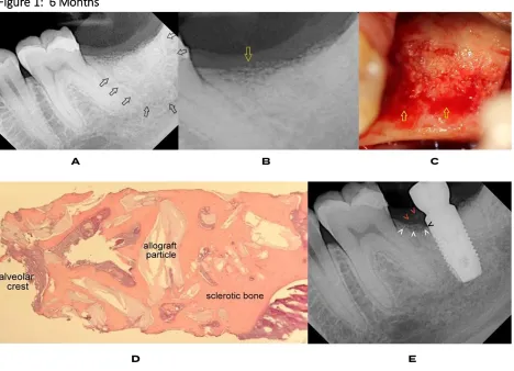
Figure 1: 6 Months
A healthy middle-aged female was grafted with mineralized freeze-dried bone allograft after extraction of her mandibular second molar. (A) This radiograph was taken 6 months after extraction and grafting. It is noted that the graft site has lost the normal contours of the
extraction socket and the grafted site is surrounded by a radiolucent boarder (black arrows). (B) A radiographic enlargement of the crest shows loose bone particles in the gingiva and soft connective tissue reaching into the crest. (C) The crest of the extraction socket shows inflammation in areas where the loose bone particles are exposed on the surface and no inflammation in areas of the crest where the bone graft particles are covered with
mineralization. (D) A core sample of the site shows the inclusion of cadaver bone graft particles with an accumulation of bone graft particles at the crest. There are no osteoclasts with no resorption of the bone graft particles or the sclerotic bone . There are no basic multicellular units active in remodeling the graft site. The newly formed bone is sclerotic.3 The alveolar crest shows
arrow. White arrows show a layer of loose bone particles at the crest. The red arrows identify bone particles that have migrated from the crest into the gingiva. As the alveolar crest has broken down around the implant, bone particles have migrated into the gingiva. Studies have documented the formation of angular defects when implants are placed in sites grafted with cadaver bone grafts.4,5
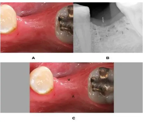
Figure 2: 2 Years
A healthy middle-aged woman had a mandibular first molar extracted and grafted with
where bone particles are exiting the gingiva into the oral cavity. The rapid exfoliation of the bone graft particles resulted in failure of the graft to preserve the mandibular ridge.
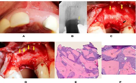
Figure 3: 5 Years
Tooth # 9 was extracted and grafted with mineralized freeze-dried bone allograft 5 years prior. (A) The photograph shows successful ridge preservation. (B) Radiographically, there is a
This histology clearly shows that the particles that exfoliate out of the alveolar crest and into the gingiva are both cadaver bone graft particles and particles of the sclerotic bone.
The association of the radiolucent zone on radiograph B with the intense bleeding found clinically, indicates that the radiolucent line represents a hypervascular zone that separates the grafted tissue from the patient’s bone.
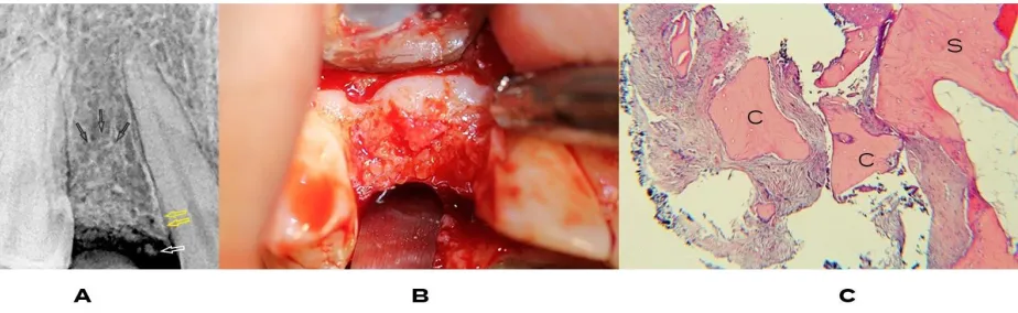
Figure 4: 7 Years
Figure 5: 10 Years
This patient was a middle-aged woman referred for a failed bridge. Tooth #20 failed under a bridge that had been in place for 10 years. (A) Prior to the bridge being placed, #19 was extracted and grafted with mineralized freeze-dried bone allograft. The extraction site of #20 was grafted with Socket Graft two months prior to this radiograph with implants planned for #19 and #20. (B) The crest of the graft site of #20 and the graft site of the freeze-dried bone allograft are shown after a full thickness flap. The crest of #20 was mineralized, however, the graft site for #19 contained loose cadaver bone graft particles in granulation tissue after 10 years. The loose graft material was removed, and a core sample was taken. (C) The core sample is composed of a large concentration of mineralized freeze-dried bone graft particles encased in sclerotic bone indicated by ‘U’. The crest is shown on the left with loose bone particles in soft connective tissue. There are no osteoclasts present and no indication of resorption of the bone graft particles or the sclerotic bone. There are no basic multicellular units active in remodeling the graft site. The bone around the graft particles is sclerotic.
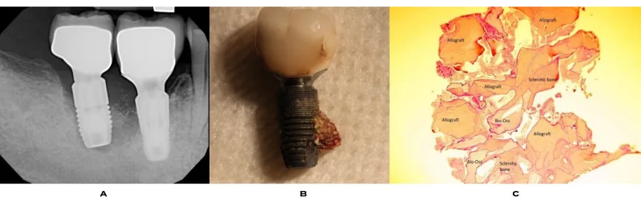
Figure 6: 15 Years
the implant is mobile. (B) Upon removal of the implant, a large portion of the bone adhered to the implant surface. However, the bone was not integrated to the implant surface and was easily removed from the implant intact. (C) Histology of the bone fragment showed mineralized freeze-dried bone allograft and Bio-Oss particles encased in sclerotic bone. Her treating oral surgeon was contacted and he reported that he removed the posterior teeth in 1998 and grafted the sockets with a combination of mineralized freeze-dried bone allograft and Bio-Oss. There are no osteoclasts present nor resorption of the bone graft particles or the sclerotic bone. There are no basic multicellular units and no active bone remodeling in the graft site.
Results:
Cadaver bone graft particles were found exfoliating out of the alveolus and into the gingiva. No resorption or remodeling of bone graft particles or sclerotic bone were found in any of the cases. The area of retained bone graft material and sclerotic bone appears to remain unchanged after mineralization but reduces in size over time. The reduction of the area of the grafted socket progresses over time as the bone graft particles and sclerotic bone breaks up at the crest and are exfoliated into the gingiva. Residual bone graft particles encased in sclerotic bone bound to an implant 15 years after grafting shows that even after loading, no remodeling of the residual bone graft particles occurs.
When using cadaver bone grafts, success or failure of ridge preservation appears to be dependent on the rate of exfoliation of the bone particles. Rapid exfoliation of the bone graft particles was found to result in failure to maintain the alveolar ridge. A slow rate of exfoliation appeared to be a requirement for successful ridge preservation. Osteoclasts were not present and no active resorption of the bone graft particles or the sclerotic bone was found in any of the histologic samples. There were no basic multicellular units found remodeling the graft site. The bone surrounding the graft particles was sclerotic. A common finding was the development of separation artifacts between the bone graft particles and the sclerotic bone that formed over them. This indicates that sclerotic bone does not integrate to the residual graft particles and likewise, the easy separation of bone from the implant surface indicates that sclerotic bone does not integrate to the implant surface.
Discussion:
The literature has reported that after mineralization there is no change in the percentage of retained bone graft particles over time.1,2 In all of the histological publications of mineralized
once mineralization of freeze-dried bone graft particles occurs in extraction sockets, no
resorption occurs afterwards. The allograft particles remain trapped in sclerotic bone until they are exfoliated into the gingiva. The radiolucent border commonly noted on radiographs
surrounding sclerotic bone and the retained cadaver graft particles has been found to be a hypervascular zone formed around the grafted tissue. It is proposed that the hypervascular zone exerts force that produces the glacial exfoliation of the grafted site. If the exfoliation of the grafted site is slow, the ridge preservation procedure is successful. However, if the exfoliation is rapid, the grafted material is rejected at a much faster rate and the ridge collapses. It is assumed that the hypervascular zone around sites grafted with mineralized freeze-dried bone allografts is an immune response to the transplanted tissue.
Studies have shown that the percentage of tissue occupied by retained graft particles does not change over-time. This study is consistent with those findings, however, while the percentage of retained bone graft particles does not change, the area of the graft site does reduce in volume. This study found that the graft site reduces in volume as the graft site migrates to the crest and is exfoliated.
As noted in the histology, exfoliation of the grafted site continues after implant placement. While there are no studies on the success of implants placed in sockets grafted with mineralized
freeze-dried bone allografts, the clinical impression is that implants in sites grafted with these materials fail at a much higher rate than sites grafted with resorbable materials that produce normal bone or implants placed in non-grafted sites. A common feature of the histology presented here was the presence of separation artifacts between the sclerotic bone and the bone graft particles. This finding indicates that the cadaver particles are not integrated into the sclerotic bone that surrounds them and, as a result, the strength of the bone in the graft site may be compromised. In addition, the finding that the grafted site never remodels allows for an accumulation of microfractures that can result in failure of the sclerotic bone and implant loss.6
The finding that sclerotic bone does not integrate with cadaver bone particles and that sclerotic bone does not integrate to the implant surface implies that implants placed in sockets that are grafted with cadaver bone grafts are retained mechanically rather than biologically. This finding reflects the fact that there are no studies that have shown implant integration of implants in sockets grafted with cadaver bone grafts.
Studies have found that many implants are associated with an increased incidence of open contacts.7 It is understandable that if the graft site that supports the implant is migrating toward
the crest, the opening of adjacent contacts is plausible. However, no studies have evaluated the correlation of implants placed in sites grafted with cadaver bone grafts and open contacts.
No long-term studies have been published on the success rates of implants placed in sockets grafted with freeze-dried allograft bone particles. Freeze-dried bone allografts are a transplant tissue and as such are not evaluated for safety and effectiveness by the FDA. It is the
the transplant recipient. It is the responsibility of the transplant team to know how the transplant will perform in the recipient and ensure that the transplant has a high likelihood of success. None of this is known about freeze-dried bone allograft particles that have been grafted into sockets that will receive an implant. Without knowing what the graft material is doing to the patient and without knowing anything about the success rates of implants placed in sockets grafted with mineralized freeze-dried bone allograft, the clinician could be held legally liable for performing a transplant without scientific justification.
Conclusion:
Mineralized freeze-dried bone allograft particles are not resorbed after mineralization. A
hypervascular zone forms around the grafted site and exfoliates the bone graft particles into the gingiva. The mineralization process produces sclerotic bone which never remodels into normal bone. Sclerotic bone covers but does not integrate to the retained bone graft particles or implant surface resulting in a weakened structure with an increased likelihood of bone failure and implant loss.
1. J Periodontol. 2010 Dec;81(12):1765-72. doi: 10.1902/jop.2010.100286. Epub 2010 Jul 27. Histologic analysis of healing after tooth extraction with ridge preservation using mineralized human bone allograft. Beck TM1, Mealey BL
2. J Periodontol. 2016 Sep;87(9):1022-9. doi: 10.1902/jop.2016.160139. Epub 2016 Apr 30. Effect of Healing Time on New Bone Formation After Tooth Extraction and Ridge Preservation With Demineralized Freeze-Dried Bone Allograft: A Randomized Controlled Clinical Trial. Whetman J, Mealey BL
3. Compend Contin Educ Dent. 2011 Nov-Dec;32(9):E146-55. Alveolar ridge augmentation: comparison of two socket graft materials in implant cases.
Tolstunov L1, Chi J
4. J Periodontol. 2016 Jan;87(1):14-20. doi: 10.1902/jop.2015.150229. Epub 2015 Sep 3. Relationship Between Osteoporosis and Marginal Bone Loss in Osseointegrated Implants: A 2-Year Retrospective Study. Corcuera-Flores JR1, Alonso-Domínguez AM1, Serrera-Figallo MÁ1, Torres-Lagares D1, Castellanos-Cosano L1, Machuca-Portillo G1
6 Preprints How Cadaver Bone Transplants Mineralize and Sclerotic Bone Fails. Preprints 2018, 2018100300 (doi: 10.20944/preprints201810.0300.v1). Steiner, G.