Characterization of the functional properties of the
neuroectoderm in mouse Cripto
-/-
embryos showing
severe gastrulation defects
GIOVANNA L. LIGUORI*
,1,#, DIEGO ECHEVARRIA
2,#, SONIA BONILLA
2,###, DANIELA D’ANDREA
1,
ANNAMARIA LIGUORO
1, MARIA G. PERSICO
1,##,†and SALVADOR MARTINEZ
2,##1Institute of Genetics and Biophysics «A. Buzzati-Traverso», CNR, Naples, Italy and 2CIBERSAM. Instituto de Neurociencias UMH-CSIC, University Miguel Hernandez, Alicante, Spain
ABSTRACT During development of the mammalian embryo, there is a complex relation between formation of the mesoderm and the neuroectoderm. In mouse, for example, the role of the node and its mesendoderm derivatives in anterior neural specification is still debated. Mouse Cripto -/- embryos could potentially help settle this debate because they lack almost all embryonic endoderm and mesoderm, including the node and its derivatives. In the present paper, we show that Cripto -/- embryos can still form functional neural stem cells that are able to differentiate and maintain a neural phenotype both in vivo and in vitro. These data suggest that signals emanating from the mesoderm and endoderm might not be essential for the formation and differentiation of neural stem cells. However, we use grafting experiments to show that the Cripto -/- isthmus (the secondary organizer located at the midbrain-hindbrain boundary) loses its inductive ability. We further show that the Cripto -/- isthmus expresses lower amounts of the isthmic signalling molecule, Fgf8. Since nearby tissues remain competent to respond to exogenously added Fgf8, this reduction in Fgf8 levels in the Cripto -/- isthmus is the potential cause of the loss of patterning ability in graft experiments. Overall, we interpret our data to suggest that the mammalian node and primitive streak are essential for the development of the regional identities that control the specification and formation of the secondary organizers within the developing brain.
KEY WORDS:
Cripto, mammalian node, neural differentiation, isthmic organizer, Fgf8, gastrulation defect
Introduction
Neural induction is the process by which embryonic cells in the epiblast acquire a neural fate rather than give rise to other structures such as epidermis and mesoderm (Stern, 2006). The relation between mesoderm and neuroectoderm is complex. Recent studies emphasise that definition of the neural plate during normal development first requires establishing a boundary between the region of the epiblast destined to ingress into mesendoderm during gastrulation and the medial edge of the presumptive neural plate (Sheng et al., 2003; Stern, 2005; Stern, 2006). On the hand, ingression of the epiblast inside the primitive streak to form mesoderm must be blocked in order to specify the
BIOLOGY
www.intjdevbiol.com*Address correspondence to: Giovanna L. Liguori. Institute of Genetics and Biophysics "A. Buzzati-Traverso", CNR, Via Pietro Castellino 111, 80131 Naples, Italy. Fax +39-081-6132-595. e-mail: [email protected]
Notes:† Deceased 5 February 2007. # These authors contributed equally to this work. ## These authors contributed equally to this work. ### Present address:
Instituto de Biomedicina de Sevilla. Hospital Universitario Virgen del Rocio, Universidad de Sevilla, Spain.
Accepted: 10 October 2008. Published online: 23 January 2009.
ISSN: Online 1696-3547, Print 0214-6282
© 2009 UBC Press Printed in Spain
Abbreviations used in this paper: A-P, antero-posterior; AVE, anterior visceral
endoderm; M-HB, mid-hindbrain boundary.
presumptive neural plate; on the other hand mesoderm itself participates in neural induction and patterning.
refine the antero-posterior (A-P) specification of the three main domains in the brain primordium: the forebrain, the midbrain and the hindbrain. These secondary organizers are the anterior neural ridge (ANR) at the anterior end of the neural plate, the zona limitans intrathalamica (ZLI) in the middle of the diencephalon, and the isthmic organizer (IsO), located at the mid-hindbrain boundary (M-HB) (Echevarria et al., 2003). However, evidence is accumulating against this two-step model. If the mouse primary organizer is ablated either surgically during gastrulation or geneti-cally by deletion of HNF3βor Cripto, a neural plate still forms (Ang and Rossant, 1994; Davidson et al., 1999; Ding et al., 1998; Klingensmith et al., 1999; Liguori et al., 2003; Weinstein et al., 1994). The mouse Cripto null mutant embryos are very informa-tive. Cripto is the founding member of the EGF-CFC genes family, coding for membrane-anchored extracellular factors essential for vertebrate embryo development (Persico et al., 2001). Cripto is expressed in embryonic stem (ES) cells and in the forming mesoderm, but has never been detected in the visceral endoderm (Dono et al., 1993; Johnson et al., 1994; Kimura et al., 2001). As expected from this expression pattern, Cripto -/- embryos have the peculiar characteristics of losing almost all the embryonic endo-derm and mesoendo-derm, including the node and its derivatives, but the embryos still possess a functional AVE (Ding et al., 1998; Liguori et al., 2003). With these particular characteristics, Cripto
null mutants constitute an extremely useful model to study the contribution of the node and primitive streak (including their derivatives) to early anterior neural plate development in mouse. We have previously analyzed the nature and topology of the neural tissues in Cripto -/-mutants, both in vivo and in vitro. We demonstrated that Cripto -/- embryos develop complex molecular patterns, suggesting that anterior neural fate and normal A-P regionalization do not require the presence of the node and primitive streak (Liguori et al., 2003).
In the present study, we extend our analysis to the functional properties of the anterior neural cells in Cripto -/- embryos, with the aim of determining which aspects of the mutant neural epithelium are principally affected by the absence of signals emanating from the node and from the embryonic mesendoderm. Our data indi-cate that the loss of the primary organizer and of most all the embryonic mesoderm and endoderm does not affect the compe-tence of neural cells to differentiate in response to surrounding stimuli. In other words, these results indicate that specification of neural lineages does not absolutely require signals from the mesoderm and endoderm. However, gastrulation failure specifi-cally impairs the correct development of the regional identities that control the specification of the secondary organizers, that, in turn, are required for the definitive refining of anterior neural patterning.
Results
Cripto is dispensable for neural differentiation both in vitro and in vivo
Our previous analysis demonstrated that Cripto -/- embryos
form anterior neural territories (Liguori et al., 2003). In the present study, we plan to investigate the effect on neural progenitors of
Cripto inactivation and the consequent loss of embryonic meso-derm and endomeso-derm, by analysing whether Cripto -/- embryos
develop neural progenitors and whether the competence of these
progenitors is significantly affected. To this purpose, we derived
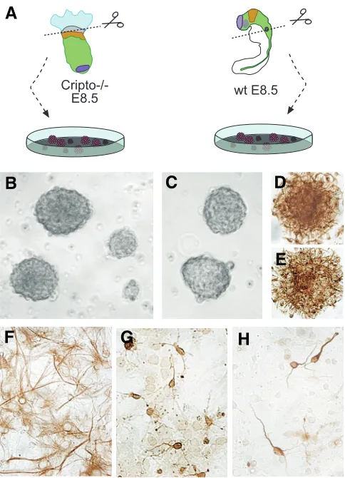
Cripto -/- neural progenitors (Fig. 1A) by dissecting the embryonic region of 8.5 dpc Cripto null mutants, which is completely com-posed of anterior neuroectoderm (Liguori et al., 2003). As a control, we dissected the corresponding region of the same stage wild type (wt) embryos, i.e. from the most rostral part of the forebrain until the otic vesicle (Fig. 1A). After three days in culture, both Cripto -/- and wt embryonic cells formed neurospheres (Fig.
1B, C). When the neurospheres were dissociated and the cells cultured in fresh medium, the isolated cells retain the ability to proliferate and to reform neurospheres. It was possible to main-tain in culture and expand the neurospheres for at least one month, without any macroscopic difference in their number and morphology between Cripto -/- and wt cultures.
After 13 days of in vitro culture, a fraction of the Cripto -/- and
wt neurospheres were fixed in PFA and analyzed by immunocy-tochemistry with anti-nestin antibody (an intermediate filament
Fig. 1. Neurospheres derived from Cripto -/- embryos behave as wild
type neurospheres in vitro.(A) Schematic representation of experi-mental assays in which anterior neural tissues from Cripto -/- and wt (control) embryos were dissociated and allowed to develop in vitro (see Materials and Methods for details). (B,C) Brightfield image of Cripto -/-neurospheres (B) compared to wt -/-neurospheres (C). (D,E) Nestin immunostaining of wt (D) and Cripto -/- (E) neurospheres. (F,G,H) Immunostaining of Cripto -/- neurospheres after differentiation (see Ma-terials and Methods). Cripto -/- neurospheres are capable of differentiating into astrocytes (GFAP positive) (F), oligodendrocytes (NG2 positive) (G) and neurons (beta-tubulin positive) (H).
G
B
C
D
E
F
H
expressed in undifferentiated neuroepithelial cells, Gritti et al., 1996; Reynolds and Weiss, 1992; Tohyama et al., 1992). The immunocytochemistry revealed that both Cripto -/- and wt
neurospheres expressed nestin (Fig. 1D, E). The remainer of the cultures were grown following a protocol to cause neurosphere cells differentiation in both neural and glial cell types (adhesion to a Matrigel substrate, removal of growth factors) (Bonilla et al., 2005). After another seven days in culture, the cells were fixed and analyzed by immunocytochemistry. The Cripto -/-
differenti-ated cultures contained cells expressing the Glial Fibrillary Acidic Protein (GFAP) (astrocytes, Fig. 1F), cells expressing the β -tubulin III (neurons, Fig. 1H) and cells expressing NG2 (oligodendrocites both mature and precursors; Fig. 1G). We obtained the same results with Cripto -/- neurospheres after 26
days of in vitro culture (data not shown). Collectively, these data show that the neuroectoderm of Cripto -/- embryos contains neural
progenitors, which are able to form neurospheres in vitro. More-over, Cripto -/- neurospheres are able to differentiate into both
neuronal and glial cell types. We were not able to detect any significant difference in the number and morphology between the
Cripto -/- and wt cultures of both neurospheres and differentiated
neural cells. However, we cannot exclude subcellular/structural or functional differences.
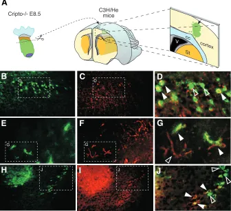
Finally, the embryonic region of Cripto null mutants at 8.5 dpc were dissected into equal parts and was grafted into the telen-cephalic region of newborn C3H/He mice (Fig. 2A). After five days, the mice were sacrificed and the brain was dissected, sectioned and analysed by immunohistochemistry. The H-2Db
antibody (specific for the C57Bl6/J background of the Cripto
-/-embryos) revealed the presence of Cripto -/- cells inside the cortex
original fate as undifferentiated progenitors or is able to mature into neuronal and glial cells.
In conclusion, in vivo Cripto inactivation and the consequent lack of mesoderm and endoderm do not significantly affect the formation and the competence of neural progenitors inside the mouse embryo. These data are in agreement with previous analyses performed on Cripto -/- ES cells, demonstrating that
Cripto is dispensable for neuronal differentiation both in vitro and
in vivo (Minchiotti et al., 2006; Parish et al., 2005; Parisi et al., 2003).
The mid-hindbrain boundary of Cripto -/- embryos does not
possess the functional properties of an organizer
We next wished to determine how Cripto influences the func-tional activity of the most studied secondary organizer, the isth-mus, which is located at the mid-hindbrain boundary (M-HB) and which controls anterior hindbrain and midbrain regionalization (Martinez, 2001). We have previously shown that Cripto null mutants do not form ANR or ZLI, but only develop the molecular pattern that characterizes the isthmic organizer (Liguori et al., 2003; 2008). In the present study, we investigate whether this putative organizer located at the M-HB possesses properties of a true functional isthmic organizer. Grafting experiments have been classically made in chick. However, the development of an experimental technique based on explanting the mouse anterior neural tube (ANT) onto polycarbonate membrane has made possible short-term neuroepithelial grafting experiments also in mice (Echevarria et al., 2001). This technique has allowed the use of wt mouse ANT as host tissue for a graft and then of tissues from knock-out mice as source for heterotopic and heterochronic
Fig. 2. Cripto -/- cells can be incorporated into
the brains of newborn mice as both undiffer-entiated and differundiffer-entiated neural cells. (A)
Schematic representation of experimental as-says in which anterior neural tissues from Cripto
-/- embryos were injected into newborn mouse
cerebral cortex (see Materials and Methods for details). (B-I) Immunohistochemistry assays on brain sections of P5 C3H/He mice injected with 8.5 dpc Cripto -/- embryo tissues. The Cripto -/-cells are identified by the anti-H2Dd antibody
(B,E,H). Some of the Cripto -/- cells incorporated into the brain expressed nestin (C,D), others expressed GFAP (F,G) and some β-tubulin (I,J). Co-expressing cells are indicated by white arrow-heads; non co-expressing Cripto -/- cells are pointed to by empty arrowheads.
G
B
C
D
E
F
H
I
J
A
of the host mice (Fig. 2B, E, H). Some Cripto
-/- cells expressed nestin (Fig. 2C, D), while
others were GFAP positive (Fig. 2F, G) being astrocytes. Finally, co-expression of H-2Db
(Fig. 2H) with anti β-Tubulin antibody (Fig. 2I, J) showed that some Cripto -/- cells also
ex-pressed neural markers. These results indi-cate that neuroepithelial cells from the Cripto
-/- embryos can be incorporated into the brain
tissue of a newborn mouse host. Moreover, the data suggest that Cripto -/-
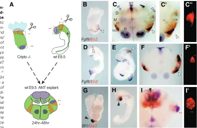
intraspecific transplants. Then, we grafted the M-HB of 8.5 dpc
Cripto -/- embryos into the diencephalic-mesencephalic boundary
region of 9.5 dpc ANT mouse explants (Fig. 3A). We cultured the explants in vitro for 24h after the graft and then analyzed, by double WISH, the expression profile of both Fgf8, coding for the isthmic signalling molecule, and En2, a known target of the Fgf8 signalling pathway, looking for ectopic induction of En2 expres-sion close to the Fgf8 positive graft (Fig. 3C-C”). As control, we also grafted the isthmus of mouse wt embryos at both 8.5 and 9.5 dpc (Fig. 3A, F, F’).
We found that approximately 50% of the explants showed Fgf8
expression within the grafts (Fig. 3C, C’, F), independent of whether the donor embryo was wt or Cripto -/-. The grafts were
labelled by DiI (Fig. 3C”, F’). The detection of Fgf8 expression confirmed that the region transplanted was at the M-HB of the
donor embryo. However, while wt isthmic grafts (see Table 1) induce En2 expression in the host tissue (Fig. 3F, F’), none of the
Cripto -/- grafts were able to induce ectopic En2 expression(Fig.
3C-C”) (see Table 1). We cultured the Cripto -/- transplants also for
48h following the graft, but even in this case we could not detect any En2 induction (data not shown; see Table1). Since the absence of inductive properties could reflect an immature condi-tion of the isthmic neuroepithelium in Cripto -/- embryos, we
explanted the Cripto -/- embryos at 8.5 dpc and let them develop
in vitro for 24h in order to allow any potential stabilization of Fgf8
expression (see also Liguori et al., 2003). Then, we grafted the
Cripto -/- M-HB in a wt host ANT at 9.5 dpc. The transplants were
cultured for 24 or 48h but none showed ectopic En2 induction (data not shown; Table 1).
We conclude that the capacity of the M-HB of the Cripto
-/-E
B
C
C'
C''
D
F
F'
A
H
I
I'
G
Graft Host Donor Time in culture after the graft Total number of grafts Fgf8 positive grafts En2 induction in the host
24 h 18 8 0
8.5 dpc Cripto-/- embryo 48h 19 6 0
8.5 dpc Cripto-/- explant cultured for 24h 48h 9 3 0
24h 18 8 0
8.5 dpc wt embryo
48h 19 6 3 (50%)
isthmus 9.5 dpc wt diencephalon
9.5 dpc wt embryo 24h 18 8 4 (50%)
TABLE 1
SUMMARY OF THE GRAFT EXPERIMENTS
Fig. 3. Cripto-/- neural tissues are
main-tained in host tissue but Cripto -/-
mid-hindbrain (M-HB) is unable to induce En2 ectopic expression.(A) Schematic representation of the embryological ex-perimental manipulation of Cripto -/- and wt IsO (*) or ANR (**) into the mes/ diencephalic boundary (m/d-B) region of wt anterior neural tube (ANT) explant culture. (B-C’’) Experimental assays where the IsO from Cripto -/- animals was transplanted into the m/d-B of ANT explant culture. (B) Expression pattern of Fgf8 (blue) and En2 (red) in Cripto -/-embryos at 8.5 dpc. (C) Double WISH expression pattern with Fgf8/ En2 in a wt 9.5 dpc ANT explant after 24 h of graft incubation. The red asterisks indi-cate the engrafted Cripto -/- IsO. The white arrowheads indicate the normal position of the IsO. (C’) Magnified area of the grafted region detected by double WISH with Fgf8/En2. (C’’) The same area as (C’) detected by fluorescence with the corresponding DiI excitation
embryos to induce En2 ectopic expression is severely affected: the M-HB of the Cripto -/- embryos appears to loose its patterning
properties and thus cannot be defined as a true organizer. Nonetheless, the transplanted region continues to express its characteristic markers (Fgf8 and En2), at least for 48h after the graft, suggesting that the tissue inside the Cripto -/- M-HB is
healthy and able to retain its molecular identity.
Finally, we performed an ectopic graft of the Cripto -/- distal
region (corresponding to forebrain) dissected at 8.5 dpc into the diencephalon of 9.5 dpc wt embryos (Fig. 3A). We cultured the grafts for 24h and then analyzed their expression profile by double WISH. The chosen Cripto -/- region expressed forebrain markers,
such as Bf1 and Six3 genes (Liguori et al., 2003; Fig. 3G) and continued to express the same genes 24h after the graft (in three out of four cases) (Fig. 3I,I’). Therefore, Cripto null mutant tissues are not severely affected by the graft and Cripto -/- cells maintain
their characteristic molecular pattern. Moreover, these data show that the ability to maintain specific gene expression (at least for 24h), when transplanted into an ectopic region, is a characteristic not only of the putative isthmus of the Cripto null mutants, but also a characteristic of other regions of the Cripto -/- embryo anterior
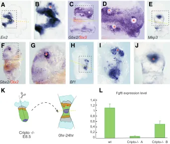
of wt and Cripto-/- embryos at 8.5 dpc by means of real-time PCR. We analysed separately the isthmus of five wt embryos and the M-HB of ten Cripto null mutants. We found that the M-HB of the
Cripto-/- embryos expresses less Fgf8 than the corresponding regions in the wt embryos. In particular, we identified two different groups of Cripto null mutants, the group A, composed by two out of ten embryos, which expresses very low doses of Fgf8 mRNA and the group B, composed by the left eight, which express almost half of the level of Fgf8 mRNA found in the wt (Fig. 4L). These results indicate that Cripto-/-embryos still express Fgf8 at the Otx2/Gbx2 molecular boundary, but that its level is at least half reduced, and in some cases dramatically reduced, respect to the wt embryos. These reduction of Fgf8 expression level in the
Cripto-/- M-HB could realistically be the cause of the loss of patterning abilities after grafting.
Discussion
The functional analysis we have performed on Cripto null mutants gives important information on the role that embryonic mesendoderm and the primary organizer play during neural
dif-Fig. 4. Cripto -/- neuroectoderm is able to respond to the inductive stimulus provided by
exogenous Fgf8b. Implanting Fgf8b-coated beads into the Cripto -/- explants causes after 24 h of
in vitro culture the expression of the midbrain marker En2(A,B), the anterior hindbrain marker Gbx2
(C,D,F,G), the forebrain marker Bf1(H,I) and the Fgf8 target Mkp3(E,J). The red asterisks indicate bead positions. The dashed squares indicate higher magnification view of examples (A,C,E,F,H) in (B,D,G,I,J) respectively. Dashed yellow lines indicate the midline of the Cripto -/- embryos flattened onto the polycarbonate membranes separating the experimental side from the control side (see Liguori et al., 2003). (K) Schematic representation of a Cripto -/- explant. (L) Histograms represent the quantitative-PCR values of Fgf8 mRNA levels between wt and Cripto -/- mid-hindbrain cases (with standard error bars).
G
B
C
D
E
F
H
I
J
K
L
A
neural plate.
Fgf8 signaling is active in Cripto -/- neu-roectoderm, but the amount of Fgf8 mRNA is lower in the M-HB of Cripto -/-compared to wt embryos
To gain insight into the lack of patterning ability of the Cripto-/- embryo isthmus, we first verified that the Fgf8 signaling pathway is active in these embryos, in particular whether the Cripto-/- neuroectoderm can respond to the Fgf8 inductive stimulus. We implanted Fgf8b-coated beads (Crossley et al., 1996; Garda et al., 2001; Liu et al., 1999; Shimamura and Rubenstein, 1997) in the
Cripto-/- embryos explanted at 8.5 dpc, cultured the explants for 24h and then ana-lyzed the expression of specific diagnostic markers by WISH (Fig. 4). In the Cripto -/-neuroectoderm Fgf8b induces the expres-sion of the midbrain marker En2 (Fig. 4A, B), the anterior hindbrain marker Gbx2 (Fig 4C-F) and also the forebrain marker Bf1 (Fig. 4G, H). The downstream modulator Mkp3
gene (Echevarria et al., 2005; Kawakami et al., 2003) is also induced in Cripto-/- neural tissue after 24 hours exposure to the Fgf8b-soaked beads (Fig. 4I, J). Collectively, these data demonstrate that Cripto-/- neural cells are able to respond to the inductive stimulus provided by exogenous Fgf8b, indicating that Fgf8 signaling pathway is active in the
Cripto-/- embryos.
ferentiation. The main conclusions of this work are essentially two. First, in Cripto null mutants, differentiation of neural stem cells towards neuronal and glial fate is not significantly affected by the absence of signals emanating from the embryonic mesoderm and endoderm. These data suggest that mesendodermic signals are dispensable not only for neural stem cells to form, but also for their differentiation into both a neuronal and glial phenotype, possibly indicating that neuroectoderm itself provides all the signals neces-sary for neuronal and glial differentiation. This view reminds very close the default model of neural induction proposed by Hemmati-Brivanlou and Melton in 1997, according to which ectoderm cells have an inherent tendency towards the neural identity, but consti-tutive BMP signalling in the ectoderm prevents realization of the neural fate. Even though challenges to the default model emerged from experiments both in frog and other vertebrates, mainly in chick, showing that Fibroblast growth factors (FGF) and canonical Wnt signals have also been implicated in the neural induction process (Delaune et al., 2005; Hongo et al., 1999; Linker and Stern, 2004; Sheng et al., 2003; reviewed in Stern, 2005, 2006), the findings that both FGF and Wnt signals can inhibit BMP signalling, might reconcile these apparent conflicting observations (Baker et al., 1999; Pera et al., 2003; Wessely et al., 2001). Recently, inhibition of Activin/Nodal/Smad2 signaling has been also reported to be involved in neural induction (Chang and Harland, 2007; Smith
et al., 2008). We note that Nodal signalling is able to crosstalk with BMP signaling and induce Bmp4 expression (Beck et al., 2002; Ben-Haim et al., 2006; Yeo and Withman, 2001). These data suggest that simultaneous suppression of BMP and Nodal-like signals, and then combined inhibitory rather than instructive sig-nals, might be crucial for neural fate determination, Cripto has a key role in this scenario, being deeply involved in Nodal signalling (Gritsmann et al., 1999). In fact, Cripto interacts with Alk receptors for the formation of the Nodal receptorial complex (Reissman et al., 2001). Thus, neural differentiation might occur in the absence of Cripto, due to impairment in the Nodal signalling and consequently in the BMP signalling.
The second main conclusion of the paper is that the Fgf8 -positive territory, corresponding to the putative isthmic organizer, in the neuroectoderm of Cripto null mutants does not develop the ability to redirect the diencephalon or rostral mesencephalon of a wt embryo explant towards a mid-hindbrain fate, as the wt isthmic region does. Our analysis also shows that the M-HB of Cripto -/-embryos expresses a lower amount of Fg8 mRNA than the wt isthmus. Being Fgf8 be recognized as the principal isthmic signal-ling molecule, we hypothesise the M-HB of Cripto-/- embryos does not possess patterning activity in graft experiments, because of the reduction in the Fgf8 expression level. It has been previously been suggested that achievement of a threshold level of Fgf8 is essential for induction of precise identity into neighbouring cells: Sato and Nakamura (2004) demonstrated that only a strong Fgf8 signal activates the Ras-extracellular signal-regulated kinase (ERK) path-way in the mesencephalon/metencephalon boundary and this is necessary and sufficient to induce cerebellar development (Sato and Nakamura, 2004). In contrast, a lower level of Fgf8 signalling seems to be required for midbrain development (Sato et al., 2004; Sato and Nakamura, 2004; Basson et al., 2008). Very recently, it has been demonstrated that transcriptional activation of Wnt1 and
En1, two targets of Fgf-signaling at the M-HB, requires different threshold levels of Ras–MAP kinase activity, thus supporting the
idea that Fgf8 functions in a dose-dependent manner at the M-HB (Vennemann et al., 2008). Furthermore, nonlinear dosage effects of Fgf8 on the expression of a subset of genes, including Bmp4 and
Msx1, have been correlated with a holoprosencephaly phenotype and with the nonlinear expression of transcription factors that regulate neocortical patterning (Storm et al., 2006). A nonlinear, threshold-like, epistatic response to an Fgf8 stimulus has also been observed during submandibular salivary gland morphogen-esis (Jaskoll et al., 2004). Overall, these data suggest that modifi-cations in the relative strength of Fgf signaling can have profound effects on the relative size and nature of anterior neural tube subdivisions (Storm et al., 2006). However, we note that most
Cripto-/- embryos can still specify mesencephalic territories, as demonstrated by the expression of Wnt1, Pax2, En1 and En2
markers (Ding et al., 1998; Liguori et al., 2003 and present data), meaning that half Fgf8 dosage remains sufficient to induce some territories. Interestingly, the distance of the cells from the Fgf8 sources might also play an important role. The amount of Fgf8 produced by the M-HB of Cripto-/-embryos might be sufficient to pattern the identity of the cells inside the graft, but not able to reach the cells of the host tissue, located outside the graft. However, we cannot exclude that other still unknown factors are affected in the
Cripto-/- M-HB and that this uncharacterized defect might contrib-ute to the phenotype we observe.
Finally, from the point of view of evolution, M-HB in Cripto null mutants might resemble the one found in some protochordates. In protochordates, the anterior part of the nerve cord is expanded into a cerebral vesicle, which is homologous to the forebrain and midbrain (Lacalli, 2006; Williams and Holland, 1998), or possibly just forebrain (Takahashi and Holland, 2004; Takahashi, 2005). Caudal to this region, the nerve cord expresses markers character-istic of the hindbrain and spinal cord (Holland and Holland, 1999). In the CNS of the cephalochordate amphioxus, Gbx and Otx genes are expressed in positions comparable to the Vertebrates, but En, Pax2/5/8 and Wnt1 are not expressed near the caudal limit of Otx,
suggesting that the genetic machinery to position the M-HB was present in the protochordate ancestors of the vertebrates, but is insufficient for induction of organizer genes (Castro et al., 2006). Therefore, the M-HB of Cripto-/- embryos might resemble this ancestral situation, possessing the M-HB molecular characteris-tics, but not fully acting as an organizer”.
Experiments in a variety of organisms (mouse, frog, zebrafish) already showed that neural tissue might be specified without an absolute requirement for primary organizer (Klingensmith et al., 1999; Liguori et al., 2003; Saude et al., 2000; Shih and Fraser, 1996; Wessely et al., 2001). However, the present study is the first time that a functional analysis is performed on the neural tissues formed in absence of such an organizer. Our data propose that the reason for the node to be essential for the development and refining of the brain architecture is its requirement for the formation of the secondary organizers.
Materials and Methods
Neurosphere preparation and differentiation
solution. Neurosphere preparation and differentiation have been per-formed according to Bonilla et al. (2005). Prior to immunochemical analysis, the cell cultures were fixed with 4% paraformaldehyde in PBS for 1h at 4°C.
Intracerebellar embryo grafts
Cripto -/- embryonic neuroectoderm pieces were dissected in cold DMEM solution and were injected into the telencephalon of cold-anaes-thetized neonatal C3H/He mice. The Cripto -/- tissues were introduced in the brain, through whole in the cranium, using a 10 µl Hamilton syringe. The injection was performed in the parietal area, 1mm caudal and lateral to the bregma point and 0.5 mm into the parenchyma from the dura mater (Franklin and Paxinos, 1997). Five days after tissue transplanta-tion, experimental mice were anaesthetized with chloroform and were fixed by intracardiac perfusion with 4% paraformaldehyde in PBS. Dis-sected brains were postfixed overnight at 4°C in the same fixative. After washing in PBS, the fixed brains were cryoprotected for 8h at room temperature in 10% sucrose-PBS and overnight at 4°C in 20% sucrose– PBS for cryotome sections. Serial sections 20 µm thick were mounted in five parallel series.
Immunocytochemistry and immunohistochemistry
Immunochemical techniques were performed as described by Bonilla and coworkers (2005), using the following primary antibodies: anti-nestin monoclonal antibody (Chemicon, Temecula, CA), anti-NG2 polyclonal antibody (Chemicon, Temecula, CA), anti Beta-III-Tubulin monoclonal antibody (Eurogenec, Belgium), and anti-glial fibrillary acid protein (GFAP) monoclonal antibody (Calbiochem, San Diego, CA).
Grafting experiments
Neural tissue explant culture of 8.5-9.5 dpc wild-type ICR and 8.5 dpc
Cripto -/- embryos were made according to Echevarria et al. (2001) and Liguori et al. (2003) respectively. For the grafting experiment, donor tissues were incubated with the lipophilic tracer DiI (100 ng/ml final concentration) (Honing and Hume, 1989) for 1h. After incubation, tissue was washed briefly in culture medium and prepared for microscopic dissection of neuroepithelium at the midbrain-hindbrain transition. Tissue grafting was performed using fine tungsten needles. The presumptive isthmus was dissected and isolated from the donor, transferred to culture medium by a glass pipette, and ectopically inserted in the host diencepha-lon into a previously made equivalent hole (heterotopic grafts). The grafts were in vitro cultured for 24-48h and then fixed with 4% paraformaldehyde in PBS overnight at 4°C. The isthmus was visualized by Fgf8 in situ
hybridization.
Bead implantation
Heparin acrylic beads (Sigma) were rinsed four to six times in PBS and then soaked in 5 µl FGF8 solution (1 mg/ml; R&D), for 1h at 4°C. The beads were then rinsed three times in PBS 0.1 M and thereafter implanted in the neuroectoderm of Cripto -/- embryos. Control beads were soaked only in PBS and implanted in the same manner. After 24 hours of culture, explants were fixed in 4% paraformaldehyde in PBS and processed for successive in situ hybridization.
In situ hybridization
RNA antisense probes were synthesized from linearized plasmid in the presence of Digoxigenin-UTP (Boehringer Manheim) or Fluorescein-UTP (Boehringer Manheim). The in situ hybridization technique was performed according to Liguori and coworkers (2003).
Real time RT-PCR analysis
Wild-type and Cripto -/- embryos were used for analysis of mRNA quantities. The region situated at the midbrain-hindbrain boundary (M-HB) of five wt and ten Cripto -/- embryos at 8.5 dpc was dissected. In the wt embryo the M-HB is easily detectable by looking at the constriction ridge of the isthmus, due to lower proliferation rate than the surrounding
tissues (Martinez et al., 1991; 1995). On the other hand, in the Cripto -/-embryo the M-HB is at the boundary between the -/-embryonic and extraem-bryonic regions, which have a different consistency and are separated by an embryonic constriction (Liguori et al., 2003). The tissue was homog-enized into Qiagen lysis buffer by passing it ten times through a 0.9 mm needle attached to a sterile plastic syringe. Total RNA from each M-HB was extracted using Rneasy Micro Kit (Qiagen) according to the manufacturer’s instructions and quantitated by NanoDrop-1000 Spectro-photometer. cDNA synthesis was achieved by using the Quanti Tect Reverse Transcription kit (Qiagen). Real Time PCR was performed using two primer sets produced by Quanti Tect Primer Assay (Qiagen) (QT00108773 for Fgf8; QT00166768 for Hprt). The primers for Fgf8 (Qiagen) amplify a fragment of 114 bp spanning the exons 5 and 6, while the primers for Hprt amplify a fragment of 168 bp spanning exons 3, 4 and 5 of the gene. The reactions were conducted according to the protocol of the Fluo Cycle SYBR Green mix (Euro Clone). The PCR protocol involved a denaturation program (95° for 15 min), followed by an amplification and quantitation program repeated 34 times (95° for 15 sec, 58° for 20 sec, 72° for 20 sec), a melting curve program (65°C – 95°C, with a heating rate of 0.5°C per second and continuous fluorescence measurement). The relative quantitation of gene expression was analyzed by the 2-ddCt method. To normalize the output for each sample, the expression of Fgf8
gene was divided by Hprt gene expression. The results are representa-tive of three independent experiments. The one-way ANOVA test was used to analyze the data from all experiments and P<0.0001 was considered significant.
Acknowledgments
We thank T. Moccia and F. Almagro for technical assistance, L. D’Orsi and both the “G. Pascale” and the Instituto de Neurociencias Animal Facility for animal care. We thank J. McGhee for critically reading of this manuscript. This work was supported by grants from the FIRB and AIRC to M.G. Persico and by the Spanish MEC BFU200509085, EC-CONSOLIDER CSD2007-00023, Imagenio2010. D. Echevarria has a contract of Program Ramón y Cajal-2004, and is supported by the Spanish Ministry of Health, Instituto de Salud Carlos III-CIBERSAM. D. D’Andrea was supported by a FIRC fellowship.
References
ANG, S. L., and ROSSANT, J. (1994). HNF-3 beta is essential for node and notochord formation in mouse development. Cell 78: 561-574.
BAKER, J. C., BEDDINGTON, R. S., and HARLAND, R. M. (1999). Wnt signaling in Xenopus embryos inhibits bmp4 expression and activates neural develop-ment. Genes Dev 13: 3149-3159.
BASSON, M. A., ECHEVARRIA, D., PETERSEN AHN, C., SUDAROV, A., JOYNER, A. L., MASON, I. J., MARTINEZ, S., and MARTIN, G. R. (2008). Specific regions within the embryonic midbrain and cerebellum require different levels of FGF signaling during development. Development 135: 889-898.
BECK, S., LE GOOD, J. A., GUZMAN, M., BEN HAIM, N., ROY, K., BEERMANN, F., and CONSTAM, D. B. (2002). Extraembryonic proteases regulate Nodal signalling during gastrulation. Nat Cell Biol 4: 981-985.
BEN-HAIM, N., LU, C., GUZMAN-AYALA, M., PESCATORE, L., MESNARD, D., BISCHOFBERGER, M., NAEF, F., ROBERTSON, E. J., and CONSTAM, D. B. (2006). The nodal precursor acting via activin receptors induces mesoderm by maintaining a source of its convertases and BMP4. Dev Cell 11: 313-323. BONILLA, S., SILVA, A., VALDES, L., GEIJO, E., GARCIA-VERDUGO, J. M., and
MARTINEZ, S. (2005). Functional neural stem cells derived from adult bone marrow. Neuroscience 133: 85-95.
CASTRO, L. F, RASMUSSEN, S. L., HOLLAND, P. W., HOLLAND, N. D., and HOLLAND, L. Z. (2006). A Gbx homeobox gene in amphioxus: insights into ancestry of the ANTP class and evolution of the midbrain/hindbrain boundary.
Dev Biol 295: 40-51.
Develop-ment 134: 3861-3872.
CROSSLEY, P. H., MARTINEZ, S., and MARTIN, G. R. (1996). Midbrain develop-ment induced by FGF8 in the chick embryo. Nature 380: 66-68.
DAVIDSON, B. P., KINDER, S. J., STEINER, K., SCHOENWOLF, G. C., and TAM, P. P. (1999). Impact of node ablation on the morphogenesis of the body axis and the lateral asymmetry of the mouse embryo during early organogenesis. Dev Biol 211: 11-26.
DELAUNE, E., LEMAIRE, P., and KODJABACHIAN, L. (2005). Neural induction in
Xenopus requires early FGF signalling in addition to BMP inhibition. Develop-ment 132: 299-310.
DING, J., YANG, L., YAN, Y. T., CHEN, A., DESAI, N., WYNSHAW-BORIS, A., and SHEN, M. M. (1998). Cripto is required for correct orientation of the anterior-posterior axis in the mouse embryo. Nature 395: 702-707.
DONO, R., SCALERA, L., PACIFICO, F., ACAMPORA, D., PERSICO, M. G., and SIMEONE, A. (1993). The murine cripto gene: expression during mesoderm induction and early heart morphogenesis. Development 118: 1157-1168. ECHEVARRIA, D., MARTINEZ, S., MARQUES, S., LUCAS-TEIXEIRA, V., and
BELO, J. A. (2005). Mkp3 is a negative feedback modulator of Fgf8 signaling in the mammalian isthmic organizer. Dev Biol 277: 114-128.
ECHEVARRIA, D., VIEIRA, C., GIMENO, L., and MARTINEZ, S. (2003). Neuroepi-thelial secondary organizers and cell fate specification in the developing brain.
Brain Res Brain Res Rev 43: 179-191.
ECHEVARRIA, D., VIEIRA, C., and MARTINEZ, S. (2001). Mammalian neural tube grafting experiments: an in vitro system for mouse experimental embryology. Int J Dev Biol 45: 895-902.
GARDA, A. L., ECHEVARRIA, D., and MARTINEZ, S. (2001). Neuroepithelial co-expression of Gbx2 and Otx2 precedes Fgf8 co-expression in the isthmic orga-nizer. Mech Dev 101: 111-118.
GRITSMAN, K., ZHANG, J., CHENG, S., HECKSCHER, E., TALBOT, W. S., and SCHIER, A. F. (1999). The EGF-CFC protein one-eyed pinhead is essential for nodal signaling. Cell 97: 121-132.
GRITTI, A., PARATI, E. A., COVA, L., FROLICHSTHAL, P., GALLI, R., WANKE, E., FARAVELLI, L., MORASSUTTI, D. J., ROISEN, F., NICKEL, D. D., and VESCOVI, A. L. (1996). Multipotential stem cells from the adult mouse brain proliferate and self-renew in response to basic fibroblast growth factor. J Neurosci 16: 1091-1100.
HEMMATI-BRIVANLOU A. and MELTON D. (1997). Vertebrate neural induction.
Annu Rev. Neurosci. 20:43-60.
HOLLAND, L. Z. and HOLLAND, N. D. (1999). Chordate origins of the vertebrate central nervous system. Curr Opin Neurobiol 9: 596-602.
HONGO, I., KENGAKU, M., and OKAMOTO, H. (1999). FGF signaling and the anterior neural induction in Xenopus. Dev Biol 216: 561-581.
JASKOLL, T., WITCHER, D., TORENO, L., BRINGAS, P., MOON, A. M., and MELNICK, M. (2004). FGF8 dose-dependent regulation of embryonic subman-dibular salivary gland morphogenesis. Dev Biol 268: 457-469.
JOHNSON, S. E., ROTHSTEIN, J. L., and KNOWLES, B. B. (1994). Expression of epidermal growth factor family gene members in early mouse development. Dev Dyn 201: 216-226.
KAWAKAMI, Y., RODRIGUEZ-LEON, J., KOTH, C. M., BUSCHER, D., ITOH, T., RAYA, A., NG, J. K., ESTEBAN, C. R., TAKAHASHI, S., HENRIQUE, D., SCHWARZ, M. F., ASAHARA, H., and IZPISUA BELMONTE, J. C. (2003). MKP3 mediates the cellular response to FGF8 signalling in the vertebrate limb.
Nat Cell Biol 5: 513-519.
KIMURA, C., SHEN, M. M., TAKEDA, N., AIZAWA, S., and MATSUO, I. (2001). Complementary functions of Otx2 and Cripto in initial patterning of mouse epiblast. Dev Biol 235: 12-32.
KLINGENSMITH, J., ANG, S. L., BACHILLER, D., and ROSSANT, J. (1999). Neural induction and patterning in the mouse in the absence of the node and its derivatives. Dev Biol 216: 535-549.
LACALLI, T. C. (2006) Prospective protochordate homologs of vertebrate midbrain and MHB, with some thoughts on MHB origins. Int J Biol Sci 2: 104-109. LIGUORI, G. L., BORGES, A. C., D’ANDREA, D., LIGUORO, A., GONCALVES, L.,
SALGUEIRO, A. M., PERSICO, M. G., and BELO, J. A. (2008). Cripto-independent Nodal signaling promotes positioning of the A-P axis in the early mouse embryo. Dev Biol 315: 280-289.
LIGUORI, G. L., ECHEVARRIA, D., IMPROTA, R., SIGNORE, M., ADAMSON, E., MARTINEZ, S., and PERSICO, M. G. (2003). Anterior neural plate regionaliza-tion in cripto null mutant mouse embryos in the absence of node and primitive streak. Dev Biol 264: 537-549.
LINKER, C., and STERN, C. D. (2004). Neural induction requires BMP inhibition only as a late step, and involves signals other than FGF and Wnt antagonists.
Development 131: 5671-5681.
LIU, A., LOSOS, K., and JOYNER, A. L. (1999). FGF8 can activate Gbx2 and transform regions of the rostral mouse brain into a hindbrain fate. Development
126: 4827-4838.
MARTINEZ, S. (2001). The isthmic organizer and brain regionalization. Int J Dev Biol 45: 367-371.
MINCHIOTTI, G., PARISI, S., and PERSICO, M. G. (2006). Cripto signaling in differentiating embryonic stem cells. Methods Mol Biol 329: 151-169. PARISH, C. L., PARISI, S., PERSICO, M. G., ARENAS, E., and MINCHIOTTI, G.
(2005). Cripto as a target for improving embryonic stem cell-based therapy in Parkinson’s disease. Stem Cells 23: 471-476.
PARISI, S., D’ANDREA, D., LAGO, C. T., ADAMSON, E. D., PERSICO, M. G., and MINCHIOTTI, G. (2003). Nodal-dependent Cripto signaling promotes cardiomyogenesis and redirects the neural fate of embryonic stem cells. J Cell Biol 163: 303-314.
PERA, E. M., IKEDA, A., EIVERS, E., and DE ROBERTIS, E. M. (2003). Integration of IGF, FGF, and anti-BMP signals via Smad1 phosphorylation in neural induction. Genes Dev 17: 3023-3028.
PERSICO, M. G., LIGUORI, G. L., PARISI, S., D’ANDREA, D., SALOMON, D. S., and MINCHIOTTI, G. (2001). Cripto in tumors and embryo development.
Biochim Biophys Acta 1552: 87-93.
REISSMANN, E., JORNVALL, H., BLOKZIJL, A., ANDERSSON, O., CHANG, C., MINCHIOTTI, G., PERSICO, M. G., IBANEZ, C. F., and BRIVANLOU, A. H. (2001). The orphan receptor ALK7 and the Activin receptor ALK4 mediate signaling by Nodal proteins during vertebrate development. Genes Dev 15: 2010-2022.
REYNOLDS, B. A., and WEISS, S. (1992). Generation of neurons and astrocytes from isolated cells of the adult mammalian central nervous system. Science
255: 1707-1710.
SATO, T., JOYNER, A. L., and NAKAMURA, H. (2004). How does Fgf signaling from the isthmic organizer induce midbrain and cerebellum development? Dev Growth Differ 46: 487-494.
SATO, T., and NAKAMURA, H. (2004). The Fgf8 signal causes cerebellar differen-tiation by activating the Ras-ERK signaling pathway. Development 131: 4275-4285.
SAUDE, L., WOOLLEY, K., MARTIN, P., DRIEVER, W., and STEMPLE, D. L. (2000). Axis-inducing activities and cell fates of the zebrafish organizer. Devel-opment 127: 3407-3417.
SHAWLOT, W., WAKAMIYA, M., KWAN, K. M., KANIA, A., JESSELL, T. M., and BEHRINGER, R. R. (1999). Lim1 is required in both primitive streak-derived tissues and visceral endoderm for head formation in the mouse. Development
126: 4925-4932.
SHENG, G., DOS REIS, M., and STERN, C. D. (2003). Churchill, a zinc finger transcriptional activator, regulates the transition between gastrulation and neurulation. Cell 115: 603-613.
SHIH, J., and FRASER, S. E. (1996). Characterizing the zebrafish organizer: microsurgical analysis at the early-shield stage. Development 122: 1313-1322. SHIMAMURA, K., and RUBENSTEIN, J. L. (1997). Inductive interactions direct early regionalization of the mouse forebrain. Development 124: 2709-2718. SIMEONE, A., and ACAMPORA, D. (2001). The role of Otx2 in organizing the
anterior patterning in mouse. Int J Dev Biol 45: 337-345.
SMITH, J. R., VALLIER, L., LUPO, G., ALEXANDER, M., HARRIS, W. A., and PEDERSEN, R. A. (2008). Inhibition of Activin/Nodal signaling promotes speci-fication of human embryonic stem cells into neuroectoderm. Dev Biol 313: 107-117.
STERN, C. D. (2005). Neural induction: old problem, new findings, yet more questions. Development 132: 2007-2021.
STERN, C. D. (2006b). Neural induction: 10 years on since the ‘default model’. Curr Opin Cell Biol 18: 692-697.
STORM, E. E., GAREL, S., BORELLO, U., HEBERT, J. M., MARTINEZ, S., MCCONNELL, S. K., MARTIN, G. R., and RUBENSTEIN, J. L. (2006). Dose-dependent functions of Fgf8 in regulating telencephalic patterning centers.
Development 133: 1831-1844.
TAKAHASHI, T. (2005) The evolutionary origins of vertebrate midbrain and MHB: insights from mouse, amphioxus and ascidian Dmbx homeobox genes. Brain Res Bull 66: 510-517
TAKAHASHI. T., and HOLLAND P. W. (2004). Amphioxus and ascidian Dmbx homeobox genes give clues to the vertebrate origins of midbrain development.
Development 13: 3285-3294.
THOMAS, P., and BEDDINGTON, R. (1996). Anterior primitive endoderm may be responsible for patterning the anterior neural plate in the mouse embryo. Curr Biol 6: 1487-1496.
TOHYAMA, T., LEE, V. M., RORKE, L. B., MARVIN, M., MCKAY, R. D., and TROJANOWSKI, J. Q. (1992). Nestin expression in embryonic human
neu-roepithelium and in human neuroepithelial tumor cells. Lab Invest 66: 303-313. VENNEMANN, A., AGOSTON, Z., and SCHULTE, D. (2008). Differential and dose-dependent regulation of gene expression at the mid-hindbrain boundary by Ras-MAP kinase signaling. Brain Res 1206: 33-43.
WEINSTEIN, D. C., RUIZ I ALTABA, A., CHEN, W. S., HOODLESS, P., PREZIOSO, V. R., JESSELL, T. M., and DARNELL, J. E., JR. (1994). The winged-helix transcription factor HNF-3 beta is required for notochord development in the mouse embryo. Cell 78: 575-588.
WESSELY, O., AGIUS, E., OELGESCHLAGER, M., PERA, E. M., and DE ROBERTIS, E. M. (2001). Neural induction in the absence of mesoderm: beta-catenin-dependent expression of secreted BMP antagonists at the blastula stage in Xenopus. Dev Biol 234: 161-173.
WILLIAMS, N. A., and HOLLAND, P. W. (1998) Gene and domain duplication in the chordate Otx gene family: insights from amphioxus Otx. Mol Biol Evol 15: 600-607.
Further Related Reading, published previously in the
Int. J. Dev. Biol.
See our recent Special Issue Fertilization, in honor of David L. Garbers and edited by Paul M. Wassarman and Victor D. Vacquier at: http://www.ijdb.ehu.es/web/contents.php?vol=52&issue=5-6
Expression of complement components coincides with early patterning and organogenesis in Xenopus laevis
Valérie A. McLin, Cheng-Hui Hu, Rina Shah and Milan Jamrich Int. J. Dev. Biol. (2008) 52: 1123-1133
Retinoic acid metabolizing factor xCyp26c is specifically expressed in neuroectoderm and regulates anterior neural patterning in Xenopus laevis
Misaki Tanibe, Tatsuo Michiue, Akira Yukita, Hiroki Danno, Masayuki Ikuzawa, Shoichi Ishiura and Makoto Asashima Int. J. Dev. Biol. (2008) 52: 893-901
Neural differentiation from human embryonic stem cells in a defined adherent culture condition
Hossein Baharvand, Narges-Zare Mehrjardi, Maryam Hatami, Sahar Kiani, Mahendra Rao and Mahdi-Montazer Haghighi Int. J. Dev. Biol. (2007) 51: 371-378
Expression and regulation of Xenopus CRMP-4 in the developing nervous system
Jacob Souopgui, Tiemo J. Klisch, Tomas Pieler and Kristine A. Henningfeld
2006 ISI **Impact Factor = 3.577** Int. J. Dev. Biol. (2007) 51: 339-343
Genetic control of dorsoventral patterning and neuroblast specification in the Drosophila Central Nervous System
Guoyan Zhao, Scott R. Wheeler and James B. Skeath Int. J. Dev. Biol. (2007) 51: 107-115
Isolation, genomic structure and developmental expression of Fgf8 in the short-tailed fruit bat, Carollia perspicillata
Chris J. Cretekos, Jian-Min Deng, Eric D. Green, NISC Comparative Sequencing Program, John J. Rasweiler and Richard R. Behringer
Int. J. Dev. Biol. (2007) 51: 333-338
Retinoic acid is required for specification of the ventral eye field and for Rathke’s pouch in the avian embryo
Malcolm Maden, Aida Blentic, Susan Reijntjes, Sophie Seguin, Emily Gale and Anthony Graham
Int. J. Dev. Biol. (2007) 51: 191-200
Developmental expression of Shisa-2 in Xenopus laevis
Ana-Cristina Silva, Mário Filipe, Marta Vitorino, Herbert Steinbeisser and José-António Belo
Int. J. Dev. Biol. (2006) 50: 575-579
Systematic screening for genes specifically expressed in the anterior neuroectoderm during early Xenopus development
Noriyuki Takahashi, Naoko Tochimoto, Shin-Ya Ohmori, Hiroshi Mamada, Mari Itoh, Masako Inamori, Jun Shinga, Shin-Ichi Osada and Masanori Taira
Int. J. Dev. Biol. (2005) 49: 939-951
The Fox gene family in Xenopus laevis:FoxI2, FoxM1 and FoxP1 in early development
Barbara S. Pohl, Antje Rössner and Walter Knöchel Int. J. Dev. Biol. (2005) 49: 53-58
Isthmus organizer and regionalization of the mesencephalon and metencephalon
Harukazu Nakamura and Yuji Watanabe Int. J. Dev. Biol. (2005) 49: 231-235
Expression of an Otx gene in the adult rudiment and the developing central nervous system in the vestibula larva of the sea urchin Holopneustes purpurescens.
Valerie B Morris, Jing-Ting Zhao, Deborah C A Shearman, Maria Byrne and Marianne Frommer