An obsession with the chick
RUTH BELLAIRS*
Department of Cell and Developmental Biology, University College London, London, UK.
ABSTRACT This paper provides a brief account of some aspects of the career of Ruth Bellairs using selected examples from her research publications, with the emphasis being placed on the early stages of chick embryo development, and in particular, on cell migration. Topics include the role of Hensen’s node, the vitelline membrane, the structure and segmentation of somites, the tail bud and the Wolffian duct. Her research approach has involved embryo culture, experimental surgery, transmission and scanning electron microscopy, time-lapse filming and immunostaining techniques.
KEY WORDS:
fibronectin, somites, cell migration, primitive streak, precardiac mesoderm.
I have always liked domestic fowls, with their beaks and their combs and their gizzards, so it is perhaps not surprising that I chose to become a zoologist. I had been much impressed as a schoolgirl with a book by Lancelot Hogben called “Science for the Citizen”, which had enjoyed a wide popularity in the pre-war years, and when I discovered that Hogben was the head of the Zoology Department in the University of Birmingham I decided that that was the place for me. But what a disappointment it was when I arrived in Birmingham in 1944 to find that Hogben was permanently away on war work. He returned to the university two years later when the war ended, but soon it became apparent to the students that Professor Hogben was not an easy man. Within a few months the entire research and teaching staff resigned. Among those who left us was Michael Abercrombie who was then soon appointed as a lecturer in Embryology at UCL (University College London).
As a student during the war I had given little thought to my future career as I knew that once I graduated I would be drafted into one of the women’s services. I rather fancied the WAAF (the women’s branch of the Royal Air Force). But with the end of the war the compulsory call-up was ended for women and I became free to choose my next step. At the critical moment Michael Ab-ercrombie wrote and invited me to join him as a PhD student to work on the chick embryo. I had looked at parasitology and inves-tigated entomology but Michael’s idea appealed much more, and so | went to the Sub-department of Embryology run by Professor G R De Beer (later Sir Gavin) in the Department of Anatomy and Embryology at UCL.
Before the war Michael had been a postgraduate student of CH Waddington at the Strangeways Research Laboratory in Cambridge. It was a period when the search was in full swing to identify the supposed chemical organiser of amphibian embryos, and Waddington had been the first to demonstrate experimentally
www.intjdevbiol.com
*Address correspondence to: Ruth Bellairs. Department of Cell and Developmental Biology, University College London, Gower Street, London WC1E 6BT, U.K. E-mail: [email protected] - http://orcid.org/0000-0002-1896-0179
Submitted: 12 January, 2018; Accepted: 12 January, 2018
ISSN: Online 1696-3547, Print 0214-6282 © 2018 UPV/EHU Press
Printed in Spain
Abbreviations used in this paper: UCL, University College London.
that Hensen’s node in the chick was in many ways comparable to the dorsal lip of the blastopore of amphibian embryos. His ap-proach was to insert grafts of Hensen’s node under the ectoderm in different regions of the area pellucida, and he found this often resulted in the induction of a secondary axis. It is almost impossible to carry out experiments of this type in ovo on the early embryo but Waddington (1932) had bi-passed this problem by devising the first successful technique for growing an entire chick blastoderm
in vitro. Essentially, it consisted of freeing the blastoderm from the
yolk and the vitelline membrane and then explanting it onto a bed of clotted blood plasma. It was still in use when I joined Michael’s lab in 1947. Suitable plasma was not yet available commercially so we were obliged to anaesthetise fowls in the lab and draw off blood from the carotids with home-made glass canulae that were awk-ward and slippery. Strict sterility was needed because the plasma was a suitable medium for bacterial infection. Waddington’s was a tricky technique but later was successfully simplified and greatly improved by New (1959) who abandoned the plasma clot and replaced it with the vitelline membrane, the embryo’s own natural substratum. Embryos grown by Waddington’s method could be expected to survive for only about 24 hours and were smaller than control specimens developing simultaneously in ovo in their natural environment. Embryos explanted by New’s technique could survive for twice as long or longer and were within the normal size range; with minor modifications this technique is still widely used today.
16 R. Bellairs
on the site of origin of the graft, indicating that the properties of Hensen’s node owed much to the influence of the surrounding area pellucida. It didn’t take long for me to realise how fortunate I was in having Michael Abercrombie as my PhD supervisor, always calm, encouraging and stimulating with an immense knowledge of the literature and a keen analytical mind. Over the years I have observed how sometimes PhD students have been left by their supervisor to flounder with impossible tasks, or have suffered from their findings being used without true acknowledgement. The “Embryologists’ Club” was initiated at this period, largely at the instigation of Michael Abercrombie and David Newth, (then a lecturer in Zoology at UCL and subsequently Regius Professor in the University of Glasgow). There were about 12 of us at the first meeting, all drawn from London Medical Schools and Zoology Departments. Later, the Embryologists’ Club was transformed into the Society for Developmental Biology, then subsequently renamed The British Society for Developmental Biology.
The fevered search for the supposed chemical organiser was already fading with the onset of the war and had virtually disap-peared as I embarked on my PhD and by the time I had finished it there were few interested in the organiser at all. A new fashion and excitement had arisen among experimental biologists - to locate the origin and distribution of alkaline phosphatase in every tissue imaginable - but as this seemed to have little significance for an understanding of the early chick embryo I decided to branch out on my own.
I was appointed as a junior lecturer at UCL in 1949 and began to investigate the origin of the chick foregut endoderm, which until then had been thought to be from cells ingressing at the posterior end of the primitive streak, rather like the gut cells of the amphibian blastopore, but my labelling experiments now indicated that it was derived from the anterior end of the primitive streak, its presumptive area lying around Hensen’s node (Bellairs, 1953a). A further paper traced the morphogenetic movements involved (Bellairs, 1953b).
It is worth mentioning here two factors that affected embry-ologists at this time. First, the methods for labelling and tracing
cell movements which are now in use were not available then. Interestingly, as early as 1898 Florence Peebles had pushed a tiny hair into Hensen’s node and later found that it had moved toward the posterior end of the area pellucida, probably the first experimental demonstration of node regression using a labelling method. But by the middle of the 20th century the two most widely
used techniques to mark areas of vertebrate embryos were either jabbing tiny particles (usually carbon) onto the surface of the cells, or dabbing patches of vital stain (eg Nile blue sulphate) on to them. There were disadvantages to each procedure (see Bellairs and Osmond, 2014). More sophisticated and reliable techniques were yet to be designed. The second handicap for chick embryologists at this time was the inadequacy of good Normal tables for recording the stage of development of an embryo. For example, with Wad-dington’s (1932) terminology, the primitive streak was described as either short, medium or long. But the now universally used HH Normal table (Hamburger and Hamilton (1951) which replaced it, was less satisfactory for the all-important primitive streak stages; for example, Waddington’s stage Short corresponds to HH stage 2, and Waddington’s stage Medium is comparable with HH stages 3 and 3+, Waddington’s stage Long is not represented in the HH table, despite the fact that this is the critical stage in early development when the primitive streak is fully formed and node regression and head process formation are about to begin. But good sense has prevailed and Waddington’s Long stage is now known by most chick workers as stage 4- (see Plate 9 of Bellairs and Osmond, 2014).
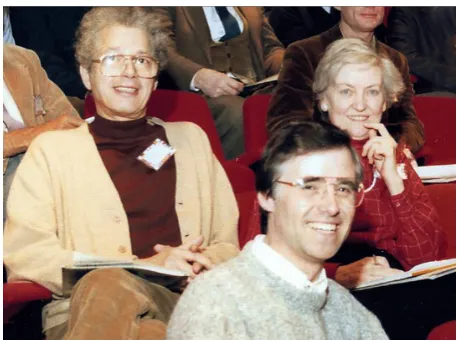
During the nineteen fifties the Anatomy department at UCL ac-quired its first transmission electron microscope (TEM), thanks to the foresight of Professor JZ Young, the head of the department. These were early days for the study of biological tissues by electron microscopy, and despite extensive searches through the literature at that time I was able to find only one paper on the chick embryo, a report by Duncan (1957) on neural tube. These were pioneering days when every scrap of tissue studied yielded new information. When I saw my first electron microscopy sections I was amazed to discover quantities of small intracellular yolk droplets, too small to be seen by light microscopy, in the supposedly yolk-free cells of the area pellucida. Many of my early investigations using transmis-sion electron microscopy and, later scanning electron microscopy, Fig. 1. Ruth Bellairs in 1973.
whether alone or with co-workers, were initially designed less to answer specific questions than to describe what we saw. It was still an unknown world and these were exciting times, though soon electron microscopy settled down, as is common with new techniques, and became just another tool in our armoury.
Much of my later work was concerned with aspects of cell migration, especially in the embryo proper, combining electron microscopy, tissue culture, labelling, experimental surgery and aspects of molecular biology. I had become much impressed by the findings of Abercrombie and his colleagues on cell migration in tissue culture (Abercrombie 1980) but felt it was important to establish if migrating cells in an embryo behaved in the same way as monolayers of fibroblasts migrating on glass in tissue culture. I was of course not the only one to raise this point; eg Trinkaus and Erickson (1981) exhibited a beautiful film of cells migrating within living and transparent teleost (Fundulus) embryos. An example of the investigations I and certain of my colleagues made during this period was into the structure and composition of the vitelline membrane, the transparent bag that surrounds the yolk of the laid egg. It had long been thought to be composed of collagen, but we found none, either by TEM or by biochemical analysis (Bellairs
et al., 1963). Instead we discovered that it consisted of two
mor-phologically distinct layers of non-collagenous fibrous connective tissue, the inner layer secreted by the follicle cells in the ovary, the outer added in the infundibulum of the oviduct. These findings were of especial interest in view of the significant role played by the inner layer of vitelline membrane in the centrifugal expansion of the chick blastoderm. New (1959) had shown that this migration depended on the edge cells of the area opaca clinging to the inner surface of the vitelline membrane and using it as a substrate for migration. These edge cells are bound to their neighbours in an epithelial sheet and we were able to demonstrate by time-lapse studies (Bellairs et al., 1969) that their mode of locomotion on the inner surface of the vitelline membrane was similar to that of epithelial sheets studied by time lapse cinematography in tissue culture (Abercrombie 1982). Our associated studies with scan-ning electron microscopy showed that the edge cells possessed the usual appearance of migrating cells with long cytoplasmic processes extending out from the leading edge.
In the early stages of incubation gap junctions and desmosomes are scarce but tight junctions are present in the area opaca and the borders of the area pellucida and play an important role in keeping the entire blastoderm under tension. This tension is brought about by the adhesion of the edge cells as they migrate under the vitelline membrane. When we reduced the tension in various ways the morphogenetic movements, such as regression, still took place, though the embryos remained small (Bellairs and Veini, 1984). Locomotion is possible only by the temporary making and breaking of cell adhesions to the substrate, brought about by changes in the extra-cellular materials and/or in the adhesivity of the cells themselves.
As development proceeds, the adhesivity of the cells changes (Bellairs et al., 1980) and the composition of the basement mem-brane and the extracellular matrix change too.This may lead to changes in the shape of cells, eg. the neural crest cells as they begin to migrate away from the neural tube settle down flatly on the cells beneath them, as was well illustrated by Bancroft and Bellairs (1976).
Any migrating cell needs to be in harmony with its substrate and
this is often related to the production of adhesion molecules by the migrating cells. For example, we have been able to demonstrate that fibronectin plays an important role in the migration of the edge cells under the inner layer of the vitelline membrane. Using both immunocytochemical staining and experimental treatment with GRGDS, which contains the specific adhesion molecule RGD, the edge cells detach and cannot advance again for some time (Lash et al., 1990). Fibronectin is also important in the migration of the pre-cardiac mesoderm, forming a haptotactic gradient of adhesiveness up which the cells migrate (Easton et al., 1990). By contrast however the nephric duct, which crawls toward the cloaca at about stage 10 of HH (at the astonishing rate of about 400mm per hour) requires a different adhesion molecule, PDSA (polysialic acid). Treatment withEndo-N, which destroys sialic acid, prevents further migration (Bellairs et al., 1995).
The formative role that cell locomotion plays in early development is not restricted to gastrulation nor ends when all the undifferentiated mesoderm has ingressed beneath the ectoderm. Cell locomotion, especially the regression of Hensen’s node, and changing cell relationships continue to play an important role in later stages. For example, presumptive somite mesoderm cells are unable to become committed until they have passed through the primitive streak (Bellairs, 1980) and become part of the segmental plate. Like others, my colleagues and I investigated a variety of aspects of the control of somite formation, from commitment, segmentation itself, to the control of somite numbers, sizes and shapes. At first the cells are loosely arranged as somitomeres (Bellairs and Sanders, 1986), but with increasing adhesivity (Bellairs et al., 1978) become more closely packed together and clearly segmented. Alongside these events there is a changing pattern of extracellular materials. Eventually the fully segmented somites extend right down the body into the tail, but a small region remains unsegmented at the tip of the tail where it is gradually removed by pyknosis; similar patches of pyknosis are visible in most amniote embryos and may play a
18 R. Bellairs
role in monitoring somite numbers. Our experimental analysis led to a new theory of how the embryo controls the sequences of somite formation (Bellairs, 1985).
Much of my research life has involved the chick embryo though I have strayed occasionally to amphibians, reptiles and even to millipedes (which have the most unusual and interesting segmenta-tion), but have always returned to the chick. During recent years I have enjoyed preparing “The Atlas of Chick Development” (Bellairs and Osmond, 2014).
I have been most fortunate in my colleagues and collaborators, not just the ones mentioned here, but all those who have shared my obsession with the formative stages of the young chick embryo. I owe much to them for their input of ideas, their commitment and, not least, their unending cheerfulness. I consider this an opportunity to thank them for all they have done for the subject and for me.
References
ABERCROMBIE M (1980). The crawling movement of metazoan cells. Proc Roy
Soc Lond B 207: 129-147.
BANCROFT M. and BELLAIRS R. (1976) The neural crest cells of the trunk region of the chick embryo studied by SEM and TEM. ZOON 4: 73-85.
BELLAIRS, R. (1953a) Studies on the development of the foregut in the chick blas-toderm 1. J Embryol Exp Morph 1: 115-124.
BELLAIRS, R. (1980). The segmentation of somites in the chick embryo. Bull Zool 47: 245-252.
BELLAIRS, R. (1985) A new theory about somite formation in the chick. In
Develop-mental Mechanisms: Normal and Abnormal (Eds JW LASH and L SAXEN): 207
-213. Alan R Liss, Inc.
BELLAIRS R, BOYDE A, HEAYSMAN J E M (1969) The relationship between the edge of the chick blastoderm and the vitelline membrane. Wilhelm Roux’ Archiv 63: 113-121.
BELLAIRS, R. CURTIS, A.S.G. and SANDERS, E.J. (1978). Cell adhesiveness and embryonic differentiation. J Embryol Exp Morph 46: 207-213.
BELLAIRS R, HARKNESS MARGARET, HARKNESS R D (1963). The vitelline membrane of the hen’s egg; a chemical and electron microscopical study. J
Utrastruc. Res. 8: 339 -359.
BELLAIRS, R., LEAR, P., YAMADA, K., RUTISHAUSER, U. and LASH, J. (1995). Posterior extension of the chick nephric (Wolffian) duct: the role of fibronectin and NCAM polysialic acid. Dev Mech 202: 333-342.
BELLAIRS R and OSMOND M (2014). The Atlas of Chick Development 3rd Edition.
Academic Press, Elsevier, New York.
BELLAIRS RUTH and SANDERS E J (1986) Somitomeres in the chick tail bud: an SEM study. Anat Embryol 175: 235 -240.
BELLAIRS R, VEINI M (1984) Experimental analysis of control mechanisms in somite segmentation in avian embryos. I. Reduction of material at the gastrula stage in
Coturnix coturnix japonica. J Embryol Exp Morph 74: 1-14.
DUNCAN D (1957) Electron microscope study of the embryonic neural tube and notochord. Texas Rep Biol Med 15: 367-377.
EASTON H S, BELLAIRS R, Lash J W (1990). Is chemotaxis a factor in the migration of precardiac mesoderm in the chick? Anat Embryol 181: 461-468.
HAMBURGER V, HAMILTON H L (1951). A series of normal stages in the develop-ment of the chick embryo. J Morph 88: 49-92.
LASH J W, GOSFIELD EDWARD III, OSTROVSKI DAVID, BELLAIRS RUTH (1990). Migration of chick blastoderm under the vitelline membrane. Dev Biol 139: 407-416. MILLS C L, BELLAIRS RUTH (1989). Mitosis and cell death in the tail of the chick
embryo. Anat Embryol 180: 301-308.
NEW D A T (1955). A new technique for the cultivation of the chick embryo in vitro.
J Embryol Exp Morph 4: 326-331.
NEW D A T (1959). The adhesive properties and expansion of the chick blastoderm.
J Embryol Exp Morph. 7: 146-164.
TRINKAUS JP, ERICKSON CA (1981). Locomotion of Fundulus deep cells during gastrulation. Am. Zoologist 21: Abstract.
The surface ectoderm of the chick embryo exhibits dynamic variation in its response to neurogenic signals
Vineeta-Bhasker Tripathi, Yasuo Ishii, Muhammad M. Abu-Elmagd and Paul J. Scotting Int. J. Dev. Biol. (2009) 53: 1023-1033
https://doi.org/10.1387/ijdb.082780vt
Cellular dynamics and molecular control of the development of organizer-derived cells in quail-chick chimeras
Jean-Baptiste Charrier, Martin Catala, Françoise Lapointe, Nicole Le Douarin and Marie-Aimée Teillet Int. J. Dev. Biol. (2005) 49: 181-191
http://www.intjdevbiol.com/web/paper/041962jc
Retinal stem cells and regeneration
Ala Moshiri, Jennie Close and Thomas A. Reh Int. J. Dev. Biol. (2004) 48: 1003-1014
http://www.intjdevbiol.com/web/paper/041870am
Notch activity is required to maintain floorplate identity and to control neurogenesis in the chick hindbrain and spinal cord
5 yr ISI Impact Factor (2016) = 2.421
Isabelle le Roux, Julian Lewis and David Ish-Horowicz Int. J. Dev. Biol. (2003) 47: 263-272
http://www.intjdevbiol.com/web/paper/12755331
Early neurogenesis in Amniote vertebrates
N M Le Douarin
Int. J. Dev. Biol. (2001) 45: 373-378
http://www.intjdevbiol.com/web/paper/11291868
Complementary roles of the insulin family of factors and receptors in early development and neurogenesis
F De Pablo, C Alarcón, B Díaz, M García-De Lacoba, A López-Carranza, A V Morales, B Pimentel, J Serna and E J De la Rosa
Int. J. Dev. Biol. (1996) 40: S109-S110 http://www.intjdevbiol.com/web/paper/9087719
Transplantations of the chick eye anlage reveal an early determination of nasotemporal polarity
D Dütting and S U Meyer
Int. J. Dev. Biol. (1995) 39: 921-931