61. Tang MX, Maestre G, Tsai WY, et al. Relative risk of Alzhei-mer disease and age-at-onset distributions, based on APOE genotypes among elderly African Americans, Caucasians, and Hispanics in New York City. Am J Hum Genet 1996;58:574 – 584.
62. Tang MX, Stern Y, Marder K, et al. The APOE-epsilon4 allele and the risk of Alzheimer disease among African Americans, whites, and Hispanics [see comments]. JAMA 1998;279:751– 755.
63. Romas SN, Mayeux R, Rabinowitz D, et al. The deletion poly-morphism and Val1000Ile in alpha-2-macroglobulin and Alz-heimer disease in Caribbean Hispanics. Neurosci Lett 2000; 279:133–136.
64. Tycko B, Feng L, Nguyen L, et al. Polymorphisms in the human apolipoprotein-J/clusterin gene: ethnic variation and
distribution in Alzheimer’s disease [published erratum ap-pears in Hum Genet 1998;102:496]. Hum Genet 1996;98:430 – 436.
65. Osborne LC, Mason JM. HLA-A/B haplotye frequencies among US Hispanic and African-American populations. Hum Genet 1993;91:326 –332.
66. Fraser PA, Yunis EJ, Alper CA. Excess admixture proportion of extended major histocompatability complex haplotypes of Caucasian origin among rheumatoid arthritis associated hap-lotypes in African Americans and Afro-Caribbeans. Ethn Health 1996;1:153–159.
67. Day JC. Population projections of the United States by age, sex, race, and Hispanic origin: 1995 to 2050. Washington, DC: US Bureau of the Census, Current Population Reports 1996; 25–1130.
Anatomic dissociation of auditory and
visual naming in the lateral
temporal cortex
Marla J. Hamberger, PhD; Robert R. Goodman, MD, PhD; Kenneth Perrine, PhD; and Tara Tamny, MA
Article abstract—Background and Objective:Visual object naming traditionally has been used to identify cortical areas essential for naming (i.e., word retrieval), and investigators have found critical naming sites in the middle and posterior temporal region in most patients. Based on clinical observation, empirical findings, and the pathophysiology of temporal lobe epilepsy, the authors hypothesized that naming sites identified from auditory cues might also be relevant, and that within the temporal region, these sites would be anatomically distinct and located anterior to naming sites based on visual cues.Methods: Twenty patients requiring resective surgery involving the left (language dominant) temporal lobe under-went pre-resection language mapping using direct cortical stimulation. Visual and auditory naming were tested at lateral temporal sites extending from 1 cm from the anterior tip to the parietal operculum. Results: Auditory naming was consistently disrupted by stimulation in the anterior temporal lobe, whereas both auditory and visual naming were impaired by stimulation in the posterior temporal region. Conclusions: This pattern may explain why word finding difficulties sometimes arise or worsen following surgical procedures in which the anterior temporal region is resected without language mapping, or when resection is based on mapping that identifies language cortex exclusively using visual tasks. These results suggest that utilization of auditory based naming tasks might improve pre-resection identification of essential language cortex during direct stimulation cortical mapping, as well as noninvasive localization of dysfunction during presurgical cognitive testing.
NEUROLOGY 2001;56:56 –61
Stimulation-based cortical language mapping is
of-ten necessary in patients with intractable epilepsy
who are candidates for surgical resection within the
language dominant hemisphere. Lateral cortical
sites at which electrical stimulation impedes
lan-guage are considered essential for normal lanlan-guage
function and, therefore, are not included in the
resec-tion in order to preserve language postoperatively.
Although there is some variability in the particular
tasks employed during language mapping (e.g.,
nam-ing, countnam-ing, reading),
1most investigators rely
pri-marily on visual object naming.
2-5This consists of
asking patients to name pictured items (e.g., bell,
escalator) during a brief electrical stimulus.
2The
ra-tionale for this approach is that visual object naming
is impaired in virtually all aphasic syndromes and,
therefore, preservation of cortex necessary for object
naming should reduce the probability of
postopera-tive aphasia.
6Results from investigations using
ob-ject naming tasks have been used to create “maps”
illustrating the cortical distribution of “essential”
language areas.
3,7Although there is considerable
From the Departments of Neurology (Dr. Hamberger) and Neurological Surgery (Dr. Goodman), College of Physicians and Surgeons, Columbia University; Department of Neurology, New York University (Dr. Perrine); and City College of New York, City University of New York (T. Tamny), New York, NY. Supported by The National Institute of Neurological Disorders and Stroke (NS35140-01A1) (M.J.H.).
Received May 10, 2000. Accepted in final form September 22, 2000.
variability in the precise localization of these areas
among individual patients, across individuals,
lan-guage sites tend to cluster in the perisylvian region,
especially in the middle and posterior portion of the
temporal lobe.
4Although naming sites have been
identified in the anterior temporal region in a small
percentage of patients,
8,9in general, visual naming
areas are not usually found near the temporal pole.
Thus, many epilepsy surgery programs perform
“standard” anteromesial temporal resections for
me-dial temporal lobe epilepsy (TLE) without undue
con-cern for postoperative aphasia.
10This consists of an
en bloc removal of the anterior 3 to 3.5 cm of the
inferior and middle temporal gyri and adjacent
fusi-form gyrus, and a radical removal of the medial
structures (including the hippocampus and
parahip-pocampal gyrus and the majority of the
uncus/amyg-dala) without cortical language mapping.
11Although visual naming is impaired in cases of
frank aphasia, we and others (Strauss, personal
com-munication, 1998; Trennery, personal
communica-tion, 1999) have observed that patients with left (i.e.,
language dominant) TLE (LTLE) rarely complain of
difficulty naming concrete objects. Conversely, they
commonly describe more subtle naming difficulties
that occur in the context of everyday auditory–verbal
discourse, both before and after surgery. In an
em-pirical study in which a heterogeneous group of left
and right TLE patients were administered both
audi-tory and visual naming tasks that were equated for
difficulty, we found that auditory naming was
signif-icantly more sensitive to the word finding deficits in
LTLE patients than visual object naming.
12In light
of this finding, it might be important to identify
cor-tical areas involved in auditory as well as visual
naming.
In contemplating the cortical areas involved in
au-ditory naming, we considered the following: 1) the
anterior and medial temporal regions are most often
involved in TLE, both ictally and interictally,
11,13,142)
auditory naming is disproportionately impaired in
LTLE patients,
12and 3) auditory cortex is anterior to
visual cortex. We therefore hypothesized that
audi-tory naming is supported by the anterior temporal
region, whereas visual naming is supported by the
posterior temporal region. Accordingly, auditory but
not visual naming would be disrupted by electrical
stimulation at anterior sites, whereas visual but not
auditory naming would be disrupted by electrical
stimulation at posterior sites.
In this study, we tested auditory and visual
nam-ing in 20 patients who underwent cortical language
mapping before left temporal lobe surgery, and
com-pared the topographic distributions of sites at which
stimulation interrupted auditory versus visual
naming.
Methods. Subjects. Twenty consecutive right-handed patients (12 women) who underwent cortical language mapping before surgery involving the left temporal region were included in this study. All patients were left
hemi-sphere language dominant, as determined by intracarotid amobarbital testing. Six had medial temporal sclerosis (MTS; defined as MRI evidence of abnormal signal and hippocampal atrophy), four had temporal lobe tumors (two in posterior, inferior temporal region, one in the posterior superior temporal gyrus, and one in the anterior, middle to inferior temporal region), two had vascular malformations (one deep within the sylvian fissure and insular cortex, one in the sylvian fissure), and eight had no abnormality on MRI. Of the eight patients with normal MRI, four had medial onset, and four had neocortical onset (one involving the anterior 3.5 cm of the middle and inferior temporal gyrus, and anterior 2.5 cm of the superior temporal gyrus, one involving the anterior 4 cm of the inferior temporal gyrus, and two involving the anterior 4.5 cm of the middle and inferior temporal gyri). In all patients, the left tempo-ral region was identified as the area of seizure onset by intracranial EEG monitoring or a combination of MRI evi-dence of MTS and scalp EEG/video recording. Twelve pa-tients underwent language mapping extraoperatively via subdural electrodes, five at Columbia Presbyterian Medi-cal Center (CPMC) and seven at New York University Medical Center (NYU). Eight patients underwent intraop-erative language mapping before resection at CPMC. De-mographic and clinical information were as follows: Wechsler Adult Intelligence Scale–Revised15Full-Scale IQ
(mean: 96.4, SD: 13.1, all ⬎ 69), age at mapping (mean: 37.9, SD: 13.8), age at seizure onset (mean: 22.2, SD: 14.3), years of education completed (mean: 14.7, SD: 3.7). There were no differences between CPMC and NYU patients on any of these variables (Mann–Whitney test, allp⬎0.05).
Electrodes. For the eight patients evaluated intraoper-atively, 12 (min) to 19 (max) sites along the superior, mid-dle, and inferior temporal gyri and the posterior perisylvian cortex were stimulated using a bipolar stimula-tor with 2 mm diameter ball contacts separated by 5 mm (Ojemann Cortical Stimulator, Radionics Inc., Burlington, MA). The sites were chosen based on gyral/vascular anat-omy and spaced less than 10 mm apart.
For the 12 patients who underwent extraoperative map-ping, an eight by eight (i.e., 64 contact) grid array, with 5 mm diameter electrodes embedded in silastic with center to center interelectrode distances of 1 cm (Ad-Tech, Racine, WI), was positioned over the frontal-parietal-temporal re-gion (trimmed as needed to conform to the covered area). The exposed cortical surface and grid position was docu-mented by digital photography and schematic diagrams. A similar approach documented the stimulation sites for the intraoperatively mapped patients. Subdural electrode posi-tions were verified by skull X-rays, postoperatively. Thir-teen (min) to 34 (max) sites were tested.
Mapping procedures. All auditory and visual naming stimuli were administered to patients within 1 to 4 months before surgery. Auditory and visual items, selected from previously published stimuli,12were equated for word
in the therapeutic range, to minimize afterdischarges and seizure activity.
Extraoperative language mapping was conducted fol-lowing video/EEG monitoring to identify the seizure onset zone. Testing was conducted during electrical stimulation applied to adjacent electrodes. When results were positive, each electrode was studied individually, referenced to a remote electrode in “silent cortex.” All available sites along lateral temporal cortex, as well as parietal sites in the perisylvian area, were stimulated.
Patients who underwent intraoperative mapping were initially anesthetized with propofol. Language mapping be-gan following craniotomy/dural opening, electrocorticogra-phy, and stimulation to determine the threshold for afterdischarges. Several practice trials were conducted to ensure an adequate level of patient responsiveness. Stimu-lation sites were primarily in the vicinity of the anticipated resection, as determined by the presence of a lesion or intracranial EEG evidence of seizure onset. If no visual naming cortex was identified, additional perisylvian sites were tested with the goal of positively identifying the vi-sual naming cortex (rather than relying on negative re-sponses alone). Sites were tested with a bipolar stimulator (see above).
Stimulation mapping followed well established meth-ods.2,4 For both intra- and extraoperative mapping at
CPMC, a constant current stimulator (Ojemann Cortical Stimulator, Radionics Inc.) delivered a biphasic square waveform at a frequency of 60 Hz with a 2 msec pulse duration and amperage ranging from 3 to 15 mA during extraoperative mapping and 1 to 6 mA during intraopera-tive mapping. Mapping at NYU was conducted using a Grass Instruments S-212 cortical stimulator (Winston, MA) with a biphasic square waveform at frequency 50 Hz with a 0.3 msec pulse duration, with amperage ranging from 3 to 15 mA. Afterdischarge levels were determined by increasing amperage until an afterdischarge was elicited, with an upper limit of 15 mA. Amperage for stimulation was set at 0.5 to 1 mA below that which elicited an after-discharge (or 15 mA), which was determined for each site individually. Results reported here are from trials during which no afterdischarges were elicited.
At least two trials each of visual and auditory naming were conducted at each site. If results were ambiguous or the patient was temporarily inattentive, additional trials were administered. For visual naming, patients were shown line drawings of common items (e.g., bench, helicop-ter), and for auditory naming, patients heard oral descrip-tions of concrete items (e.g., “What a king wears on his head”). For visual naming, patients began with the phrase, “It is a” to enable differentiation between speech arrest and anomia, whereas, for auditory naming, patients were instructed to name the target item. To reduce differences in duration of cortical stimulation across tasks, the audi-tory stimuli were limited to those that contained a maxi-mum of eight words and could be presented clearly within 4 seconds. Additionally, the requirement for patients dur-ing visual namdur-ing to articulate the carrier phrase (i.e., This is a —) before naming the pictured object further balanced the stimulus processing and stimulation duration times among tasks. For each task, electrical stimulation began immediately before presentation of pictures or audi-tory descriptions, and lasted for a maximum of 10 seconds,
but terminated immediately upon the patient’s production of a correct response. For both tasks, patients were in-structed to respond as quickly as possible. Sites were con-sidered critical for task performance if the patient could not name target items during stimulation, but provided correct responses upon cessation of stimulation. When one of two trials was performed inaccurately, another two tri-als were administered. Sites were considered critical for task performance only when responses to both of these two trials were incorrect. Sites at which this further testing resulted in 50% accuracy were not considered critical for task performance.
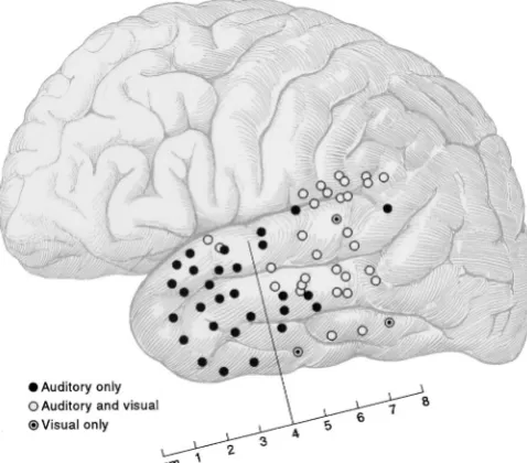
Analyses of naming areas. For each patient, the loca-tion of electrode sites was determined by intraoperative digitized photographs and schematic drawings, and sup-plemented by postoperative skull X-rays. Naming sites from each patient were plotted on a schematic of the tem-poral lobe region and coded to indicate whether auditory, visual, or both auditory and visual naming were disrupted by stimulation. In keeping with conventions of other inves-tigators,16and considering the posterior boundary of
stan-dard resections at most epilepsy surgery programs, we defined the anterior temporal region as ⱕ4 cm from the temporal pole, and the posterior temporal region as⬎4 cm from the temporal pole. The topographic distribution of auditory and visual naming sites was analyzed via Fisher’s exact test.
Results. Results of mapping revealed three types of pos-itive naming sites: 1) sites at which stimulation impaired auditory but not visual naming (“auditory-only”), 2) sites at which stimulation impaired both auditory and visual naming (“dual sites”), and 3) sites at which stimulation impaired visual but not auditory naming (“visual-only”). Of the 20 patients who underwent mapping, at least one type of naming site was identified in 18 patients. Of the two patients in whom no naming sites were found, one had a temporal lobe tumor and one had a vascular malformation; both were mapped intraoperatively. The number of sites tested per patient ranged from 12 to 34 (mean: 20.5, SD: 6.1). The mean number of sites tested per patient was 16.1 (SD⫽2.8) in the intraoperative group, and 23.4 (SD⫽6.0) in the extraoperative group. This difference (t18⫽3.18,p⬍
0.01) likely reflected greater time constraints and more rapid patient fatigue associated with awake surgery. Nonetheless, results of Fisher’s exact test to determine whether the likelihood of finding auditory sites was influ-enced by intra- versus extraoperative mapping were not significant.
Of the 18 patients in whom naming sites were found, 15 exhibited auditory-only sites, three exhibited visual-only sites, and 11 exhibited both auditory-only and dual (or visual-only) sites. In patients in whom naming sites were identified, the number of naming sites (auditory-only, visual-only, or dual) identified within an individual patient ranged from one to eight per patient (auditory-only sites: one to four per patient, visual-only sites: one per patient, dual sites: one to five per patient).
and visual naming. At only three sites did stimulation disrupt visual but not auditory naming. Results of Fisher’s exact test assessing the anterior/posterior distinction (i.e., anterior/auditory-only versus posterior/dual or visual-only) was significant (p⬍0.001).
Of the 11 patients who demonstrated both auditory and visual naming sites (i.e., auditory-only plus visual-only or dual sites), auditory sites were anterior to visual only and dual sites in all but three patients. Each of these patients, however, demonstrated several positive sites, and most sites were consistent with the anterior/posterior pattern. Specifically, in Patient 13, of five naming sites identified, the three posterior sites were dual sites and two of three anterior sites were auditory-only sites. In Patient 3, five of six posterior sites were dual sites, and two of two anterior sites were auditory. In Patient 15, two of four posterior sites were dual sites, and one was a visual-only site.
Naming sites as a function of pathology. Although sub-groups of patients based on brain pathology were too small for statistical analysis, there were no apparent differences in presence or distribution of naming sites as a function of pathology. Of the five MTS patients, two exhibited auditory-only sites, whereas the other three patients had both auditory-only and dual sites. Of the nine non-MTS patients, two exhibited only a single auditory-only naming site, whereas the remaining seven exhibited auditory-only and dual sites (one had a visual-only site as well). Of the four tumor patients, three exhibited auditory-only and dual sites, and in one patient, no naming sites were found. Of the two vascular patients, a dual site and a visual-only site were identified in one, and no sites were found in the other. The far posterior temporal auditory-only site was elicited in a single patient with a tumor in the inferior posterior temporal region.
Discussion.
Utilization of both auditory and
vi-sual naming tasks during cortical mapping revealed
a modality related distribution of naming sites
across the lateral temporal region. As anticipated,
stimulation at most anterior temporal sites (
ⱕ
4 cm
from temporal pole) disrupted auditory naming, but
did not impede visual naming. Although we
hypoth-esized that posterior temporal stimulation would
im-pair
visual
naming
exclusively,
stimulation
disrupted both visual and auditory naming at most
sites in this region. In patients in whom both
audi-tory and dual (i.e., audiaudi-tory and visual) sites were
identified, auditory sites were anterior to dual sites
in most, with only three patients deviating from this
pattern. Even in these three patients, however, most
positive sites conformed to this general pattern. In
one patient, however, an auditory naming site was
identified in the far posterior temporal region,
sug-gesting that in some patients, auditory naming areas
may not be limited to the anterior temporal region.
Identification of an anterior temporal auditory
naming area carries both theoretical and clinical
im-plications and may explain, at least in part, several
previously reported phenomena. As naming has
tra-ditionally been mapped with visual stimuli, which
will not detect auditory naming sites, the
topo-graphic pattern revealed in this study might explain
why some investigators have claimed that “naming”
is represented in the posterior portion of the
tempo-ral lobe.
3,16It might also explain why many patients
who undergo “standard” anterior resections often
experience increased word finding difficulty
post-surgically.
17-20The dissociation of auditory and visual
naming sites might also explain why some patients
experience word finding decline after surgery,
de-spite having been mapped with visual naming tasks
before resection.
5,8Interestingly, results of a recent
multicenter study demonstrated no postsurgical
naming differences between patients who were
mapped with visual naming compared to those who
had standard resections without language mapping,
suggesting no benefit of preresection mapping.
5It
may be that mapping with visual stimuli enabled
identification of cortex essential for picture naming;
however, auditory naming sites remained
unidenti-fied, and may have been removed in some patients. If
so, utilization of auditory naming during
preresec-tion mapping could potentially assist in preserving
word retrieval postsurgically.
Although the current findings support the notion
that poorer visual compared to auditory naming is
consistent with posterior temporal pathology, it could
be argued that anterior temporal pathology might not
impede auditory naming as auditory as well as visual
naming sites were found in the posterior temporal
re-gion. Although this certainly is possible, we believe
that under normal circumstances—i.e., everyday
au-ditory–verbal discourse—the anterior temporal
re-gion is most critical for auditory-based word
retrieval. Conceivably, the most parsimonious
expla-nation for impaired auditory naming during
poste-rior temporal stimulation is that the auditory
descriptions, which were visually descriptive, and
Figure. Topographic distribution of naming sites across patients indicating whether auditory, visual, or both audi-tory and visual naming were disrupted during
target items, which were all concrete, highly
image-able items, frequently elicited visual processing. In
fact, some patients reported that they “couldn’t help
but
picture
the items being described.” Thus, visual
association areas were probably recruited during the
auditory task due to the nature of the stimuli in this
contrived situation. Yet, this is less likely to occur in
the context of everyday conversation, which typically
has a more auditory/conceptual basis.
On a related note, that auditory naming was
dis-rupted with posterior temporal stimulation in some
patients raises the question as to why the anterior
temporal region failed to support word retrieval
dur-ing the auditory namdur-ing task independently. One
possibility is that the anterior and posterior regions
support different, yet equally essential, components
of auditory-based word retrieval. However, in
keep-ing with the explanation noted above, visual
associa-tion cortex was probably essential when patients
utilized strategies that relied primarily on visual
im-agery. Although concrete nouns were selected
inten-tionally with the goal of developing comparable
tasks, this may have contributed to the partial
over-lap of auditory and visual naming sites.
Another possibility to consider is that compared to
visual naming, auditory naming may require
partic-ipation of a wider distribution of cortical areas.
Al-though visual and auditory tasks were equated for
word frequency, which is known to affect facility of
word retrieval,
21and healthy controls performed
sim-ilarly on the two tasks, the auditory task may have
required more elaborate processing.
Comparisons with other studies.
To our
knowl-edge, there is one other published study (although
smaller scale) in which the topography of stimulation
based auditory and visual naming sites was
com-pared.
22These investigators mapped auditory and
vi-sual naming in six left hemisphere language
dominant LTLE patients, six to 12 sites per patient,
and found poorer auditory naming than visual
nam-ing durnam-ing stimulation in the anterior and posterior
lateral region. It was unclear if, and to what extent,
the region within the first 4 cm of the temporal pole
was tested; however, the figures shown suggest that
coverage was less extensive in the anterior region.
Consequently, it is difficult to compare these
find-ings with ours, yet the differential performance as a
function of topography and modality is consistent
with our results. A study comparing results of PET
activation and cortical stimulation showed some
evi-dence of greater posterior activation during visual
naming compared to that observed during auditory
naming.
23With cortical stimulation, however,
sev-eral regions in the latsev-eral temporal lobe were
identi-fied where stimulation produced errors in auditory
but not visual naming, and auditory errors were
elic-ited at lower thresholds than visual naming errors.
More detailed information regarding the precise
lo-cation of these sites was not provided, as the purpose
of the study was to compare results of the two
tech-niques rather than the topography generated by
test-ing in different modalities. Another study compartest-ing
PET images generated during auditory naming
25with PET images obtained during visual naming
from a different study (i.e., using different subjects)
26showed activation in primary and secondary visual
brain regions, similar to the activation generated by
visual images. Similar to the current study, the
vi-sual nature of the target stimuli, as well as the
im-agery created by descriptions themselves (e.g., “tall
pink bird”), very likely elicited visual processing.
De-spite problems with task comparability, it might be
necessary to eliminate or at least reduce the confound
of visual processing during imaging and stimulation
studies to determine whether visual association areas
are, in fact, essential for auditory naming.
Several investigators have reported disruptions in
expressive and receptive language processing during
stimulation of the basal temporal area.
16,26In a PET
study comparing activation patterns in LTLE
pa-tients and healthy controls, however, the left
ante-rior fusiform activation observed in controls was
absent among patients.
27It is unknown, at this
point, how our findings regarding lateral temporal
cortex might relate to this region. In light of these
observations, it might be of interest to pursue
map-ping using both disruptive (e.g., stimulation) and
ac-tivation (e.g., PET, fMRI) techniques.
The heterogeneity of the patient sample and the
small number of patients in various subgroups
lim-ited our ability to explore potential relationships
be-tween particular topographic patterns and the
nature or location of pathology. Among the patients
studied, however, no such trends were apparent. In
fact, most patients, regardless of the nature or
loca-tion of pathology, demonstrated auditory naming
sites that were anterior to visual or dual naming
sites. The consistent pattern observed across
pa-tients suggests it might generalize across pathologic
conditions. Nonetheless, there may be significant
dif-ferences in the cortical representation of auditory
and visual naming as a function of age at onset,
location, and nature of pathology, and we are
pursu-ing further studies to address these questions.
As is often the case with patient populations,
gen-eralization of findings to the normal population must
be made cautiously. Other investigators have noted
that an epileptogenic lesion or abnormal brain
activ-ity could potentially alter the cortical distribution of
cognitive functions.
8,9,28The fact that most patients
in this study developed seizures during adulthood
might control for this to some extent, although a
larger study in which age at onset or age at first risk
could be examined systematically would better
ad-dress this issue. Additionally, functional MRI or
other imaging techniques using healthy volunteers,
although less precise with respect to spatial
resolu-tion than stimularesolu-tion based mapping, could be used
to more directly study the distribution of naming
areas in normal individuals.
theoreti-cally and may have important clinical applications
as well. From a heuristic perspective, localization of
a modality specific naming region potentially
en-hances our understanding of how the temporal lobe
mediates semantic and lexical processing. At the
same time, results of this study raise questions
re-garding the clinical significance of auditory naming
sites in lateral temporal cortex. Extensive
investiga-tions, involving rigorous pre- and postsurgical
test-ing, accounting for the location of auditory naming
sites relative to the boundaries of the resection as
well as patient variables (i.e., age at onset, nature
and location of pathology) are in progress in our
laboratory.
Acknowledgment
The authors thank Drs. Martha J. Morrell, Timothy A. Pedley, and William T. Seidel for their editorial comments; Dr. Stephen Chan for his radiologic readings; Drs. Werner Doyle and Guy McKhann for neurosurgical information; and Kehinde Odedosu for assistance with data management.
References
1. Gordon B, Hart J, Fisher RS, Lesser RP. Language during electrical cortical stimulation: variety of errors and dissocia-tions and reliability of results. Epilepsia 1988;29:668 – 669. Abstract.
2. Ojemann GA. Brain organization for language from the per-spective of electrical stimulation mapping. Behav Brain Res 1983;2:189 –230.
3. Ojemann G, Ojemann J, Lettich E, Berger M. Cortical lan-guage localization in left-dominant hemisphere. J Neurosurg 1989;71:316 –326.
4. Ojemann G. Cortical organization of language. J Neurosci 1991;11:2281–2287.
5. Hermann BP, Perrine K, Chelune GJ, et al. Visual confronta-tion naming following left ATL: a comparison of surgical ap-proaches. Neuropsychology 1999;13:3–9.
6. Goodglass H, Wingfield A. Word-finding deficits in aphasia: brain-behavior relations and clinical symptomatology. In: Anomia: neuroanatomical and cognitive correlates. San Diego, CA: Academic Press, 1997:3–27.
7. Penfield W, Roberts L. The evidence from cortical mapping. In: Speech and brain mechanisms. Princeton, NJ: Princeton University Press, 1959: 119 –137.
8. Devinsky O, Perrine K, Llinas R, Luciano DJ, Dogali M. Ante-rior temporal language areas in patients with early onset of TLE. Ann Neurol 1993;34:727–732.
9. Schwartz TH, Devinsky O, Doyle W, Perrine K. Preoperative predictors of anterior temporal language areas. J Neurosurg 1998;89:962–970.
10. Spencer SS, McCarthy G, Spencer DD. Diagnosis of medial temporal lobe seizure onset: relative specificity and sensitivity of quantitative MRI. Neurology 1993;43:2117–2124.
11. Spencer DD, Spencer SS, Mattson RH, Williamson PD, No-velly RA. Access to the posterior medial temporal lobe struc-tures in the surgical treatment of temporal lobe epilepsy. Neurosurgery 1984;15:667– 671.
12. Hamberger M, Tamny T. Auditory naming and temporal lobe epilepsy. Epilepsy Res 1999;35:229 –243.
13. Fernandez Torre JL, Alarcon G, Binnie CD, et al. Generation of scalp discharges in temporal lobe epilepsy as suggested by intraoperative electrocorticographic recordings. J Neurol Neu-rosurg Psychiatry 1999;67:51–58.
14. Hamer HM, Najm I, Mohamed A, Wyllie E. Interictal epilep-tiform discharges in temporal lobe epilepsy due to hippocam-pal sclerosis versus medial temporal lobe tumors. Epilepsia 1999;40:1261–1268.
15. Wechsler D. WAIS-R manual. New York: Psychological Corpo-ration, 1981.
16. Burnstine TH, Lesser RP, Hart J Jr., et al. Characterization of the basal temporal language area in patients with left tem-poral lobe epilepsy. Neurology 1990;40:966 –970.
17. Bennett–Levy J, Polkey CE, Powell GE. Self-report of memory skills after temporal lobectomy: The effect of clinical vari-ables. Cortex 1980;16:543–557.
18. Milner B. Amnesia following operation on the temporal lobes. In: CWM Whitty, OL Zangwill, eds. Amnesia. London: Butter-sworth, 1966:109 –133.
19. Saykin AJ, Stafiniak P, Robinson LJ, et al. Language before and after temporal lobectomy: specificity of acute changes in relation to early risk factors. Epilepsia 1995;36:1071–1077. 20. Langfitt J, Rausch R. Word-finding deficits persist after
an-terotemporal lobectomy. Arch Neurol 1996;53:72–76.
21. Johnson CJ, Pavio A, Clark JM. Cognitive components of pic-ture naming. Psychol Bull 1996;120:113–120.
22. Malow B, Blaxton T, Sato S, et al. Cortical stimulation elicits regional distinctions in auditory and visual naming. Epilepsia 1996;37:245–252.
23. Bookheimer SY, Zeffiro TA, Blaxton et al. A direct comparison of PET activation and electrocortical stimulation mapping for language localization. Neurology 1997;48:1056 –1065. 24. Bookheimer SY, Zeffiro TA, Blaxton TA, Gaillard, Malow B,
Theodore WH. Regional cerebral blood flow during auditory responsive naming: evidence for cross-modality neural activa-tion. NeuroReport 1998;9:2409 –2413.
25. Bookheimer SY, Zeffiro TA, Blaxton TA, Gaillard W, Theodore W. Regional cerebral blood flow changes during object naming and word reading. Hum Brain Mapp 1995;3:93–106.
26. Luders H, Lesser R, Hahn J, et al. Basal temporal language area demonstrated by electrical stimulation. Neurology 1986; 6:505–510.
27. Henry TR, Buchtel HA, Koeppe RA. Absence of normal activa-tion of the left anterior fusiform gyrus during naming in left temporal lobe epilepsy. Neurology 1998;50:787–790.