Copyright © 2017 Ilina NG, et al. This is an Open Access article distributed under the terms of the Creative Commons Attribution-Non Commercial 4.0 International License (http://creativecommons.org/licenses/by-nc/4.0/), permitting all non-commercial use, distribution, and reproduction in any ORIGINAL RESEARCH ARTICLE
Clinical and dermoscopic features of nevi in patients with psoriasis
Ilina Natalia Glebovna
1*, Krinitsuna Yulia Mihailovna
1,2, Taganov Aleksey Viktorovich
3,
Tulupov Andrey Aleksandrovich
1,4, Kolesnikova Irina Stanislavovna
5, Yudkin Dmitry
Vladimirovich
5, Sergeeva Irina Gennadievna
1,41 Medical Department, Novosibirsk State University, Novosibirsk, Russia
2 Novosibirsk Research Institute of Regional Pathology and Pathomorphology, Novosibirsk, Russia
3 Scientific Research Centre ‗Clinic of Dermatology‘, Moscow, Russia
4 Laboratory of translational brain research, The Institute International Tomography Center of the Russian Academy of Sciences, Novosibirsk, Russia
5 Chromosome Pathology Group, Institute of Molecular and Cellular Biology SB Russian Academy of Science, Novosibirsk, Russia
Abstract: The aim of the present study is to display the clinical and dermoscopic features of melanocytic nevi of more than 5 mm in diameter in psoriasis patients. A total of 32 patients with psoriasis (21 male, 11 female; average age 37.4) formed the first study group. In 22 patients (68.8%), melanocytic nevi of 5 mm in diameter and bigger (total of 68 nevi) were clinically found. For a randomized trial, 100 people (21 male, 79 female; average age 27.8) without psoria-sis were invited to form the second study group. Only 37 of them had nevi ≥5 mm in diameter (total of 60 nevi). Complete questionnaire, full body photometric skin examination, dermoscopy examination on the dermatoscope HEINE MINI 10X with 70% ethyl alcohol immersion, skin type identification according to the Fitzpatrick classifica-tion, and nevi assessment according to ABCD and ABC rules were obtained for all recruited people. Our study showed that patients with psoriasis are more susceptible to melanocytic nevi of >5 mm in diameter (68.8%), while the result for the second group was 37%. As for nevi of dysplastic criteria, we found 32% in psoriasis patients vs. 42% in the monitoring group. Moreover, we should bear in mind the influencing factors of skin phototype II and artificial insola-tion (e.g., tanning, PUVA (Psoralen and ultraviolet A), and narrowband phototherapy 311 nm) obtained by the patients from the first group.
Keywords: Melanocytic nevi; psoriasis; dermoscopy; melanoma
Citation: Ilina NG, Krinitsuna YM, Taganov AV, Tulupov AA, Kolesnikova IS, et al. Clinical and dermoscopic fea-tures of nevi in patients with psoriasis. J Surg Dermatol 2017; 2(1): 50–57; http://dx.doi.org/10.18282/jsd. v2.i1.50.
*Correspondence to: Ilina Natalia Glebovna, Medical Department, Novosibirsk State University, Novosibirsk, Russia, [email protected].
Received: 23rd February 2016; Accepted: 25th October 2016; Published Online: 10th January 2017
Introduction
Melanocytic nevi (MN) are benign melanogenic tumors, where melanocytes tend to develop into melanoma. Melanomas can arise from melanocytic nevi but much more often on intact skin. Multiple dysplastic or atypical
main risk factor of melanocytic dysplasia appearance and further melanoma development[4]. Patients with psoriasis are of greatest interest because they receive UV therapy such as narrowband phototherapy 311 nm, PUVA (Pso-ralen plus UVA), and excimer laser therapy[5]. Besides that, they take retinoids (which increase their sensitivity to UV radiation) and methotrexate (which has an immu-nosuppressing and cytostatic effect), further increasing the risk of skin melanoma appearance. Melanoma and Epstein-Barr virus-associated lymphomas in psoriasis patients taking methotrexate have been reported[6].
Recently, a systematic literature review with me-ta-analysis was performed. The conclusion was that there is a slightly increased risk of some cancers (e.g., upper aero-digestive tract-, liver-, lung-, pancreatic-, and uri-nary tract cancers), with the most increased risk for skin carcinoma but no increased risk for melanoma[7].
A literature presented one case of a psoriasis patient suffering from metastatic melanoma which regressed spontaneously without any treatment. The patient had psoriasis associated with HLA-Cw6 (human leukocyte antigen). So, there is a possible relationship between psoriasis, HLA-Cw6, and spontaneous melanoma regres-sion; in that, psoriasis as an immune-mediated dise-ase can play a protective role against malignant mela-noma[8].
Nowadays, dermoscopy is considered to be the most efficient tool for early diagnosis of melanoma. It reduces the frequency of unreasonable biopsy[9,10] and is efficient for nevi monitoring.
The purpose of the present study is to display clinical and dermoscopic features of melanocytic nevi (of diameter 5 mm and more) among patients with psoriasis.
Materials and methods
The first step of the study was to recruit 32 patients with psoriasis (21 male and 11 female patients with an aver-age aver-age of 37.4 years old). They formed the first study group. Out of the 32 patients, 22 of them (68.8%) were clinically found with melanocytic nevi of 5 mm in diameter and bigger (total of 68 nevi).
For a randomized trial, 100 people (21 male and 79 female with an average age of 27.8 years old) without psoriasis diagnosis were invited. They underwent a de-tailed examination to form the second monitoring group. Only 37 of them had nevi ≥5 mm in diameter (total of 60 nevi). All patients from both groups were followed-up only with a one-time visit.
Complete questionnaire and full body photometry, examination on the dermatoscope HEINE MINI 10X
with 70% ethyl alcohol immersion, skin type identifica-tion, and nevi assessment with ABCD and ABC rules were obtained for all patients of the two groups. ABCD assessment included the following criteria: asym-metry, border, color, and dermoscopic features.
Ethics statement
This study was approved by the ethics committee. Writ-ten informed consent for participation in the study was obtained from all participants. There were no conflicts of interests reported.
Results
The questionnaire revealed the number of risk factors for melanoma skin cancer. It was filled by the patients themselves during their one-time visit.
The 1st and 2nd skin phototypes associated with a greater risk of melanoma were identified in 1 (4.5%) and 16 (72.7%) patients with psoriasis, respectively, and 2 (5.4%) and 22 (59.5%) people in the second study group, respectively. The 3rd skin type was identified in 5 (22.7%) psoriasis patients and 13 (35.1%) people in the second group. Thus, patients with psoriasis have the most cases of 2nd skin type and are vulnerable to sun exposure.
Sunburn episodes, at the age up to 18 years old, in-creased the melanoma appearance factor in 11 (50.0%) psoriasis patients and 20 (59.5%) people from the second group. Only 2 (9.1%) and 3 (8.1%) people from each respective group reported of having ever lived in coun-tries with higher insolation than Novosibirsk. Presently, due to job or hobby factor, a total of 6 (27.3%) and 8 (21.6%) recruited people in each group, respectively, spend so much time in the open air, and most of the patients in both groups (13 (59.1%) and 25 (67.6%), respectively) are highly exposed to sunlight for more than two weeks a year. Moreover, 5 (22.7%) and 5 (13.5%) people from each group, respectively, had at-tended sun tanning sessions.
Additional melanoma risk factors for psoriasis patients were due to methotrexate and phototherapy treatments, of which 27.3% of recruited psoriasis patients received narrowband phototherapy 311 nm treatment, 9.1% received PUVA therapy, and 9.1% received metho-trexate. For some patients, several methods of treatment were applied and therefore these patients had one or more additional risk factors. Thus, 36.4% of patients with psoriasis have additional factors negatively associated with melanoma risk.
melanoma development[11]. Only 1 (4.5%) patient suf-fering with psoriasis and 3 (8.1%) recruited people from the second group used SPF products when exposed to the open air for more than 1 h, whereas only 2 (9.1%) pa-tients from the first group and 10 (27.1%) people from the second group used SPF products when intermittently exposed to sunlight (for example, sunbathing). It is worth mentioning that patients used sun protective products with low SPF coefficient of up to 30. The low frequency of SPF products usage among psoriasis patients could be explained by insolation as a treatment unit.
When being assessed subjectively, 50% of patients from the first group and 84% of people from the second group had a big nevi number. Besides that, 5 (22.7%) and 18 (48.6%) of them, respectively, reported on the nevi‘s change over the course of time, but none of them consulted a doctor for an examination. Only 1 person from both groups (4.5% and 2.7%, respectively) had family melanoma anamnesis.
Clinical characteristics
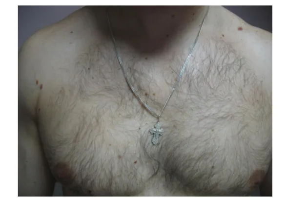
When being examined, the number, size, and location of nevi were assessed (Table 1).The figures show that the size and location of nevi in individuals from the first and second groups did not change greatly, but patients with psoriasis showed a greater number of multiple MN (up to 15 per person) (Figure 1).
Dermoscopic characteristics
Nevi asymmetry was assessed according to three param-eters: color, structure, and border. In the first group, most of the examined nevi (52, or 76.5%) were symmet-ric by color while about 16 nevi (23.5%) indicated an axial symmetry of the color. A total of 49 nevi (72.1%) were symmetric by structure, 15 nevi (22.1%) showed axial structure symmetry, and 4 nevi (5.8%) were asym-metric. The border symmetry assessment of the nevi re-vealed the following: 47 symmetric nevi (69.1%), 13 nevi with an axial symmetry (19.1%), and 8 asymmetric nevi (11.8%).
As for the second group, symmetry along both axes was observed by: color, 43 (71.7%); structure, 28 (46.7%); and borders, 30 nevi (50.0%).
In both groups, none of the examined nevi had rough pigmentation cutoff at the lesion border.
When assessing lesions, the following color variations were observed: white, red, light-brown, and dark-brown.
Lesions of blue-black or black color were not found. Polychromy was found in 5 cases (7.4%), with 56 cases (82.4%) having two colors and 7 cases (10.2%) having only one color of nevi in patients from the first group, whereas 12 (20.0%), 33 (55.0%), and 15 (25.0%) of the abovementioned nevi cases were found in people from the second group, respectively.
While assessing dermoscopic features, pigment network was found in all examined nevi. Typical pig-ment network—light-to-dark-brown colored, regularly meshed and narrowly spaced network distributed regu-larly, thinning out at the periphery—was found in 46 cases (67.6%) of the first group and in 37 cases (61.7%) of the second group of people. Atypical pigment network is characterized by a black, brown, or gray col-ored, irregularly meshed network distributed irregularly
Table 1.Clinical characteristics of melanocytic nevi >5 mm in diameter among psoriasis patients and patients of the monitored group
1st group N = 22 2nd group N = 37
Location
Back 28 (41.2%) 19 (31.7%)
Chest 12 (17.6%) 15 (25.0%)
Shoulder 10 (14.7%) 3 (5.0%)
Abdomen 9 (13.2%) 5 (8.3%)
Huckle 3 (4.4%) 8 (13.3%)
Crus 3 (4.4%) 1 (1.7%)
Face 2 (2.9%) 4 (7.0%)
Forearm 1 (1.6%) 1 (1.7%)
Neck - 2 (3.0%)
Buttock - 1 (1.7%)
Foot - 1 (1.7%)
Number
1 nevus 10 (45.5%) 25 (67.5%)
2 nevi 6 (27.3%) 7 (18.9%)
≥3 nevi 6 (27.3%) 5 (13.6%)
Diameter
5–7 mm 53 (77.9%) 44 (73.3%)
8–9 mm 9 (13.2%) 8 (13.3%)
Figure 1. The chest area rated as a second location among psoriasis patients and patients of the monitored group
throughout the lesion. Lines of an atypical pigment net-work are often thickened and end abruptly at the periph-ery. Atypical network was found in 22 cases (32.4%) from the first group and 22 cases (36.7%) of the people from the second group, respectively.
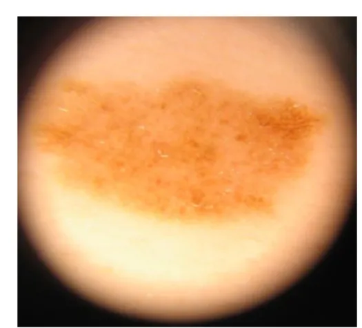
A structureless area of light-brown or pink color was found in 29 cases (42.6%) from the first group and in 30 cases (50.0%) from the second group. Structureless areas were less pigmented compared to other areas, and often combined with pigment network areas or globules (Figure 2).
Figure 2. Dermal nevi with the combination of globules and structureless areas
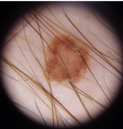
Another frequent dermoscopic pattern is globules of dark-brown or light-brown color with round or oval structures, equal in size and distribution in the skin lesion. Globules were found in 47 nevi (69.1%) of patients with psoriasis and 29 nevi (48.3%) of the monitoring group. A net-globule combination was often observed (Figure 3). It is known that acquired nevi have an age-related dy-namic morphological pattern. First, an epidermal-dermal melanocytic nevus is formed due to the nevus cell prolif-eration along the border of epidermis and derma. In the course of time, nevi cells move further into the derma, thus forming a compound MN. When nevi cells immerse into the structure of the derma intensively, dermal MN is formed. A higher frequency of the globule structure nevi as a marker of the dermal MN among the patients of the first group can be explained by the average age of the recruited people, 37.4 years old, against the average age of 27.8 years old in the second group. Thus, a higher percentage of acquired nevi were observed in the phase of dermal MN.
Figure 3. Large melanocytic nevi with globular pattern
Figure 4. Large melanocytic nevi with pseudopods
Blotches—represented by areas of dark and dark-bro-wn pigmentation, being quite major and structureless, and located in the center of the skin lesion—were found in 3 nevi (4.4%) of the patients from the first group.
Forty-two nevi (61.8%) in the first study group had two dermoscopic features, with 19 nevi (27.9%) show-ing three dermoscopic features, and 6 nevi (8.8%) re-vealing one feature. As for the second group, 29 (48.3%), 19 (31.7%), and 12 (20.0%) cases were found, respec-tively. Only one nevus in the first group revealed four dermoscopic features. Combined structureless areas or a combination of pigment network and globules were often observed.
Using the ABCD rule, we calculated the Dermoscopy Score (DS) and stated that the index value of >4.75 cor-responded to benign lesions, the value from 4.75 to 5.45 corresponded to potentially malignant lesions, and value of >5.45 to malignant lesions. We obtained
index values of from 1 to 4.6 in 62 cases (91.2%), 5.1 in 4 nevi (5.9%), and 5.6 in 2 nevi (2.9%) from the first study group, and 54 (90.0%), 4 (6.7%), and 2 (3.3%) nevi in the monitoring group, respectively.
ABC assessment included the same diagnostic criteria as in the ABCDE Rule (asymmetry, border, color, der-moscopic features, and skin lesion evolution), but another score system. The ≥4 points assessment cor- responds to malignant lesion. While assessing the nevi of patients with psoriasis, we obtained the following data:
≤3 points for 66 cases (97.1%), and thus their skin le-sions were classified as benign; and 4 points for 2 nevi (2.9%), hence these skin lesions were of malignant na-ture. In the second group, these values were 59 (98.3%) for benign and 1 (1.7%) for malignant lesions.
Only one nevus was diagnosed as a malignant lesion according to both ABCD and ABC rules in the group of psoriasis patients, and none in the monitoring group.
As for the Pehamberger analysis, melanocytic vi corresponded to the following criteria:
The criterion of an epidermal-dermal nevus with an even pigment network was observed in 5 (7.4%) and 9 (15.0%) cases of the first and second group, respectively.
Compound melanocytic nevus, which appears as reticular/granular model with a typical pigment network and hypopigmentation, were found in 36 (52.9%) and 25 (41.6%) cases, respectively.
Dermal nevus with a typical dot/granular location, hypopigmentation, glomerular vessels were found in 5 (7.4%) and 1 (1.7%) cases, respectively.
Dysplastic nevus which appears as reticular/gra-nular model with a typical or atypical pigment network, regular or irregular dots/granules, regu-lar streaks, blots and diffuse hypopigmentation were found in 22 (32.4%) and 25 (41.6%) cases, respectively (Figure 5).
Discussion
Figure 5. Dysplastic nevi characterized by Pehamberger crite-ria
precursor lesions for melanoma because of the rare transformation of any individual nevus to a melanoma[12].
Overall, dysplastic nevi are relatively common with a frequency of about 10% (range: 7%–24%) in Northern European populations[12]. Nevus with geographic shape and muddy borders, dominantly macular, red-to-brown-colored with 5 mm or more in diameter is a clinically dysplastic nevus. ABCD rules count for dys-plastic nevi as well as for melanoma but the preferable diagnostic criteria for dysplastic nevi has been the ―ugly duckling sign‖ (the only atypical lesion or the most atypical among other lesions). Pathohistological analy-sis can be the only diagnostic tool in the differential di-agnosis between dysplastic nevus and melanoma in situ. So, surgical excision is the only therapy that should be done for dysplastic nevus with ―ugly duckling sign‖[13].
Some authors consider the term ―dysplastic nevus‖ to be a misnomer and that it has to be abandoned. Pref-erably a specific diagnosis should be made based on dermoscopic pattern and, if this is not possible, on cli-nical or dermatoscopic signs alone, and the term ―nevus, not otherwise specified‖ or ―nevus with non-specific signs‖ should be used[14,15]
. For further diagnosis, other clinical-diagnostic tools should be used and indi-vidual‘s melanoma risk should also be taken into account in decision of further treatment management.
According to all these up-to-date concepts of dysplas-tic nevi, we should consider 32% of ―dysplasdysplas-tic nae-vi‖ by Pehamberger as not precursors for melanoma any more but only as nevi with structural changes, and use ―ugly duckling sign‖ as the main dermoscopy rule to exclude melanoma.
Our study showed patients with psoriasis being more vulnerable to melanocytic nevi of >5 mm in diameter (68.8%), whereas the second group showed the result of 37%. Dermoscopic picture of nevi in patients with
psori-asis is symmetrical by color (76.5%), border (69.1%), and structure (72.1%). In addition, 82.4% of nevi had two colors of light and dark-brown, and 69.1% had a globular structure of pigment.
Patients suffering from psoriasis had 32.4% cases of MN corresponding to Pehamberger dysplastic nevi, and only one case had the ABC and ABCD malignant symp-toms.
In a very early study on 76 patients suffering from psoriasis, histological characteristics of melanocytic nevi within and near psoriatic lesions were investigated. His-topathologically examined nevi did not show any psori-atic alterations of the epidermis; the adjacent epidermis was free of psoriasis. Hence, that melanocytic nevi is a possible ―protective‖ factor against psoriasis[16], was discussed for the first time.
Further investigation showed that some of the cyto-kines such as interleukin (IL)-1α, IL-6, and tumor necro-sis factor (TNF)-α gene expression have effects on melanogenesis, and they are also reported to be involved in psoriasis. In this study, patients with psoriasis showed a lower number of areas with nevi compared to the con-trol group. This suggests the probable inhibiting effect of the proinflammatory cytokines in psoriasis skin on mel-anocyte growth, and/or progression to nevi[17].
This statement was also supported by another study among patients with psoriasis under therapy with bio-logics. It showed that, the number of biologics used in treatment was a risk factor for a higher nevus count, whereas the psoriasis severity did not correlate with the number of nevi[18]. So, treatment with biologics influence the proinflammatory cytokine level and therefore can cause further melanocyte growth. This study also showed that the frequency of nevi in psoriatic patients was lower than in the control group (patients without psoriasis) as in our study.
Recent publication about development of new nevi in patients using immunosuppressive agents showed that therapy with biologics was associated with increased nevi counts and appearance of dermoscopy changes in nevi, but none of the changes and the subsequently ex-cised nevi were malignant[19].
It is accepted, that PUVA is one of the risk factors for non-melanocytic skin cancer (squamous cell carcinoma), especially after 350 or more phototherapy sessions over a lifetime. Unlike risk of melanocytic skin cancer and gen-ital skin cancer are not increased by PUVA alone. But additional factors as previous UV damage due to chronic sun exposure and sun burns, immunosuppressive treat-ment, increase cutaneous carcinogenesis through PUVA. In contrast, other artificial UV treatment as broadband UVB, narrowband UVB, and UVA1 has not yet been linked to cutaneous carcinogenesis probably due to lack of large follow-up studies[21]. One retrospective study including 92 patients affected by psoriasis under treat-ment with narrowband UVB or PUVA-therapy showed noteworthy number of non-melanoma skin cancer[22].
Our research showed that 36% patients with psoriasis have additional risk factors for melanoma due to other treatments, with 27% received narrowband phototherapy 311 nm, 9% received methotrexate, and 9% received PUVA-therapy. We have found no cases of malignancy in our study, but further follow up of the patients is neces-sary, as careful monitoring is crucial.
A systematic literature search performed on PubMed, Embase, and Cochrane databases using the keywords ―Psoriasis AND Neoplasms‖ from 1980 to January 2012 showed possible increased risk of some solid cancers in psoriasis such as respiratory tract-, upper aerodigestive tract-, urinary tract-, and liver cancer, as well as increased risk of non-melanocytic skin cancer (squam-ous cell carcinoma and basal cell carcinoma), whereas the risk of melanoma is not increased. A higher risk of non-melanoma skin cancers, especially squa-mous cell carcinoma, that could be explained as cumula-tive effect of UV and photosensicumula-tive agents and other immunosuppressive therapy[23,24].
Another study also showed no increased risk for mel-anoma, but for non-melanoma skin cancer. It was associ-ated with PUVA, cyclosporine and anti-TNF treatment in psoriasis. It also suggests to take into consideration inci-dences of cancer in patients history before using biologic or imunnosuppressive therapy because of lack of studies in these patient groups[7].
Only 9% of all patients with psoriasis who used sun-screen creams or underwent total skin examination with dermoscopy were classified as having prophylaxis of melanoma and other skin cancer. Low frequency of SPF products usage among psoriasis patients could be explained by insolation as a treatment unit.
Conclusion
Patients with psoriasis tend to have no increased risk of melanoma and probably tend to have less melanocytic nevi because of possible protective role of inflammato-ry cytokines on melanogenesis.
Under biological treatment or other immunosuppres-sive therapy any dermoscopic changes in melanocytic nevi should be carefully monitored as alternative to exci-sion. Nevertheless, we should always pay attention to individual risks of both melanocytic and non-melano-cytic skin cancers from previous treatment of UV or im-munosuppressant such as methotrexate. Despite the fact that the influence of long term UV therapy on melano-cytic nevi is yet to be studied, we should provide rou-tine clinical examination and dynamic dermoscopy for melanocytic lesions in patients with psoriasis during UV and systemic treatment. We also should keep in mind the importance of prophylaxis and the use of the sunscreen products among patients of this group.
Acknowledgements
The study was financially supported by the Russian Science Foundation (project # 14-35-00020) in the decision to submit the manuscript for publication.
Conflict of interest
The authors declare no potential conflict of interest with respect to the research, authorship, and/or publication of this article.
References
1. Rezze GG, Leon A, Duprat J. Nevo displásico (nevo atípico) (Portugese) [Dysplastic nevus (atypical nevus)]. An Bras Dermatol 2010; 85(6): 863–871. doi: 10.1590/ S0365-05962010000600013.
2. Cyr PR. Atypical moles. Am Fam Physician 2008; 78(6): 735–740.
3. Csoma Z, Erdei Z, Bartusek D, Dosa-Racz E, Dobozy A, et al. The prevalence on melanocytic naevi among schoolchildren in South Hungary. J Eur Acad Dermatol Venereol 2008; 22(12): 1412–1422. doi: 10.1111/j.1468- 3083.2008.02887.x.
4. Narayanan DL, Saladi RN, Fox JL. Review: Ultraviolet radiation and skin cancer. Int J Dermatol 2010; 49(9): 978–986. doi: 10.1111/j.1365-4632.2010.04474.x. 5. Wong T, Hsu L, Liao W. Phototherapy in psoriasis: A
6. Gross RL, Schwartzman-Morris JS, Krathen M, Reed G, Chang H, et al. A Comparison of Malignancy Incidence among Psoriatic and Rheumatoid Arthritis Patients in a Large US Cohort Arthritis Rheumatol. 2014 Jun; 66(6): 1472–1481. doi: 10.1002/art.38385
7. Beyaert R, Beaugerie L, Van Assche G, Brochez L, Renauld JC, et al. Cancer risk in immune-mediated in-flammatory diseases (IMID). Mol Cancer 2013; 12(1): 98. doi: 10.1186/1476-4598-12-98.
8. Pérez Ramírez S, Parra V, Avilés Izquierdo JA, Vicario JL, Martín M, et al. Metastatic melanoma with spontaneous regression, psoriasis and HLA-Cw6: Case report and a hypothesis to explore. Tumori 2014; 100(4): 144e–147e. doi: 10.1700/1636.17932.
9. Marghoob AA, Scope A. The complexity of diagnosing melanoma. J Invest Dermatol 2009; 129(1): 11–13. doi: 10.1038/jid.2008.388.
10. Goodson AG, Grossman D. Strategies for early melanoma detection: Approaches to the patient with nevi. J Am Acad Dermatol 2009; 60(5): 719–735. doi: 10.1016/ j.jaad.2008.10.065.
11. Mancebo SE, Hu JY, Wang SQ. Sunscreens: A review of health benefits, regulations, and controversies. Dermatol Clin 2014; 32(3): 427–438. doi: 10.1016/j.det.2014.03.011. 12. Goldstein AM, Tucker MA. Dysplastic nevi and mel-anoma. Cancer Epidemiol Biomarkers Prev 2013; 22(4): 528–532. doi: 10.1158/1055-9965.EPI-12-1346.
13. Dediol I, Bulat V, Živković MV, Marković BM, Šitum M. Dysplastic nevus–Risk factor or disguise for melanoma. Coll Antropol 2011; 35 (Suppl 2): 311–313.
14. Kittler H, Tschandl P. Dysplastic nevus: Why this term should be abandoned in dermatoscopy. Dermatol Clin 2013; 31(4): 579–588. doi: 10.1016/j.det.2013.06.009. 15. Hurwitz RM, Tavel ME. The mythical concept and
unto-ward consequences of a diagnosis of dysplastic nevus: An overdue tribute to A. Bernard Ackerman, MD. Dermatol Pract Concept 2015; 5(1): 31–34. doi: 10.5826/dpc.0501a05. 16. Wollina U, Schaarschmidt H, Henkel U, Roth H, Barta U,
et al. (German) [A combination of psoriasis vulgaris and nevus cell nevus–Clinical, histopathologic and histoche-mical findings]. Z Hautkr 1990; 65(4): 381–322, 385– 387.
17. Balato N, Di Costanza L, Balato A, Patruno C, Scalvenzi M, et al. Psoriasis and melanocytic naevi: Does the first confer a protective role against melanocyte progres-sion to naevi? Br J Dermatol 2011; 164(6): 1262–1270. doi: 10.1111/j.1365-2133.2011.10271.x.
18. Di Cesare A, Riitano A, Suppa M, Fidanza R, Zangrilli A, et al. Frequency of melanocytic nevi in psoriatic patients is related to treatment and not to disease severity. J Am Acad Dermatol 2013; 69(6): 947–953. doi: 10.1016/j.jaad. 2013.08.017.
19. Koseoglu G, Akay BN, Kucuksahin O, Erdem C. Dermo-scopic changes in melanocytic nevi in patients receiving immunosuppressive and biologic treatments: Results of a prospective case-control study. J Am Acad Dermatol 2015; 73(4): 623–629. doi: 10.1016/j.jaad.2015.07.013. 20. Kowalzick L, Eickenscheidt L, Komar M, Schaarschmidt
E. Prolongierte Psoriasistherapie mit TNF-α-Antago-nisten. Entwicklung des malignen Melanoms (German) [Long term treatment of psoriasis with TNF-alpha antag-onists. Occurrence of malignant melanoma]. Hautarzt 2009; 60(8): 655–657. doi: 10.1007/s00105-008-1695-8. 21. Hofbauer G. Phototherapie und karzinogenese (German)
[Phototherapy and carcinogenesis]. Hautarzt 2013; 64(5): 349–353. doi: 10.1007/s00105-013-2587-0.
22. Maiorino A, De Simone C, Perino F, Caldarola G, Peris K. Melanoma and non-melanoma skin cancer in psoriatic patients treated with high-dose phototherapy. J Derma-tolog Treat 2016; 27(5): 443–447. doi: 10.3109/09546-634.2015.1133882.
23. Pouplard C, Brenaut E, Horreau C, Barnetche T, Misery L, et al. Risk of cancer in psoriasis: A systematic review and meta-analysis of epidemiological studies. J Eur Acad Dermatol Venereol 2013; 27(Suppl 3): 36–46. doi: 10.1111/jdv.12165.