Original Research Article
Haemostatic abnormalities in solid malignancies
Supriya D. Joshi
1*, Hemant G. Murdeshwar
2, Gopal A. Pandit
2INTRODUCTION
The association between malignancy and thromboembolic disease is well established and has been recognized in the medical literature for at least 135 years, since Professor Armand Trousseau had described it.1 The
underlying pathomechanism of thrombosis in cancer is complex. It is generally thought that both the tumour, through production of procoagulant factors; and the host, through its inflammatory response, takes part in the processes. The role of tissue factor (TF) has gathered the most attention. It is shown that this prothrombotic and antithrombotic property of tumour is also responsible for
its growth and metastasis.2 A better understanding of the
pathophysiology of thrombophilia in cancer should provide clinicians with an improved rationale for more aggressive and specific anticoagulant strategies in selected patients.
The present study is undertaken with the aim to find out the coagulation disorders in patients with solid malignancies and to compare their levels according to the stage of the cancer. If these alterations are recognized in time it will be beneficial for better management of patients.
1Department of Pathology, SMBT Medical College, Nashik, Maharashtra, India 2Department of Pathology, VM Govt. Medical College, Solapur, Maharashtra, India
Received: 09 August 2018
Accepted: 01 November 2018
*Correspondence:
Dr. Supriya D. Joshi,
E-mail: [email protected]
Copyright: © the author(s), publisher and licensee Medip Academy. This is an open-access article distributed under the terms of the Creative Commons Attribution Non-Commercial License, which permits unrestricted non-commercial use, distribution, and reproduction in any medium, provided the original work is properly cited.
ABSTRACT
Background: Though the actual symptoms of any haemostatic abnormalities in patients of solid malignancies are not seen commonly screening in all such cases can guide us to correct those abnormalities in time and improve the outcome. The present study is undertaken with an objective to find out coagulation disorders in patients of solid malignancies and compare their levels according to the stage of the cancer.
Methods: A prospective study was undertaken in a tertiary care centre in Maharashtra, India from December 2010 to September 2012. Total 102 cases with malignancies diagnosed on histopathology/cytological examination were tested for BT, CT, Platelet count, PT, APTT, TT and D-dimer levels. These tests were repeated on first postoperative or post chemotherapy day wherever possible. Early and advanced stages of cancer were divided according to the spread of the tumor. Results were compared between the two. DIC cases were also noted.
Results: Out of 102 cases studied, haemostatic abnormalities were more common in adenocarcinomas that too in mucin secreting adenocarcinomas. The percentage of cases with increased D-dimer values was higher in the advanced disease compared to early disease. The PT, APTT, TT and platelet count showed statistically significant differences between the early and advanced disease groups. Compared to preoperative values, postoperative values were abnormal but the change was not statistically significant.
Conclusions: Screening for coagulation profile in all solid malignancies can help to predict the chances of complication and therapeutic interventions can be done.
Keywords: D-dimer, DIC, Haemostatic, Malignancy
Aims and objectives was to find out the platelet disorders, coagulation disorders and abnormalities of fibrinolytic system in patients with solid malignancies and to recognize haemostatic abnormalities in absence of its clinical manifestations for early detection and prevention of further complications. Also, to compare the haemostatic abnormalities in solid malignancies in pre operative and post operative cases
.
METHODS
A prospective study titled ‘Haemostatic abnormalities in solid malignancies’ was conducted in the department of Pathology at a tertiary care center, from December 2010 to September 2012.
A total 102 cases with solid malignancies (malignancy that forms a discrete tumour mass) diagnosed on histopathological/cytological examination during Dec. 2010 to Septmber 2012 were included. Informed consent was obtained from each of the patients. Tests were done preoperatively and were repeated on the first postoperative or post chemotherapy day wherever possible. Following patients were excluded from the study:
• Collagen vascular diseases,
• History of thromboembolic events,
• Patients on heparin or any other anticoagulant,
• Patients on antiplatelets,
• Patients having central venous catheter.
Detailed history, important clinical findings and relevant investigations were noted. Early stage was considered when malignancy was limited to the organ involved. Advanced stage was considered when tumor had spread beyond the organ.
Patients were considered to have DIC if in addition to the presence of petechiae, ecchymoses, oozing from venipuncture sites, gastrointestinal or central nervous system hemorrhage and/or thromboembolic phenomena, the coagulation profile revealed thrombocytopenia, D-dimer >200ng/ml and abnormalities of any of the other coagulation tests like prothrombin time, activated partial thromboplastin time and thrombin time.
Control cases who were healthy and matching the patients with respect to age and sex without any obvious signs of bleeding or thrombosis were chosen. Complete blood counts including platelet count were obtained using a Coulter. Platelet poor plasma (PPP) was prepared by centrifuging at 2000g for 15min (approximately 4000 revolution/minute). Controls are included alongside patient samples in a batch of tests.
The coagulation studies were performed on plasma prepared from whole blood anticoagulated with 3.8% sodium citrate (9:1)
The one stage PT and the aPTT were performed at 370C
utilizing semiautomated coagulometer (Trinity Biotech). For the PT Thromboplastin-Liquiplastin reagent (Tulip diagnostics) was used. For the aPTT, Liquicelin reagent (tulip diagnostics) and .025 M calcium chloride were used as reagents. TT was performed by thrombin solution (tulip diagnostics)-A lyophilised preparation of bovine thrombin solution of approximately 5NIH/ml. The preparation was reconstituted before use with 1 ml of distilled water. D-dimer was determined by semiquantitative latex agglutination method using Tulip diagnostic kit.
RESULTS
Total 102 cases with solid malignancy diagnosed histopathologically from December 2010 to September 2012 were studied along with 35 healthy controls. Tests were done preoperatively and were repeated on the first postoperative or post chemotherapy day wherever possible. In the present study maximum number of cases were of carcinoma cervix (26.47%) followed by carcinoma breast (15.69%). Sixty (58.62%) cases were in the early stage of malignancy and 42 (42.18%) cases were in the advanced stage of malignancy.
In the present study squamous cell carcinoma comprised 51.96% of cases followed by adenocarcinoma which comprised (42.16%) of cases. Abnormal results in preoperative state were seen in 49 (48.04%) cases. Out of 60 cases in early stage of malignancy, 20 (33.33%) cases showed abnormal results. Out of 42 cases in advanced stage of malignancy, 29 (69.05%) cases showed abnormal results. Thus, advanced stage of malignancies showed higher percentage of haemostatic abnormalities than early stage of malignancies irrespective of organ involved. When compared to squamous cell carcinoma (15.09%); coagulation profile was mostly deranged in adenocarcinomas (83.72%) especially in mucin secreting ones.
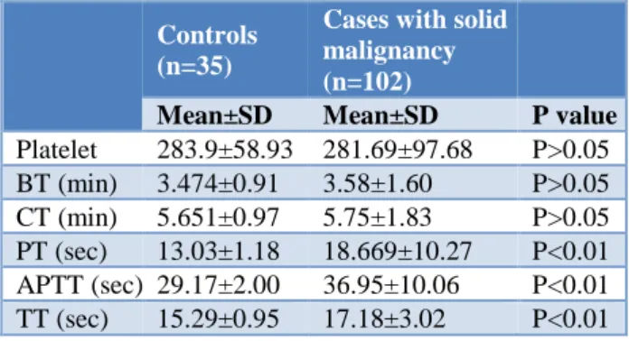
Table 1: Statistical analysis of haemostatic parameters in cancer patients and control group.
Controls (n=35)
Cases with solid malignancy (n=102)
Mean±SD Mean±SD P value
statistically significantly prolonged in cancer patients than those of the respective controls (Table 1).
Plasma levels of D-dimer were found to be raised in study group (>200ng/ml in 10.78 % cases) as compared to control group (>200ng/ml in 0% cases). Also, D-dimer was increased more in patients with advanced stage (>200ng/ml in 19.05% cases) than early stage of cancer (>200ng/ml in 3.33% cases).
Postoperative hemostatic profile
Tests were repeated in 53 cases that had undergone major surgery or chemotherapy. All tests were done on the first post-operative or post chemotherapy day. In the postoperative state, 31 (58.49%) cases showed abnormal results. Compared to preoperative values of all six coagulation parameters, post-operative/post
chemotherapy values on day one was increased (48.04% vs. 54.71%) but this change was not statistically significant.
These abnormalities were more common in patients undergoing surgery in advanced stage of malignancy (68.97%) as compared to the patients in early stage of malignancy (45.83%) (Table 2).
Table 2: Distribution of result of coagulation profile according to the stage of malignancy in
postoperative state.
Stage of malignancy
No. of cases
No. of cases showing
abnormal result %
Early 24 11 45.83
Advanced 29 20 68.97
Total 53 31 58.4
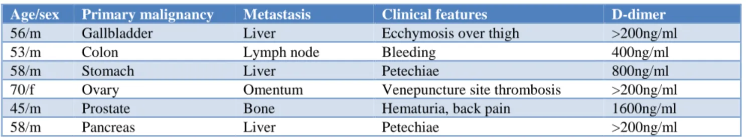
Table 3: Clinical data of patients with disseminated intravascular coagulation (DIC).
Age/sex Primary malignancy Metastasis Clinical features D-dimer
56/m Gallbladder Liver Ecchymosis over thigh >200ng/ml
53/m Colon Lymph node Bleeding 400ng/ml
58/m Stomach Liver Petechiae 800ng/ml
70/f Ovary Omentum Venepuncture site thrombosis >200ng/ml 45/m Prostate Bone Hematuria, back pain 1600ng/ml
58/m Pancreas Liver Petechiae >200ng/ml
Six patients were considered to have DIC as defined by methods section. Clinical Characteristics of these patients are given in Table 3. The D-dimer values in patients range from 200 to 1600 ng/ml. All patients show prolongation of PT, APTT and TT with thrombocytopenia.
DISCUSSION
Platelet count
It was observed that platelets were increased in study group as compared to control, but this difference was not statistically significant. Similar findings were observed by Ali Enshaei et al and Komurcuoglu et al.3,4
However, Karagoz and colleagues in their study reported significantly increased platelet count in lung cancer patients (p=0.011).5 Xuan et al, found this difference to be
statistically significant in their study on primary liver cancer patients in 2011.6
Kies et al, found thrombocytopenia in 16% of cases as opposed to thrombocytosis in 11%. All other authors however found thrombocytosis to be more common, as found in our study.7
Prothrombin time
Mean value of prothrombin time in our study was 18.67±10.27. When compared to control value it was found to be significantly prolonged (p<0.001).
Turna et al, found significant shortening of PT (0.04) when compared to control value, while Ikram et al and Xuan et al found significant prolongation of PT (0.26) similar to our study.8,9,6
Activated partial thromboplastin time
The mean activated partial thromboplastin time in present study was 36.95±10.06 in cancer patients as compared to controls (29.17±2.00), which was statistically highly significant. This finding was similar to Xuan et al and Ikram et al.9,6 Turna et al, however found this
prolongation to be not significant in 104 patients with various malignancies.8 So, the value of APTT in our
study matches with that of Xuan et al and Ikram et al.
Thrombin time
Xuan et al, studied thrombin time in 228 liver cancer patients and found it significantly prolonged.6 Similarly
Kovakova studied it in gastric cancer patients and found prolongation of thrombin time to be significant.10
D-dimer
D-dimer is the smallest degradation product of fibrin resulting from the proteolytic action of plasmin. It is a sensitive marker of fibrinolytic process. Various studies were undertaken which showed increased level of D-dimer in cancer patients and increasing levels in advanced stage of cancer.
Estimation of D-dimer has been done in various units of measurement as per the availability of reagents. We have
performed a semiquantitative method and measured D-dimer in ng/ml and divided as <200, 200-800 and >800ng/ml for comparison.
The value of D-dimer was <200ng/ml in 91 (89.21%) cases, between 200 to 800 in 10(9.89%) cases and >800 in one (0.98%) case, in our study. It was <200ng/ml in all 35 controls. So, the value was increased in 11 (10.78%) cases.
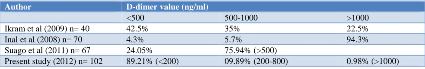
Some studies, which measured D-dimer semi quantitatively, have divided the results as <500, 500-1000 and >1000ng/ml or as < and >500 ng/ml.11,12 Taguchi et
al have divided it as < and >150ng/ml.13 It was classified
as high and low in an Indian study by Subramanian et al (Table 4).14
Table 4: Comparison of mean D-dimer values with other studies.
Author D-dimer value (ng/ml)
<500 500-1000 >1000
Ikram et al (2009) n= 40 42.5% 35% 22.5%
Inal et al (2008) n= 70 4.3% 5.7% 94.3%
Suago et al (2011) n= 67 24.05% 75.94% (>500)
Present study (2012) n= 102 89.21% (<200) 09.89% (200-800) 0.98% (>1000)
The reason for low percentage of cases with increased D-dimer in our study may be the larger number of cases in early stage of malignancy and more cases of squamous cell carcinoma. In contrast, most of the other studies have performed the tests in lung cancer and other adenocarcinomas which are known to be associated with increased tendency of fibrinolysis.
Incidence of DIC and thromboembolic events
Belt et al reported very low (1%) incidence of DIC in a group of 718 solid tumor patients over a seven-year period, whereas Sun et al found clinical bleeding or clotting in 46 of their 61 cases.15 Sallah et al, evaluated
the occurrence of DIC in 1117 patients with solid tumors. Of these patients, 76 (6.8%) were diagnosed with DIC.16
Our study also found only 6 of 102 (5.9%) cases with overt DIC and only 4 of 102 cases without DIC showing clinical evidence of bleeding similar to Sallah et al. True rates of thromboembolic events are still unknown owing to lack of information in clinical trials and difficulties in performing full diagnostic workup in every patient.
Postoerative/postchemotherapy state
In our study we found slight prolongation of both PT and APTT and increased platelet count after 24hours of treatment. While Kirwan et al found decreased platelet
count and shortening of APTT and prolongation of PT. The value of D-dimer was not significantly affected.17
Wang et al, studied perioperative changes in liver cancer patients receiving liver transplant. They found significantly prolonged PT and decreased platelet count on day one. APTT and TT were not significantly changed.18
Xu et al, studied pre and post-operative changes of coagulation profile in lung cancer patients. PT in lung cancer group was prolonged, APTT was reduced, D-dimer was increased, and platelets were reduced. The difference between pre- and post-surgery was significant (P<0.05).19
Gang et al, found the mean preoperative plasma levels of D-dimer in patients with colorectal cancer (1.06±0.24mg/L) were significantly higher than those of controls (0.33±0.12mg/L,P<0.01).20
CONCLUSION
be done to predict the prognosis. Also, therapeutic interventional studies are needed for better outcome.
Funding: No funding sources Conflict of interest: None declared
Ethical approval: The study was approved by the Institutional Ethics Committee
REFERENCES
1. Trousseau A. Phlegmasia alba dolens. In: Lectures on clinical medicine. Hotel-Dieu, Paris, P. Victor Bazire byTrousseau, Armand, 1801-1867; 1865:654-712.
2. Varki A. Trousseau’s syndrome: multiple definitions and multiple mechanisms. Blood. 2007;110(6):1723-9.
3. Enshaei A, Moradi A, Mikaili P, Rezaei S. Correlation of preoperative level of D-dimer with pathological staging of colorectal cancers. Am J Sci Res. 2012;(52):5-12.
4. Komurcuoglu B, Ulusoy B, Gayaf M, Guler A, Ozden E. Prognostic value of plasma D-dimer levels in lung carcinoma. Tumori. 2011;97(6):743-8. 5. Karagöz B, Alacacioğlu A, Bilgi O, Demirci H,
Ozgün A, Alev Akyol Erikçi, et al. Platelet count and platelet distribution width increase in Lung cancer patients. Anatol J Clin Investing. 2009:3(1):32-4.
6. Guo X, Chen M, Ding L, Zhao S, Wang Y, Kang Y, Liu Y. Application of cox model in coagulation function in patients with primary liver cancer. AJOSG. 2011;58(106):326-30.
7. Kies MS, Posch JJ, Gioma JP, Rubin RN. Hemostatic function in cancer patients. Cancer. 1980;46:831-7.
8. Turna H, Ozguroglu M, Bolayirli M, Orhanoglu T, Balci H. Is there any effect of tumor burden on hemostatic parameters in cancer patients? a case-control study of hemostatic abnormalities and anticardiolipin antibodies in solid tumors. Clin Appl Thromb Hemost. 2009;15:454.
9. Ujjan ID, Khokhar NA, Shaikh MA, Shaikh IA, Memon RA, Maheshwari N. Evaluation of coagulation abnormalities in lung cancer patients. JLUMHS. 2009;8(2):118-20.
10. Kovacova E, Kinova S, Duris I, Remcova A. General changes in hemostasis in gastric cancer. Bratisl Lek Listy. 2009;110(4):215-21.
11. Inal S, Tasci C, Karadurmas N, Kuzhan O, Balkan A, Ozkan M, et al. The association of D-dimer levels with other prognostic factors in patients with lung cancer. Turk J Med Sci. 2008;38(3):209-17. 12. Suega K, Bakta IM. Correlation between clinical
stage of solid tumor and D dimer as a marker of coagulation activation. Acta Med Indones. 2011 Jul;43(3):162-7.
13. Taguchi O, Gabazza EC, Yasui H, Kobayashi T, Yoshida M, Kobayashi H. Prognostic significance of plasma D-dimer levels in patients with lung cancer. Thorax. 1997;52(6):563-5.
14. Sitalakshmi S, Rameshkumar K, Damodar P. Significance of haemostatic markers in ovarian carcinoma. Ind J Med Paedia Oncol. 2008;29(2):6-10.
15. Sun NC, McAfee WM, Hum GJ, Weiner JM. Hemostatic abnormalities in malignancy, a prospective study of one hundred eight patients. Part I. Coagulation studies. Am J Clin Pathol. 1979;71(1):10-6.
16. Sallah S. Wan JY, Nguyen NP, Hanrahan LR, Sigounas G. Disseminated intravascular coagulation in solid tumors: clinical and pathologic study. Thrombosis and hemostasis. 2001;86(3):828-33. 17. Kirwan CC, McDowell G, McCollum CN, Kumar
S, Byrne GJ. Early changes in the haemostatic and procoagulant systems after chemotherapy for breast cancer. Brit J Cancer. 2008;99:1000-6.
18. Wang HY, Zhao QY, Yuan YF. Perioperative changes of coagulation functions in the local advanced liver cancer patients receiving liver transplantation. Chin J Cancer. 2008 Jul;27(7):65-9. 19. Xu C, Fu X. The changes of blood coagulation in
surgical patients with lung cancer. Zhongguo Fei Ai Za Zhi. 2010;13(2):136-9.
20. Gang Xu, Ya-Li Zang, Wen Huang. Relationship between plasma D-dimer levels and clinicopathologic parameters in resectable colorectal cancer patients. World J Gastroenterol. 2004;10(6):922-3.