R E S E A R C H A R T I C L E
Open Access
Comparative genomic analysis of
Polypodiaceae chloroplasts reveals fine
structural features and dynamic insertion
sequences
Shanshan Liu
1, Zhen Wang
1, Yingjuan Su
1,2*and Ting Wang
3*Abstract
Background:Comparative chloroplast genomics could shed light on the major evolutionary events that
established plastomic diversity among closely related species. The Polypodiaceae family is one of the most species-rich and underexplored groups of extant ferns. It is generally recognized that the plastomes of Polypodiaceae are highly notable in terms of their organizational stability. Hence, no research has yet been conducted on genomic structural variation in the Polypodiaceae.
Results:The complete plastome sequences ofNeolepisorus fortunei,Neolepisorus ovatus, andPhymatosorus
cuspidatuswere determined based on next-generation sequencing. Together with published plastomes, a
comparative analysis of the fine structure of Polypodiaceae plastomes was carried out. The results indicated that the plastomes of Polypodiaceae are not as conservative as previously assumed. The size of the plastomes varies greatly in the Polypodiaceae, and the large insertion fragments present in the genome could be the main factor affecting the genome length. The plastome ofSelliguea yakushimensisexhibits prominent features including not only a large-scale IR expansion exceeding several kb but also a unique inversion. Furthermore, gene contents, SSRs, dispersed repeats, and mutational hotspot regions were identified in the plastomes of the Polypodiaceae. Although dispersed repeats are not abundant in the plastomes of Polypodiaceae, we found that the large insertions that occur in different species are mobile and are always adjacent to repeated hotspot regions.
Conclusions:Our results reveal that the plastomes of Polypodiaceae are dynamic molecules, rather than
constituting static genomes as previously thought. The dispersed repeats flanking insertion sequences contribute to the repair mechanism induced by double-strand breaks and are probably a major driver of structural evolution in the plastomes of Polypodiaceae.
Keywords:Plastome, Polypodiaceae, Insertions, Dynamic evolution
© The Author(s). 2021Open AccessThis article is licensed under a Creative Commons Attribution 4.0 International License, which permits use, sharing, adaptation, distribution and reproduction in any medium or format, as long as you give appropriate credit to the original author(s) and the source, provide a link to the Creative Commons licence, and indicate if changes were made. The images or other third party material in this article are included in the article's Creative Commons licence, unless indicated otherwise in a credit line to the material. If material is not included in the article's Creative Commons licence and your intended use is not permitted by statutory regulation or exceeds the permitted use, you will need to obtain permission directly from the copyright holder. To view a copy of this licence, visithttp://creativecommons.org/licenses/by/4.0/. The Creative Commons Public Domain Dedication waiver (http://creativecommons.org/publicdomain/zero/1.0/) applies to the data made available in this article, unless otherwise stated in a credit line to the data.
* Correspondence:[email protected];[email protected] 1
School of Life Sciences, Sun Yat-sen University, Guangzhou, Guangdong, China
3College of Life Sciences, South China Agricultural University, Guangzhou, Guangdong, China
Background
Chloroplasts are the plant organelles in which photosyn-thesis takes place. Each chloroplast contains its own gen-ome (plastgen-ome), which usually occurs in multiple copies within the organelle [1]. In recent years, the chloroplast (cp) genome has become a preferred target for compara-tive genomics because of its mostly uniparental inherit-ance, compact size, lack of recombination, and moderate evolutionary rate compared to the two other genomes found in plant cells (nuclear and mitochondrial ge-nomes) [2–4]. Advances in DNA sequencing technology have provided highly efficient, cost-effective sequencing platforms, and the properties of the plastome made it one of the first candidates for high-throughput sequen-cing and assembly. Plastomes have now been extensively used for exploring phylogenetic relationships and under-standing evolutionary processes of plants [5–7].
Ferns, which are the second largest group of vascular plants, play important ecological roles and hold a pivotal phylogenetic position [8]. The sequencing of fern plas-tomes has greatly increased our understanding of the plastomic diversity and evolution of this lineage. The sizes of the plastomes of ferns are highly conserved, and they usually exhibit a circular structure ranging from 120 to 170 kb [9]. The plastomes of ferns typically con-sist of four parts, including a pair of large inverted re-peats (IRs), a large single copy (LSC) region, and a small single copy (SSC) region. Almost all fern IRs contains a core gene set of four ribosomal RNAs (16S, 23S, 4.5S, and 5S) and several tRNA genes (trnA-UGC,trnI-GAU,
trnN-GUU, and trnR-ACG). Structurally, the plastome of fern lineages has evolved in a conservative manner. Most fern plastomes are largely collinear, requiring only a few inversions and IR expansions to account for the large-scale structural rearrangements observed among major lineages. For example, the unique chloroplast gen-omic rearrangement of core leptosporangiate ferns (Sal-viniales, Cyatheales, and Polypodiales) and Schizaeales can be explained by an expansion of the IRs and“two in-versions” [10], which mainly affect the orientation and gene content of the IRs [11]. The conservative nature of the plastome makes it homogeneous enough to allow comparative studies to be conducted across higher-level taxa, but it is also sufficiently divergent to capture vari-ous evolutionary events.
The Polypodiaceae family is one of the most species-rich groups of extant ferns [12], displaying remarkable morphological and systematic diversity. Leptosporangi-ate ferns diversified in an angiosperm-dominLeptosporangi-ated can-opy during the Cenozoic radiation period, thus establishing the diversity of the Polypodiaceae [13]. Most species within Polypodiaceae are epiphytes, and this family represents one of the most diverse and abundant groups of pantropical vascular epiphytes in
tropical and subtropical forests [14,15]. The plastomes of Polypodiaceae have undergone a variety of complex genomic reconstructions over evolutionary time, mak-ing them significantly different from those of the basal ferns (Marattiales, Ophioglossales, Psilotales, and Equi-setales). Few studies have analyzed the gene content and structural changes of Polypodiaceae plastomes in detail because plastome evolution in Polypodiaceae is considered relatively dormant compared with that in other lineages. Most of the relevant research has fo-cused on phylogenetic topics. Recent studies, however, have shown that the plastomes of polypods contain not only hypervariable regions but also widespread mobile sequences [16, 17]. Unfortunately, the corresponding studies have rarely involved Polypodiaceae because of the limited available plastome data for this group. This indicates that the currently available plastome informa-tion may be insufficient to elucidate the evoluinforma-tionary patterns of the fern genome, and the exploration of the structural diversity of Polypodiaceae plastomes is also far from sufficient. Comparative genomic analyses of the chloroplasts among closely related species can gen-erate genetic markers and provide a more exhaustive understanding of the evolutionary trajectory of the gen-ome [18,19]. Nevertheless, the plastome structural and sequence homogeneity among low-level taxa makes it difficult to identify various evolutionary events. There-fore, it is necessary to compare the fine structural char-acteristics of Polypodiaceae plastomes to increase the understanding of the diversity and dynamic evolution of fern plastomes. Upon this premise, we performed the first family-scale comparative analysis of plastome structure and content in Polypodiaceae.
We utilized a high-throughput sequencing platform to assemble three new plastomes, two fromNeolepisorusand one from Phymatosorus, and used them to perform detailed comparative analysis with the set of all previously published Polypodiaceae plastomes in an effort to: 1) characterize the genomic structure and gene content of newly sequenced plastomes; 2) examine Polypodiaceae plastome variation at the fine structural level; and 3) gain insight into the plastome evolution of Polypodiaceae. Results
Genome assembly and annotation
Illumina paired-end sequencing generated 6,760,171, 6, 821,122, and 6,764,140 raw reads from Neolepisorus fortunei, Neolepisorus ovatus, and Phymatosorus cuspi-datus, respectively (Table1). A total of 635,763 to 1,054, 231 clean reads were mapped to the reference plastome and 11 to 16 contigs were assembled for three species, reaching over 160× coverage on average over the plas-tomes. The draft plastome of N. fortunei,N. ovatus, and
The gap regions for each resulting plastome were filled by using PCR-based sequencing with corresponding pairs of primers (Table S1). The length of the complete plastome sequences ranged from 151,915 to 152,161 bp, with an average GC content of 41.7% (range 41.3–42.3%; Table 1). All plastomes exhibited the typical quadripar-tite structure, harboring a pair of large IRs (24,609–24, 756 bp). The two IR regions divide the plastomes into an LSC region (80,670–81,175 bp) and an SSC region (21, 601–21,733 bp) (Fig. 1, Table 2). The three newly sequenced plastomes have a similar gene content, with a few notable distinctions. Compared to the other two plastomes, loss oftrnR-UCGis observed inP. cuspidatus
(Fig. 1).rps16harbors an approximately 470-bp intronic deletion inN. fortunei(Fig.S1). Furthermore,N. fortunei
has extra complete copies ofndhBin IRb because of its IRa/LSC border adjacent to ndhB, whereas N. ovatus
and P. cuspidatus only contains a second fragment of
Fig. 1Plastome gene maps ofN. ovatus,N. fortunei, andP. cuspidatus. The plastome map represents all three species since their gene numbers, orders and names are the same, except thatN. fortuneihas lost thetrnR-UCG gene. Genes located outside and within the black circle are transcribed in the clockwise and counterclockwise directions, respectively. Different colors represent genes belonging to different functional groups
Table 1Details of plastome sequencing and assembly
Sample N. ovatus N. fortunei P. cuspidatus Raw Reads 6,760,171 6,821,122 6,764,140 Clean reads 5,765,899 5,907,868 5,880,444 Raw data 2.03G 2.05G 2.03G Clean data 1.73G 1.77G 1.76G Contig 13 11 16 Genome coverage (×) 313 292 160 Size (bp)/GC% 151,936/42.3 151,915/41.3 152,161/41.4
ndhB in IRb due to their IRa/LSC border lies within
ndhB.
Whole-chloroplast genome comparison among Polypodiaceae
The three newly obtained plastomes of Polypodiaceae (N. ovatus, N. fortunei, and P. cuspidatus) were com-pared with nine previously published plastomes repre-senting three subfamilies of Polypodiaceae, i.e., Microsoroideae, Platycerioideae, and Drynarioideae (Table 2). The Polypodiaceae plastomes appeared to be structurally similar to each other, showing a typical quadripartite structure consisting of two IRs separated by LSC and SSC. Overall, the analysis showed that the size of the plastome varies widely among Polypodiaceae, ranging from 151,936 bp in N. ovatus to 164,857 bp in
Selliguea yakushimensis. The lengths of the LSC and SSC regions of most species are approximately 81 kb and 21 kb, respectively, but these two regions of the
Drynaria roosii plastome are significantly larger, reach-ing 86 kb and 24 kb, respectively. Furthermore, the plas-tome of Leptochilus hemionitideusalso contains a larger SSC (25,492 bp). The IR regions of the Polypodiaceae plastomes show significant variation in length compared to the SC regions, varying from 23,416 to 32,017 bp. The base composition of the Polypodiaceae plastomes is more conservative relative to their size variation. The distribution of GC content is heterogeneous, with the highest being observed in IR regions (42.1–45.9%), followed by LSC (37.9–43.6%), while the lowest is found in the SSC region (34.4–43.0%) (Table2).
The gene order of the plastomes of Polypodiaceae is almost collinear. However, the structure of the S.
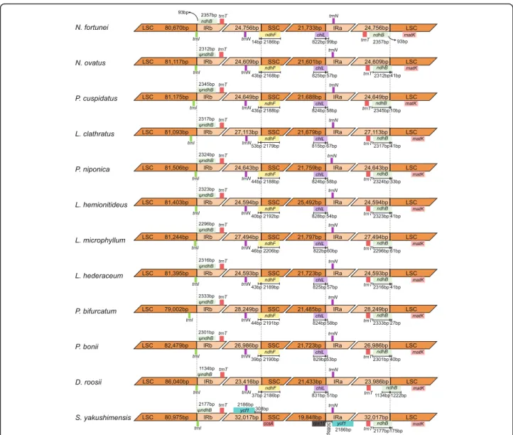
yakushimensis plastome is considerably different from the typical structure of Polypodiaceae. A notable differ-ence in the plastome of S. yakushimensis is a transpos-ition of an ~ 6 kb segment spanning ndhF to ccsA from the SSC to the IR; this inversion appears at the junction of IRb/SSC (Fig. 2). At another junction, SSC/IRa, we observed an extensive IR expansion resulting in the duplication of ycf1,chlL, and chlN at the end of IRa. In addition, a minor inversion (~ 2 kb) was detected between Lepidomicrosorum hederaceum and D. roosii
plastomes, which was located in rps15-ycf1 in the SSC region of L. hederaceum and rrnL-trnR in the LSC region of D. roosii, respectively (Fig. 2). Genomic struc-tural changes that occur in intergenic regions may play an additional evolutionary role, but they are difficult to detect because intergenic regions have no coding func-tion. This unique inversion may be related to inserted sequenced or is an intermediate form of the evolution of other plastomes. As the number of published Polypodia-ceae plastomes increased, the evolutionary processes of Polypodiaceae should become clearer.
The size variations of the plastome may be a result of the dynamic changes of IR boundaries [20]. However, the Polypodiaceae plastomes exhibit high similarity at SC/IR boundaries, except in S. yakushimensis (Fig. 3). The trnN-GUU gene is located in the IR adjacent to eitherndhForchlLat the SSC/IR border. ThendhFand
chlL genes cross the SSC/IR border, extending from 14 to 53 bp and 51–99 bp in the IR, respectively. The IRa/ LSC border is located within the coding region ofndhB and generates a pseudogene of 1,134–2,345 bp at the LSC/IRb border in 11 of the plastomes, except for that of N. fortunei. The LSC/IRb border is located within or Table 2Comparison of general features of the plastomes of Polypodiaceae
Subfamily Microsoroideae Platycerioideae Drynarioideae
Species NO NF PC PN LC LHem LM LHed PBi PBo DR SY
LSC (bp) 81,117 80,670 81,175 81,506 81,093 81,403 81,244 81,395 79,002 82,479 86,040 80,975 SSC (bp) 21,601 21,733 21,688 21,759 21,679 25,492 21,797 21,756 21,485 21,723 24,433 19,848 IR (bp) 24,609 24,756 24,649 24,643 27,113 24,594 27,494 24,593 28,249 26,986 23,416 32,017 Size (bp) 151,936 151,915 152,161 152,551 156,998 156,083 158,029 152,337 156,985 158,174 154,305 164,857 PGGs 88 89 88 88 88 87 88 88 88 88 88 91 tRNAs 35 35 34 35 35 34 35 35 33 35 35 35 rRNA 8 8 8 8 8 8 8 8 8 8 8 8 Pseudo 1 0 1 1 0 2 1 1 1 1 1 1 Genes 132 132 131 132 131 131 132 132 130 132 132 135 LSC (GC%) 40.2 42.1 40.4 41.5 40.46 43.6 40.5 41.9 37.9 40.1 39.8 39.5 SSC (GC%) 37.5 38.8 37.6 39.0 37.9 43.0 37.9 39.7 34.4 37.4 36.6 39.8 IR (GC %) 44.9 45.1 44.8 45.3 45.4 45.9 45.3 45.2 44.9 45.6 45.1 42.7 GC (Total%) 41.3 42.3 41.4 42.4 41.8 44.2 41.8 42.7 39.9 41.6 40.9 40.8
NON. ovatus,NFN. fortunei,PCP. cuspidatus,PNP. niponica,LCL. clathratus,LHemL. hemionitideus,LML. microphyllum,LHedL. hederaceum,PBiP. bifurcatum,
next to the trnI-CAU gene. In contrast to the subtle changes in IR boundaries, the IR ofS. yakushimensishas experienced extensive expansion, capturing the ycf1,
chlL, andchlN genes in the SSC region. This expansion, combined with the inversion of the ndhF-ccsA region, causes a unique SSC/IR boundary in theS. yakushimen-sisplastome (Fig.3). The SC/IR boundaries of the Poly-podiaceae plastomes are highly similar but not identical, indicating that the expansion and contraction of IR is an independent and recurrent phenomenon in evolution. Furthermore, the change in IR boundaries is not suffi-cient to cause a large difference in genome size, and we believe that microstructural changes (such as insertions and deletions) may be responsible for the difference.
There are some variations in the gene content of Polypodiaceae plastomes due to varying degrees of IR boundary changes and inversions.trnR-UCG exists in all Polypodiaceae species except for L. hemionitideus, P. cuspidatus, and Platycerium bifurcatum. trnV-UAC is
also absent in the plastome of P. bifurcatum. Another difference is that the plastome ofN. fortuneicontains an additional intact ndhB gene in the IR region, whereas only onendhBfragment exists in other species. Further-more, close inspection of the gene annotations of the 12 Polypodiaceae plastomes indicated that the rpoC1 gene of the L. hemionitideus has been pseudogenized by a frameshift mutation.
Sequence diversity and mutational hotspots of the Polypodiaceae plastome
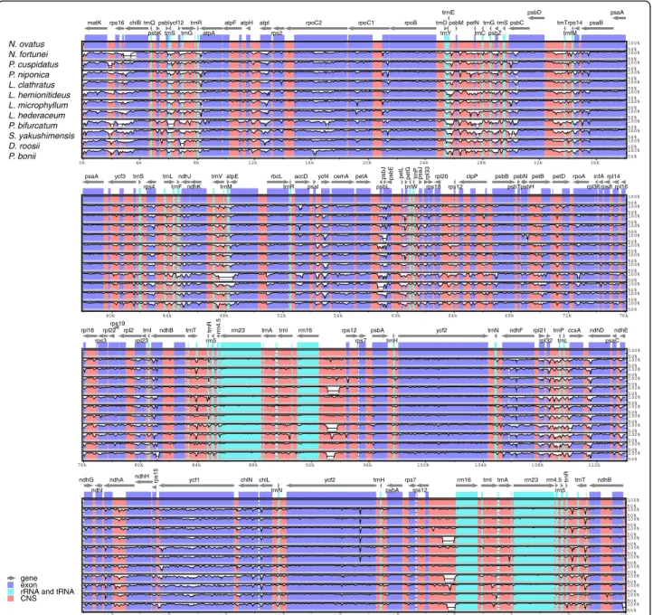
The multiple sequence alignments performed in mVISTA software showed the similarity of the whole sequences of the plastomes of the 12 Polypodiaceae species analyzed (Fig. 4). Lower divergence was found in the IR and protein-coding regions than in the single-copy and non-coding regions. Nevertheless, we found that obvious large inserted fragments were present in therrn16-rps12 spacer of the IRs in the plastomes of Lepisorus clathratus,
Fig. 2Synteny and rearrangements detected in the plastome sequences of Polypodiaceae using Mauve alignment. Local collinear blocks are represented as boxes of the same color connected with lines, which indicate syntenic regions. Histograms within each block represent the sequence identity similarity profile. The colored blocks, which are above and below the center line, represent sequences transcribed in reverse directions
Pyrrosia bonii, and P. bifurcatum. To detect mutational hotspots in the plastomes of Polypodiaceae, sliding-window analysis was performed on the whole-genome alignments of the sequences using DnaSP v5.0. The results showed that the sequence variation between the plastomes of Polypodiaceae was relatively low, with nucleotide diver-sity (Pi) ranging from 0.00232 to 0.20028. Overall, the SSC region exhibited the highest sequence variation, with an average Pi of 0.10103, followed by the LSC and IR regions, with average Pi values of 0.07317 and 0.02676, respect-ively. A total of nine highly divergent loci were identified in the plastomes of Polypodiaceae, includingmatK-rps16,
rps16, trnC-trnG, psbZ-psbC, psbD-trnT, trnP-psaJ, and
rpl2-trnI, located in LSC; rrn16-rps12, located in the IR region; and one protein-coding gene,ycf1, located in SSC. The ndhF-ccsA region in SSC showed a higher Pi value
than the other loci, most likely due to the inversion occur-ring in this region, which resulted in higher sequence vari-ation (Fig.S2). Therefore, it may not be categorized as a common mutational hotspot in the Polypodiaceae.
Analysis of SSRs and repeat sequence in Polypodiaceae plastomes
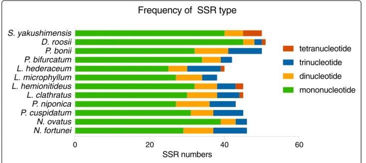
Simple sequence repeats (SSRs), or microsatellites are short, tandemly repeated DNA motifs of 1–6 nucleotides [21]. They can exhibit high polymorphism and mutation rates, which contribute to estimates of genetic variation [22]. In this study, very similar numbers of potential cpSSRs were identified from the plastomes of the 12 Polypodiaceae species by using MISA. The total number of SSRs in Polypodiaceae ranged from 38 to 51. Four kinds of SSRs were detected: mononucleotides,
Fig. 3Comparison of the border positions of LSC, SSC, and IR regions among the plastomes of Polypodiaceae species.ψrefers to the pseudogene ofndhB at the LCS/IRb border
dinucleotides, trinucleotides, and tetranucleotides (Fig.5). However, tetranucleotide repeats were discovered in only the plastomes of L. clathratus, L. hemionitideus, L. hederaceum, S. yakushimensis, and D. roosii. Different SSR motifs appeared at different frequencies in these plastomes. The most abundant observed repeats were mononucleotides, accounting for approximately 62.8– 88.3% of the total number of SSR loci, followed by smaller numbers of dinucleotide (8.7–20.9%) and trinu-cleotide (6.6–22.5%) repeats, whereas tetranucleotide repeats were the least common (0–4.4%).
We found that the predominant mononucleotide repeats in all analyzed species with the exception of D. roosii, S. yakushimensis, and P. bifurcatum were G/C tandem motifs, which accounted for 53.3 to 100% of the mononucleotide repeats in the Polypodiaceae plastomes (Fig.6). The Polypodiaceae species included in this study all exhibited similar SSR distribution patterns in the plastome. SSRs were much more frequently located in the LSC region (48.0–71.1%) than in IR (10.5–36.0%) and SSC regions (9.3–22.0%). Furthermore, the majority of the identified SSRs were located in intergenic spacers,
100% 50% 100% 50% 100% 50% 100% 50% 100% 50% 100% 50% 100% 50% 100% 50% 100% 50% 100% 50% 100% 50% 100% 50% 0k 4k 8k 12k 16k 20k 24k 28k 32k 36k matK rps16 chlB trnQ psbK psbI trnS ycf12 trnG trnR atpA
atpF atpH atpI rps2
rpoC2 rpoC1 rpoB trnD trnY trnE psbM petN trnC trnG psbZ trnS psbC psbD trnT trnfM rps14 psaB psaA 100% 50% 100% 50% 100% 50% 100% 50% 100% 50% 100% 50% 100% 50% 100% 50% 100% 50% 100% 50% 100% 50% 100% 50% 40k 44k 48k 52k 56k 60k 64k 68k 72k 76k psaA ycf3 trnS rps4 trnL trnF ndhJ ndhK ndhC trnV trnM atpE atpB rbcL trnR accD psaI
ycf4 cemA petA psbJ
psbL
psbF
psbE petL petG
trnW trnP psaJ rpl33 rps18 rpl20 rps12 clpP psbB psbT psbN psbH
petB petD rpoA
rps11 rpl36 infA rps8 rpl14 rpl16 gene exon rRNA and tRNA CNS 100% 50% 100% 50% 100% 50% 100% 50% 100% 50% 100% 50% 100% 50% 100% 50% 100% 50% 100% 50% 100% 50% 100% 50% 76k 80k 84k 88k 92k 96k 100k 104k 108k 112k rpl16 rps3 rpl22 rps19 rpl2 rpl23 trnI ndhB trnT trnR rrn5 rrn4.5 rrn23 trnA trnI rrn16 rps12 rps7 psbA trnH ycf2 trnN ndhF rpl21 rpl32 trnP trnL ccsA ndhD psaC ndhE 100% 50% 100% 50% 100% 50% 100% 50% 100% 50% 100% 50% 100% 50% 100% 50% 100% 50% 100% 50% 100% 50% 100% 50% 116k 120k 124k 128k 132k 136k 140k 144k 148k 152k ndhG ndhI ndhA ndhH rps15 ycf1 chlN chlL trnN ycf2 trnH psbA rps7 rps12 rrn16 trnI trnA rrn23 rrn4.5 rrn5 trnR trnT ndhB N. ovatus N. fortunei P. cuspidatus P. niponica L. clathratus L. hemionitideus L. microphyllum L. hederaceum P. bifurcatum S. yakushimensis D. roosii P. bonii
Fig. 4Visualization of the alignment of the Polypodiaceae plastome sequences, withN. fortuneias the reference. Gray arrows above the alignment indicate genes, including their orientation and position. The vertical scale represents the percentage of identity, ranging from 50 to 100%. Genome regions are color-coded as protein-coding genes, tRNAs, rRNAs and conserved noncoding sequences (CNSs)
0 20 40 60
N. fortunei
N. ovatus
P. cuspidatum
P. niponica
L. clathratus
L. hemionitideus
L. microphyllum
L. hederaceum
P. bifurcatum
P. bonii
D. roosii
S. yakushimensis
SSR numbers
Frequency of SSR type
mononucleotide
dinucleotide
trinucleotide
tetranucleotide
Fig. 5Types and numbers of cpSSRs in the Polypodiaceae plastomes
accounting for 66.7–83.3% of all SSRs detected. SSRs dispersed in intronic regions were the second most com-mon category (9.5–28.9%). The fewest SSRs were located in coding genes, which accounted for only 4.0–14.0% of all SSR loci (TableS2).
Forward, palindromic and reverse repeats of more than 30 bp with a sequence identity≥90% were detected in the Polypodiaceae plastomes using REPuter. The results showed that the numbers of repeats in the plas-tomes of Polypodiaceae varied considerably, ranging from 9 in L. hemionitideus to 146 in P. bifurcatum. These long repeats ranged from 30 to 307 bp in length and were repeated twice. Species showed some variation in the number of long repeat sequences located in inter-genic spacers and coding genes. Most repeats were distributed in intergenic spacer regions, and the rest were distributed in coding genes and intronic regions (TableS3). We detected some species-specific intergenic spacers with rich repeats, including the rbcL-trnR-UCG intergenic spacer of the LSC region of D. roosii, the
rps12-rrn16 intergenic spacer of the IR regions of L. clathratus and P. bifurcatum, the rps7-psbA intergenic spacer of the IR region ofLemmaphyllum microphyllum, and the ndhA intronic/chlL-chlN intergenic spacer of the SC/IR junction ofP. bonii.
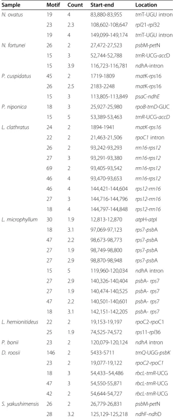
Tandem Repeats Finder v4.09 [23] was further used to identify the tandem repeats present in Polypodiaceae plastomes, with the minimum identity and size of the repeats set to 90% and 15 bp in unit length. A small number of tandem repeats were detected in all species except forP. bifurcatum andL. hemionitideus(Table 3). Among the species in which tandem repeats were detected, L. microphyllum exhibited the most repeats, and P. bonii exhibited the fewest. The intergenic spacers rrn16-rps12, rps7-psbA, and rbcL-trnR-UCG were regions containing abundant repeat sequences in the plastomes of L. clathratus, L. microphyllum, and
D. roosii, respectively. All detected tandem repeats were distributed in noncoding regions, and the pro-portions of tandem repeats located in intergenic spacers were higher than those in intronic regions in Polypodiaceae species (Table 3).
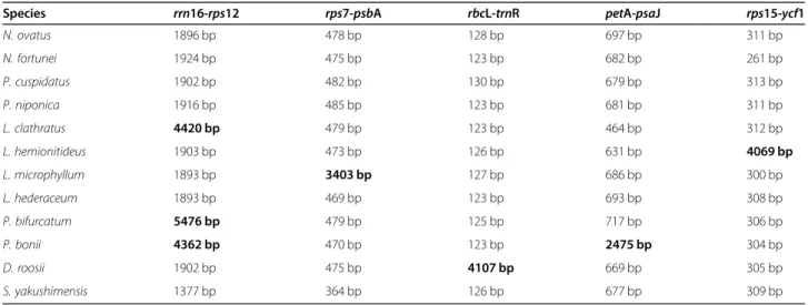
Dynamic insertion sequences in Polypodiaceae plastomes
Through a detailed whole-genome alignment, we found that there are large insertions in some regions of Polypodiaceae plastomes, including the rrn16-rps12 spacers of L. clathratus, P. bifurcatum, and D. roosii
(~ 2400–3500 bp insertions); the rps7-psbA spacer of
L. microphyllum (~ 3000 bp insertion); the rbcL-trnR spacer of D. roosii (~ 4000 bp insertion); the petA-psaJ spacer of P. bonii (~ 1700 bp insertion); and the
rps15-ycf1 spacer of L. hederaceum (~ 3700 bp inser-tion) (Table 4). The identity of the insertion
Table 3Size, number, and distribution of tandem repeats in the plastomes of Polypodiaceae
Sample Motif Count Start-end Location N. ovatus 19 4 83,880-83,955 trnT-UGU intron
23 2.3 108,602-108,647 rpl21-rpl32 19 4 149,099-149,174 trnT-UGU intron N. fortunei 26 2 27,472-27,523 psbM-petN 15 3 52,744-52,788 trnR-UCG-accD 15 3.9 116,723-116,781 ndhA-intron P. cuspidatus 45 2 1719-1809 matK-rps16 26 2.5 2183-2248 matK-rps16 15 3 113,805-113,849 psaC-ndhE P. niponica 18 3 25,927-25,980 rpoB-trnD-GUC
15 5 53,389-53,463 trnR-UCG-accD L. clathratus 24 2 1894-1941 matK-rps16 22 2 21,463-21,506 rpoC1intron 26 2 93,242-93,293 rrn16-rps12 27 3 93,291-93,380 rrn16-rps12 69 2 93,405-93,542 rrn16-rps12 46 4 93,470-93,653 rrn16-rps12 46 4 144,421-144,604 rps12-rrn16 27 3 144,716-144,796 rps12-rrn16 18 4 144,797-144,848 rps12-rrn16 L. microphyllum 30 1.9 12,813-12,870 atpH-atpI
18 3.1 97,069-97,123 rps7-psbA 47 2.2 98,673-98,773 rps7-psbA 27 1.9 98,749-98,800 rps7-psbA 27 2.9 98,870-98,948 rps7-psbA 15 5 119,960-120,034 ndhA intron 27 2.9 140,326-140,404 psbA-rps7 27 1.9 140,474-140,525 psbA-rps7 47 2.2 140,501-140,601 psbA-rps7 18 3.1 142,151-142,205 psbA-rps7
L. hemionitideus 22 2 19,153-19,197 rpoC2-rpoC1 25 1.9 74,525-74,572 rps11-rpl36
P. bonii 23 2 120,079-120,124 ndhAintron
D. roosii 146 2 5433-5711 trnQ-UGG-psbK 23 2 19,077-19,122 rpoC2-rpoC1 18 3 54,433–54,486 rbcL-trnR-UCG 47 3 54,550-55,871 rbcL-trnR-UCG 42 2 54,644-54,727 rbcL-trnR-UCG S. yakushimensis 26 2 26,779-26,831 psbM-petN 28 3.2 125,129-125,218 ndhF-ndhD
sequences in the rrn16-rps12 spacers of L. clathratus,
P. bifurcatum and D. roosii was calculated using
MegAlign v8.1.3 [24]. The pairwise alignments showed that the identity of the insertions in the three plastomes was only 48.3–50.1%, indicating that these insertions may have different origins (Fig. S3).
Robison et al. [17] previously proposed the concept of MORFFO (Mobile Open Reading Frames in Fern Organelles), which are a set of mobile insertion sequences that are widely present in fern organelles. To verify whether the insertions detected in the Poly-podiaceae plastomes are consistent with the MORFFO sequences, MORFFO sequences were determined by local BLAST searches [25, 26] using the database established from the 12 Polypodiaceae plastomes, with the consensus sequences of morffo1, morffo2, and
morffo3 as queries. Furthermore, to examine whether the insertions identified in this study present mobile properties, these sequences were subjected to local BLAST searches. Our results showed that morffo1
presents similarity to the petA-psbJ fragment of P. bonii (71.2%) and the rrn16-rps12 fragment of P. bifurcatum (70.3%). Morffo2 was detected in rrn
16-rps12 of P. bifurcatum and ycf1-ccsA of S. yakushi-mensis, with identities of 71.0% and 67.9%, respect-ively (Table 5).
Surprisingly, the insertions contained in different spe-cies show significant BLAST hits against each other, which suggests that DNA fragments may have been transferred from one plastome to another. For example, a fragment of the L. clathratus insertion shows high sequence similarity to therps7-psbA insertion located in the IR ofL. microphyllum, rbcL-trnR located in the LSC of D. roosii, and rps15-ycf1 located in the SSC of L. hemionitideus. Morffo1 is located within the P. bifurca-tum-P. bonii consensus insertion fragment, butmorffo2
does not show overlap with the insertions detected in this study (Table 5). Previous studies have revealed that MORFFO-like sequences are often associated with struc-tural changes in the genome and may be the main driv-ing force for structural evolution in the plastome. In this study, a number of movable insertions were found in the relatively static plastomes of Polypodiaceae, indicating that the different insertion fragments arising during the evolution of genome structure may have different functions.
Discussion
Plastome variation across Polypodiaceae
Our comparative analysis of 12 species from Microsoroi-deae, PlatycerioiMicrosoroi-deae, and Drynarioideae showed that the length of the plastomes varies greatly in Polypodiaceae, even within the same subfamily. Generally, dynamic ex-pansions or contractions in IR boundaries are consid-ered a primary mechanism leading to the size variation of land plant plastomes [20]. Although there are differ-ences in the IR boundaries between Polypodiaceae plas-tomes, they also exhibit obvious similarities. Therefore, the minor shifts in the IR boundaries of the Polypodia-ceae plastomes are expected to be insufficient to account for marked differences in genome size. For example, in Microsoroideae, we found that despite the IR boundaries being deeply conservative, the IR regions ofL. clathratus
and L. microphyllum are approximately 2500 bp longer than those of other species, and the SSC region of L. hemionitideus is approximately 3700 bp longer than those of other species. A fine-scale analysis of the plas-tome sequence data of Polypodiaceae revealed several large insertions in the specific intergenic spacers of IR or SC regions that correspond to the observed genome size differences. This situation corresponds well with those in the species of the other two subfamilies, with the Table 4Comparison of the length of intergenic regions containing large insertions in the plastomes of Polypodiaceae
Species rrn16-rps12 rps7-psbA rbcL-trnR petA-psaJ rps15-ycf1
N. ovatus 1896 bp 478 bp 128 bp 697 bp 311 bp N. fortunei 1924 bp 475 bp 123 bp 682 bp 261 bp P. cuspidatus 1902 bp 482 bp 130 bp 679 bp 313 bp P. niponica 1916 bp 485 bp 123 bp 681 bp 311 bp L. clathratus 4420 bp 479 bp 123 bp 464 bp 312 bp L. hemionitideus 1903 bp 473 bp 126 bp 631 bp 4069 bp L. microphyllum 1893 bp 3403 bp 127 bp 686 bp 300 bp L. hederaceum 1893 bp 469 bp 123 bp 693 bp 308 bp P. bifurcatum 5476 bp 479 bp 125 bp 717 bp 306 bp P. bonii 4362 bp 470 bp 123 bp 2475 bp 304 bp D. roosii 1902 bp 475 bp 4107 bp 669 bp 305 bp S. yakushimensis 1377 bp 364 bp 126 bp 677 bp 309 bp
exception ofS. yakushimensis. Therefore, in most species of Polypodiaceae, the main reason for the difference in plastome size is not the variation in SC/IR boundaries but the large fragment insertions that occur in different species. Selliguea yakushimensis of Drynarioideae ex-hibits the largest IRs identified in the fern lineage to date, mainly because its IR boundary has extended to-ward the SSC, causing the ycf1,chlL, and chlN genes to be captured within the IR region. Therefore, we cannot discuss the differences in the size of the plastomes only from the perspective of the expansion and contraction of the IR boundary because the conservative nature of the plastome itself will cause researchers to ignore other factors.
It is generally recognized that plastome evolution of Polypodiaceae has mostly stabilized, and structural changes such as rearrangements occur rarely. By con-trast, the results presented here indicate that the plas-tome of S. yakushimensis is highly unusual in some respects, containing not only a large-scale IR expansion exceeding several kb but also a unique inversion. The large-scale expansion of the IR presumably occurred through double reciprocal recombination between IR segments during replication [27]. Numerous dispersed repetitive sequences located at the original SSC/IR
junction (chlL/trnN-GUU) were detected in the S. yakushimensis plastome, coinciding exactly with the mechanism of IR expansion; that is, repeat sequences provide the potential for genome rearrangement within or between molecules by homologous recombination [27,28]. In general, the homogeneity of plastome struc-tural changes is low relative to the sequence data. The structural changes in the S. yakushimensisplastome can therefore serve as specific genetic markers in species dis-crimination or phylogenetic analyses. Unfortunately, there is no way to determine which event took place first because IR expansion has occurred at the SSC/IRa boundary, while inversion has occurred near the IRb/ SSC boundary. Consequently, it is necessary to sequence more plastomes in Polypodiaceae to improve our under-standing of the evolutionary trajectory of the plastome.
Whole-plastome alignments can elucidate the level of sequence divergence and easily identify large indels, which are extremely useful for phylogenetic analyses and plant identification. In the present study, our results showed that the IRs present lower sequence divergence than the SC regions. This phenomenon is considered to be a result of copy correction between IR sequences and the elimination of deleterious mutations by gene conver-sion [29]. Moreover, sequence differences among the Table 5Results of matches for insertion sequences within Polypodiaceae identified using local BLAST searches
Query Subject % Identity Alignment length Start End Location
morffo1 P. bonii 71.2 1217 59,496 58,295 petA-psbJ
P. bifurcatum 70.3 1254 93,132 91,893 rrn16-rps12 morffo2 P. bifurcatum 71.0 1668 95,199 93,541 rrn16-rps12 S. yakushimensis 67.9 1121 117,503 118,561 ycf1-ccsA L. clathratus (rrn16-rps12) L. microphyllum 91.5 1276 97,718 96,461 rps7-psbA L.microphyllum 88.1 796 98,773 97,983 rps7-psbA D. roosii 92.0 647 54,866 55,512 rbcL-trnR D. roosii 86.7 668 55,779 56,443 rbcL-trnR L. hemionitideus 81.4 737 121,798 121,063 rps15-ycf1 L. hemionitideus 71.3 523 120,796 120,281 rps15-ycf1 P. bifurcatum (rrn16-rps12) P. bonii 88.8 1502 58,225 59,724 petA-psbJ P. bonii 84.7 698 59,636 60,323 petA-psbJ S. yakushimensis 70.5 831 118,643 117,834 ycf1-ccsA L. microphyllum (rps7-psbA) D. roosii 87.0 1527 56,416 54,895 rbcL-trnR L. clathratus 91.5 1276 95,574 94,305 rrn16-rps12 L. clathratus 88.1 796 94,304 93,538 rrn16-rps12 L. hemionitideus 81.8 1425 120,281 121,697 rps15-ycf1 D. roosii (rbcL-trnR) L. microphyllum 87.0 1527 98,595 97,078 rps7-psbA L. hemionitideus 79.3 1388 121,661 120,281 rps15-ycf1 L. clathratus 92.0 647 93,658 94,304 rrn16-rps12 L. clathratus 86.7 668 94,307 94,969 rrn16-rps12
Polypodiaceae plastomes were evident in the intergenic spacers, suggesting greater conservation in coding re-gions than in noncoding rere-gions. Nine divergence hot-spots between Polypodiaceae species were identified, including matK-rps16, rps16, trnC-trnG, psbZ-psbC,
psbD-trnT, trnP-psaJ, rpl2-trnI, rrn16-rps12, and ycf1, among which nine loci were located in SC and one was located in an IR. Although the ndhF-ccsA regions also present higher Pi values, they may not be suitable as general mutational hotspots for Polypodiaceae due to the existence of inversion in this region of theS. yakush-imensis plastome. Thus, we must conduct careful data exploration when screening universal mutational hot-spots to avoid confusion by plastome structural changes.
In this study, we found many repeat regions, includ-ing forward repeats, palindromic repeats, and reverse and tandem structures, which could be important hotspots for genome reconfiguration [30, 31]. In par-ticular, the occurrence of large repeats in plastomes, such as the 307 bp palindromic repeat observed in P. cuspidatus, has been speculated to result in an un-stable genome structure due to inappropriate re-arrangement [32]. In addition, these repeats provide many informative loci for the development of molecu-lar markers for phylogenetics and population genetics [33]. As a very powerful type of molecular marker, SSRs are widely used in different research fields. They possess obvious advantages such as high polymorph-ism and cost effectiveness [34]. Most studies have shown that the predominant cpSSRs of land plants are consistent with their AT-biased plastomes. By contrast, the cpSSRs present in Polypodiaceae show considerable dissimilarity from the previously reported patterns. In this study, more than 38 SSRs were iden-tified in every Polypodiaceae plastome, among which the majority were C/G mononucleotides and were distributed in noncoding regions. The previously held idea that cpSSRs are generally composed of A/T re-peats has now been challenged [35]. Gao et al. [36] have shown in denaturation experiments that repeti-tive structures with a higher GC content contribute to increasing the thermal stability of the Dryopteris fragrans plastome and maintaining its structure in the face of thermal changes. Species of Polypodiaceae evolved diversified morphological traits and lifestyles putatively in response to changes in terrestrial ecosys-tems caused by the radiation of angiosperms during the Cretaceous period [37]. Thus, we speculate that these repeating structures with a high GC content may be one of the molecular foundations of the adap-tation of Polypodiaceae to the environment, which also provides new insights for understanding the en-vironmental adaptation mechanism of plants. Further-more, the cpSSRs developed in our study provide
unique information for investigating genetic structure and genetic variation. In particular, these cpSSRs will be complementary and comparable to nuclear SSRs from ferns.
Dynamic insertions in Polypodiaceae plastomes
Although genome-wide alignment indicates that Polypo-diaceae plastomes are rather conservative, we found abnormally large insertions in certain intergenic spacers. The large fragment insertion observed in the rrn16
-rps12intergenic spacer ofL. clathratusplastome is con-sistent with previous findings [16]. Similar insertion se-quences have been detected in the LSC regions of some distantly related species and in the mitogenome of
Asplenium nidus, implying that such sequences can move between genomic compartments [16]. Robison et al. [17] discovered a similar suite of dynamic mobile elements through an extensive investigation on fern plastomes, shedding light on the presence of MORFFO elements relative to inversions, intergenic expansions, and changes to inverted repeats. In this study, we char-acterized a completely different set of insertion se-quences in the plastomes of Polypodiaceae. There are only two insertions that overlap with the morffo1 se-quence, which are located in thepetA-psaJ spacer in the LSC region ofP. boniiand therrn16-rps12 spacer in the IR region of P. bifurcatum. The detected morffo2 se-quences are located in the ycf1-ccsA region at the IR/ LSC border inS. yakushimensisand therrn16-rps12 spa-cer in the IR region ofP. bifurcatum, showing no hom-ology with other insertions. Our results further confirm the universality of MORFFO sequences in fern plas-tomes, and the presence of such a sequence at the inver-sion endpoint in S. yakushimensis suggests that the MORFFO sequences may be related to inversion events.
Although MORFFO sequences were not detected in the remaining Polypodiaceae species, there is another set of highly mobile insertions present in their plastomes. It is worth noting that plastid genes are rarely gained or lost, whereas our study indicates that the identified in-sertion sequences have been gained and lost frequently during the evolution of Polypodiaceae plastomes. This fluidity could indicate that these insertions in plastomes act as mobile elements. Furthermore, these insertions are frequently found adjacent to regions where more dis-persed repeats occur. Many studies have shown that genomic rearrangement is related to small dispersed re-peats (SDRs), which contribute to the repair mechanism induced by double-strand breaks [38, 39]. SDRs usually make an important contribution to the repetitive space in highly rearranged genomes and increase structural polymorphism even in closely related lineages, and they are mainly present in noncoding DNA fragments and re-lated to small hairpin structures [38]. We speculate that
the presence of rich repetitive motifs combined with highly mobile insertions may constitute the “trigger mechanism”for genome rearrangement in the plastomes of Polypodiaceae species, which can induce structural changes in the plastome under certain conditions. The limitations of the current sequence data for ferns mean that it is difficult to determine the exact source of these insertion sequences. As more genomic data are pub-lished in the future, the source and migration mecha-nisms of these insertion sequences should become clear. Conclusions
As additional plastomes from Polypodiaceae are charac-terized, we obtain a clearer picture of plastome evolution for the family. It is generally considered that the evolu-tion of Polypodiaceae plastomes is conservative, and their structural features are almost invariant in this fam-ily. Against this conservative background, however, the
S. yakushimensis plastome stands out as unusual. The large-scale expansion of IRs and a unique inversion dis-tinguish theS. yakushimensisplastome from those of all other Polypodiaceae studied thus far. In addition, many large mobile insertions are found in the plastomes of Polypodiaceae species, which often flank the dispersed repeated elements in the different Polypodiaceae plas-tomes. These unusual features are found in the structur-ally stable plastomes of Polypodiaceae and may therefore be implicated in the dynamic evolution of the plastomes of the family. In other words, unlike the static plastomes of Polypodiaceae characterized previously, the plastomes characterized herein are structurally unstable, as evi-denced by the large mobile insertions found in Polypodiaceae.
Methods
Sample collection, DNA extraction, and sequencing
In this study, fresh leaves of N. fortunei, N. ovatus,and
P. cuspidatus were sampled from the living collection from the South China Botanical Garden, Chinese Academy of Sciences (CAS), quickly frozen in liquid nitrogen, and stored at ultra-low- temperature refriger-ator at −80 °C until use. Voucher specimens were de-posited in the Herbarium of Sun Yat-sen University (SYS; voucher:S. Liu201,630,S. Liu201,654, andS. Liu
201,701 for N. fortunei, N. ovatus, and P. cuspidatus, respectively). Genomic DNA was extracted using the Tiangen Plant Genomic DNA Kit according to the manufacturer’s instructions (Tiangen Biotech Co., Beijing, China). DNA quality was inspected in 0.8% agarose gels, and DNA quantification was performed using a NanoDrop spectrophotometer (Thermo Scientific, Carlsbad, CA, USA). After quality assess-ment, 500 ng of DNA was sheared to an average frag-ment size of 300 bp with a Covaris M220 ultrasonicator
(Covaris Inc., MS, USA). An Illumina paired-end (PE) sequencing library was constructed using the NEBNext, Ultra DNA Library Prep Kit (New England BioLabs Inc., Ipswich, MA). Sequencing took place on the HiSeq 2500 platform (Illumina Inc., San Diego, USA). Illumina sequencing produced approximately 2 Gb of raw data for each species.
Genome assembly and annotation
Raw reads were assessed for quality with FastQC v0.10.0 [40]. Low-quality bases (Q < 20) and adapter sequences were trimmed by using Trimmomatic v0.32 [41]. Clean reads were mapped against the reference plastome of
Lepisorus clathratus (NC_035739) to filtered chloroplast data. All mapped reads were de novo assembled into contigs with Velvet [42], and were further aligned and oriented with the reference genome. Remaining gaps were filled by direct PCR using specific primers that were designed based on contig sequences or homolo-gous sequence alignments. Chloroplast gene annotation was conducted using DOGMA [43], followed by manual correction of the start and stop codons and intron/exon boundaries based on homologous genes from other pub-lished closely related fern plastomes. Transfer RNA (tRNA) genes were verified using ARAGORN [44] and tRNAscan-SE in organellar search mode with default parameters [45]. A circular map of the plastome was drawn with OGDRAW [46]. The accession numbers of
N. fortunei, N. ovatus, and P. cuspidatus were
MT373087, MT364352, and MT364353, respectively.
Comparative analysis of Polypodiaceae plastomes
The availability of multiple plastomes from Polypodiaceae provides an opportunity to explore the diversity of the gen-ome within the family, including gengen-ome size and structure, GC content, gene order, and IR expansion/contraction. Therefore, we performed comparative analyses of three as-sembled plastome sequences and the nine other available plastomes of Polypodiaceae species, from Polypodiodes niponica (NC_040221), Lepisorus clathratus (NC_035739),
Leptochilus hemionitideus (NC_040177), Lemmaphyllum
microphyllum (MN623356), Platycerium bifurcatum
(MN623367), Lepidomicrosorum hederaceum (MN623364),
Pyrrosia bonii (NC_040226), Selliguea yakushimensis
(MN623352), andDrynaria roosii(KY075853). The plastome ofD. roosiiwas reannotated using gene prediction tools and manual adjustments before our analyses because of errors that we noticed in the D. roosii annotations. The genome size, gene content, IR boundaries, and base composition were compared based on the sequence and annotation informa-tion of these plastomes. Whole-genome alignments among the 12 Polypodiaceae species were performed to identify in-versions using the ProgressiveMauve algorithm in Mauve
v2.4.0 [47] after one inverted repeat (IR) copy was removed from each plastome.
The overall similarities of the 12 Polypodiaceae plastomes were plotted using the mVISTA [48] online program in Shuffle-LAGAN mode with the annotations of N. fortunei as a reference. To estimate nucleotide diversity (Pi) and mutational hotspots among Polypodia-ceae species, we performed pairwise alignments of 12 plastomes in MAFFT v7.310 software [49], adjusted manually with BioEdit software [50] if necessary. The nucleotide diversity values (Pi) of the aligned sequences were calculated via sliding window analysis by using DnaSP v5.0 [51] with window lengths and step sizes of 600 and 200 bp, respectively.
Characterization of SSRs and repeat sequences
Further comparisons between Polypodiaceae species were performed with the repetitive elements found in their chloroplast sequences. Simple sequence repeats (SSRs) were detected using the Perl script MISA [52] (MIcroSAt-ellite; http://pgrc.ipk-gatersleben.de/misa/), with minimal iterations of ten repeat motifs for mononucleotides, five for dinucleotide repeats, and four for tri-, tetra-, penta-and hexa-nucleotides. Tpenta-andem repeats in eight Polypodia-ceae species were recognized using Tandem Repeats Finder v4.09 [23], with matches, mismatches, and indels set at 2, 7, and 7, respectively. The parameter settings were 90 for the minimum alignment score and 500 for the max-imum period size. REPuter [53] (http://bibiserv.techfak. uni-bielefeld.de/reputer/) was used to visualize the loca-tion and size of the dispersed repeats (forward, reverse, complementary, and palindromic repeat sequences) with a minimal repeat size of 30 bp and a hamming distance of 3. Supplementary Information
The online version contains supplementary material available athttps://doi. org/10.1186/s12870-020-02800-x.
Additional file 1: Table S1.Primers used for plastomes gap closing.
Table S2.Distribution of SSRs in different plastome regions of Polypodiaceae.Table S3.Information on dispersed repeats among Polypodiaceae plastomes.
Additional file 2: Figure S1.Alignment ofrps16 exons/introns for the three plastomes that we sequenced. The intron is deleted only inN. fortunei.
Additional file 3: Figure S2.Nucleotide diversity (Pi) in the plastomes of 12 Polypodiaceae species.
Additional file 4: Figure S3.Identity of large insert fragments inrrn
16-rps12 among the plastomes ofL. clathratus, P. bifurcatum, andD. roosii.
Abbreviations
cp:Chloroplast; IR: Inverted repeat; rRNA: Ribosomal RNA; SSC: Small single copy; tRNA: Transfer RNA; LSC: Large single copy; SSR: Simple sequence repeat; SDR: Small dispersed repeat; MORFFO: Mobile Open Reading Frames in Fern Organelles
Acknowledgements
Not applicable.
Authors’contributions
TW conceived and designed the experiments; SL and YS collected and contributed sample material; ZW performed the experiments; SL analyzed the data and wrote the manuscript; TW and YS reviewed the manuscript critically. All authors read and approved the final manuscript.
Funding
This work was supported by the National Natural Science Foundation of China [31670200, 31770587, 31872670, and 32071781], the Natural Science Foundation of Guangdong Province, China [2016A030313320 and 2017A030313122], Science and Technology Planning Project of Guangdong Province, China [2017A030303007], Project of Department of Science and Technology of Shenzhen City, Guangdong, China [JCYJ20160425165447211, JCYJ20170413155402977, JCYJ20170818155249053, and
JCYJ20190813172001780], and Science and Technology Planning Project of Guangzhou City, China [201804010389]. The funders had no role in the design of the study, analysis of the data nor in writing the manuscript.
Availability of data and materials
Annotated sequences of plastomes ofN. fortunei, N. ovatus,andP. cuspidatus
were submitted to GenBank (https://www.ncbi.nlm.nih.gov/genbank/) under MT373087, MT364352, and MT364353 accession number, respectively. All data generated or analyzed during this study are included in the article and its additional files.
Ethics approval and consent to participate
Not applicable.
Consent for publication
Not applicable.
Competing interests
The authors declare that they have no competing interests.
Author details
1School of Life Sciences, Sun Yat-sen University, Guangzhou, Guangdong, China.2Research Institute of Sun Yat-sen University in Shenzhen, Shenzhen, Guangdong, China.3College of Life Sciences, South China Agricultural University, Guangzhou, Guangdong, China.
Received: 30 April 2020 Accepted: 16 December 2020
References
1. Douglas SE. Plastid evolution: origins, diversity, trends. Curr Opin Genet Dev. 1998;8:655–61.
2. Wolfe KH, Li WH, Sharp PM. Rates of nucleotide substitution vary greatly among plant mitochondrial, chloroplast, and nuclear DNAs. Proc Natl Acad Sci U S A. 1987;84:9054–8.
3. Drouin G, Daoud H, Xia J. Relative rates of synonymous substitutions in the mitochondrial, chloroplast and nuclear genomes of seed plants. Mol Phylogenet Evol. 2008;49:827–31.
4. Smith DR. Mutation rates in plastid genomes: they are lower than you might think. Genome Biol Evol. 2015;7:1227–34.
5. Palmer JD, Soltis DE, Chase MW. The plant tree of life: an overview and some points of view. Am J Bot. 2004;91:1437–45.
6. Lehtonen S. Towards resolving the complete fern tree of life. PLoS One. 2011;6:e24851.
7. Chen ZD, Yang T, Lin L, Lu LM, Li HL, Sun M, et al. Tree of life for the genera of Chinese vascular plants. J Syst Evol. 2016;54:277–306.
8. Page CN. The diversity of ferns, an ecological perspective. In: Dyer AF, editor. The experimental biology of ferns. London: Academic; 1979. p. 9–56. 9. Mower JP, Vickrey TL. Structural diversity among plastid genomes of land
plants. Adv Bot Res. 2018;85:263–92.
10. Wolf PG, Rowe CA, Sinclair RB, Hasebe M. Complete nucleotide sequence of the chloroplast genome from a leptosporangiate fern,Adiantum capillus-venerisL. DNA Res. 2003;10:59–65.
11. Hasebe M, Iwatsuki K. Gene localization on the chloroplast DNA of the maiden hair fern;Adiantum capillus-veneris. Bot Mag Tokyo. 1992;105:413–9. 12. PPG I. A community-derived classification for extant lycophytes and ferns. J
13. Schuettpelz E, Pryer KM. Evidence for a Cenozoic radiation of ferns in an angiosperm-dominated canopy. Proc Natl Acad Sci U S A. 2009;106:11200–5. 14. Gentry AH, Dodson C. Diversity and biogeography of neotropical vascular
epiphytes. Ann Mo Bot Gard. 1987;74:205–33.
15. Benzing DH. Vascular epiphytes. In: Lowman MD, Rinker BH, editors. Forest canopies. San Diego: Elsevier; 2004. p. 175–211.
16. Logacheva MD, Krinitsina AA, Belenikin MS, Khafizov K, Konorov EA, Kuptsov SV, et al. Comparative analysis of inverted repeats of polypod fern (Polypodiales) plastomes reveals two hypervariable regions. BMC Plant Biol. 2017;17:255.
17. Robison TA, Grusz AL, Wolf PG, Mower JP, Fauskee BD, Sosa K, et al. Mobile elements shape plastome evolution in ferns. Genome Biol Evol. 2018;10: 2558–71.
18. Shaw J, Lickey EB, Beck JT, Farmer SB, Liu W, Miller J, et al. The tortoise and the hare II: relative utility of 21 noncoding chloroplast DNA sequences for phylogenetic analysis. Am J Bot. 2005;92:142–66.
19. Shaw J, Lickey EB, Schilling EE, Small RL. Comparison of whole chloroplast genome sequences to choose noncoding regions for phylogenetic studies in angiosperms: the tortoise and the hare III. Am J Bot. 2007;94:275–88. 20. Kim KJ, Lee HL. Complete chloroplast genome sequences from Korean ginseng (Panax schinsengNees) and comparative analysis of sequence evolution among 17 vascular plants. DNA Res. 2004;11:247–61. 21. Li YC, Korol AB, Fahima T, Beiles A, Nevo E. Microsatellites: genomic
distribution, putative functions and mutational mechanisms: a review. Mol Ecol. 2002;11:2453–65.
22. Guichoux E, Lagache L, Wagner S, Chaumeil P, Léger P, Lepais O, et al. Current trends in microsatellite genotyping. Mol Ecol Resour. 2011;11: 591–611.
23. Benson G. Tandem repeats finder: a program to analyze DNA sequences. Nucleic Acids Res. 1999;27:573–80.
24. Burland TG. DNASTAR’s Lasergene sequence analysis software. In: Misener S, Krawetz SA, editors. Bioinformatics methods and protocols. Totowa: Humana; 2000. p. 71–91.
25. Altschul SF, Gish W, Miller W, Myers EW, Lipman DJ. Basic local alignment search tool. J Mol Biol. 1990;215:403–10.
26. Madden T. The BLAST sequence analysis tool. 2nd ed: National Center for Biotechnology Information (US); 2013.
27. Yamada T. Repetitive sequence-mediated rearragements inChlorella ellipsoideachloroplast DNA: completion of nucleotide sequence of the large inverted repeat. Curr Genet. 1991;19:139–47.
28. Palmer JD. Comparative organization of chloroplast genomes. Annu Rev Genet. 1985;19:325–54.
29. Khakhlova O, Bock R. Elimination of deleterious mutations in plastid genomes by gene conversion. Plant J. 2006;46:85–94.
30. Asano T, Tsudzuki T, Takahashi S, Shimada H, Kadowaki KI. Complete nucleotide sequence of the sugarcane (Saccharum officinarum) chloroplast genome: a comparative analysis of four monocot chloroplast genomes. DNA Res. 2004;11:93–9.
31. Gao L, Yi X, Yang YX, Su YJ, Wang T. Complete chloroplast genome sequence of a tree fernAlsophila spinulosa: insights into evolutionary changes in fern chloroplast genomes. BMC Evol Biol. 2009;9:130. 32. Maréchal A, Brisson N. Recombination and the maintenance of plant
organelle genome stability. New Phytol. 2010;186:299–317.
33. Chen C, Zheng Y, Liu S, Zhong Y, Wu Y, Li J, et al. The complete chloroplast genome ofCinnamomum camphoraand its comparison with related Lauraceae species. PeerJ. 2017;5:e3820.
34. Wang ML, Barkley NA, Jenkins TM. Microsatellite markers in plants and insects. Part I Appl Biotechnol G3. 2009;3:54–67.
35. Kuang DY, Wu H, Wang YL, Gao LM, Zhang SZ, Lu L. Complete chloroplast genome sequence ofMagnolia kwangsiensis(Magnoliaceae): implication for DNA barcoding and population genetics. Genome. 2011;54:663–73. 36. Gao R, Wang W, Huang Q, Fan R, Wang X, Feng P, et al. Complete
chloroplast genome sequence ofDryopteris fragrans(L.) Schott and the repeat structures against the thermal environment. Sci Rep. 2018;8:1–11. 37. Schneider H, Schuettpelz E, Pryer KM, Cranfill R, Magallón S, Lupia R. Ferns
diversified in the shadow of angiosperms. Nature. 2004;428:553–7. 38. Milligan BG, Hampton JN, Palmer JD. Dispersed repeats and structural
reorganization in subclover chloroplast DNA. Mol Biol Evol. 1989;6:355–68. 39. Odom OW, Baek KH, Dani RN, Herrin DL. Chlamydomonas chloroplasts can
use short dispersed repeats and multiple pathways to repair a double-strand break in the genome. Plant J. 2008;53:842–53.
40. Andrews S. FastQC a quality control tool for high throughput sequence data.http://www.bioinformatics.babraham.ac.uk/projects/fastqc/. 41. Bolger AM, Lohse M, Usadel B. Trimmomatic: a flexible trimmer for Illumina
sequence data. Bioinformatics. 2014;30:2114–20.
42. Zerbino DR, Birney E. Velvet: algorithms for de novo short read assembly using de Bruijn graphs. Genome Res. 2008;18:821–9.
43. Wyman SK, Jansen RK, Boore JL. Automatic annotation of organellar genomes with DOGMA. Bioinformatics. 2004;20:3252–5.
44. Laslett D, Canback B. ARAGORN, a program to detect tRNA genes and tmRNA genes in nucleotide sequences. Nucleic Acids Res. 2004;32:11–6. 45. Schattner P, Brooks AN, Lowe TM. The tRNAscan-SE, snoscan and snoGPS
web servers for the detection of tRNAs and snoRNAs. Nucleic Acids Res. 2005;33:W686–W9.
46. Lohse M, Drechsel O, Kahlau S, Bock R. OrganellarGenomeDRAW—a suite of tools for generating physical maps of plastid and mitochondrial genomes and visualizing expression data sets. Nucleic Acids Res. 2013;41:W575–W81. 47. Darling AC, Mau B, Blattner FR, Perna NT. Mauve: multiple alignment of
conserved genomic sequence with rearrangements. Genome Res. 2004;14: 1394–403.
48. Frazer KA, Pachter L, Poliakov A, Rubin EM, Dubchak I. VISTA: computational tools for comparative genomics. Nucleic Acids Res. 2004;32:W273–W79. 49. Katoh K, Standley DM. MAFFT multiple sequence alignment software
version 7: improvements in performance and usability. Mol Biol Evol. 2013; 30:772–80.
50. Hall TA. BioEdit: a user-friendly biological sequence alignment editor and analysis program for windows 95/98/NT. Nucleic Acids Symp Ser. 1999;41: 95–8.
51. Librado P, Rozas J. DnaSP v5: a software for comprehensive analysis of DNA polymorphism data. Bioinformatics. 2009;25:1451–2.
52. Beier S, Thiel T, Münch T, Scholz U, Mascher M. MISA-web: a web server for microsatellite prediction. Bioinformatics. 2017;33:2583–5.
53. Kurtz S, Choudhuri JV, Ohlebusch E, Schleiermacher C, Stoye J, Giegerich R. REPuter: the manifold applications of repeat analysis on a genomic scale. Nucleic Acids Res. 2001;29:4633–42.
Publisher’s Note
Springer Nature remains neutral with regard to jurisdictional claims in published maps and institutional affiliations.