621
© 2018 by the Serbian Biological Society How to cite this article: Uskoković A, Dinić S, Grdović N, Arambašić-Jovanović J, Vidaković M, Poznanović G, Mihailović M. Beneficial effects of α-lipoic acid in diabetes- and drug- induced liver injury. Arch Biol Sci. 2018;70(4):621-8.
Review Article
Beneficial effects of α-lipoic acid in diabetes- and drug-induced liver injury
Aleksandra Uskoković*, Svetlana Dinić, Nevena Grdović, Jelena Arambašić Jovanović, Melita Vidaković, Goran Poznanović and Mirjana Mihailović
* Dept. of Molecular Biology, Institute for Biological Research Siniša Stanković, University of Belgrade, Bulevar despota Stefana 142, 11060 Belgrade, Serbia
*Corresponding author: [email protected]
Received: May 3, 2018; Revised: May 24, 2018; Accepted: May 29, 2018; Published online: June 7, 2018
Abstract: This review summarizes the effects of α-lipoic acid (LA) on liver damage and complications in diabetes and drug toxicity. LA is a naturally occurring dithiol compound that plays an essential role in mitochondrial metabolism in its protein-bound form. In contrast, free LA in supplements has diverse biological actions, and its antioxidant effect is its most studied and important activity. Due to its strong antioxidant potential, LA could have a promising role in the treatment of pathologies resulting from an imbalance in redox homeostasis. This includes diabetes, which produces deleterious effects on many organs, including the liver. In diabetes specifically, LA prevents β-cell destruction, enhances glucose uptake, and its antioxidant effects may be particularly useful in slowing down the development of diabetic complications. Diabetes-related liver damage is a serious complication in which oxidative stress is the main contributor to tissue injury. Oxidative stress is regarded as one of the main pathological mechanisms underlying liver pathologies provoked by other insults, such as drug toxicity, where LA could also be a useful agent in therapeutic intervention. However, before wider application of LA in a clinical setting, experimental and clinical research needs to be extended..
Key words: α-lipoic acid; diabetic complications; liver injury; oxidative stress; nonalcoholic fatty liver disease; drug toxicity
List of abbreviations: Advanced glycation end products (AGEs); alanine aminotransferase (ALT); α-lipoic acid (LA);; aspartate aminotransferase (AST); catalase (CAT); dihydrolipoic acid (DHLA); free fatty acids (FFA); glutathione (GSH); glutathione oxidized (GSSG); glutathione peroxidase (GPx); heat shock proteins (HSPs); 4-hydroxy-2-nonenals (4-HNE); inhibitor of B kinase (IKK); inhibitor of NF-ĸB (IκB); insulin receptor substrate-1 (IRS-1); c-Jun NH2-terminal kinase (JNK); Kelch-like ECH-associated protein 1 (Keap1); nitric oxide (NO); nonalcoholic fatty liver disease (NAFLD); nuclear factor erythroid 2-related factor 2 (Nrf2); nuclear factor kappa-B (NF-κB); phosphatidylinositol 3-kinase (PI3K); reactive oxygen species (ROS); superoxide dismutase (SOD); streptozotocin (STZ)
INTRODUCTION
LA is an eight-carbon disulfide moiety-containing molecule with a single chiral center, which was first isolated and chemically identified in 1951 [1]. This naturally occurring dithiol compound is an essential cofactor for several mitochondrial enzyme complexes, including pyruvate dehydrogenase, branched chain α-keto-acid dehydrogenase and α-ketoglutarate de-hydrogenase that catalyze critical reactions related to energy production and catabolism of α-keto acids and amino acids. In humans, LA is synthesized in the liver and other tissues to a sufficient extent, meeting
its protein-bound form. Unlike endogenously synthe-sized protein-bound LA, supplemented LA is present in a free nonprotein-bound form. Orally supplied LA does not serve as a metabolic factor; instead it elicits biological activities, being a potent modulator of the cell’s redox status, which is its most prominent activity [5]. After oral intake, LA is rapidly absorbed by the gastrointestinal tract, transported to different organs and subjected to renal excretion. LA primarily accu-mulates in the liver, heart and skeletal muscle, but it is also found in other tissues. Following its uptake into tissues, LA is subjected to extensive catabolism and is rapidly reduced to dihydrolipoic acid (DHLA), which is excreted from cells [5,6]. DHLA is a potent reduc-ing agent with the capacity to reduce and regenerate intracellular antioxidants from their oxidized forms [6]. The chemical reactivity of LA/DHLA arises from the high reduction potential under physiological con-ditions, making this redox couple highly reactive, just below the NAD(P)H/NAD(P)+ pair [7]. In spite of the
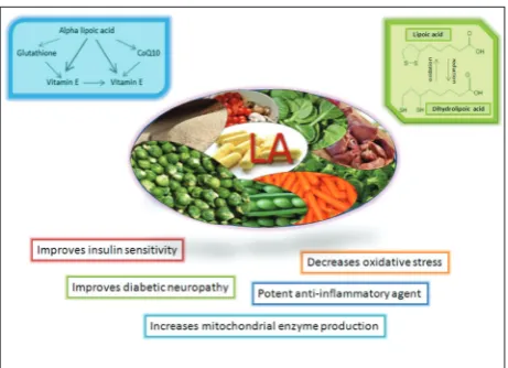
rapid gastrointestinal uptake of LA and appearance in the plasma which is followed by its rapid clearance, a large amount of evidence has revealed an unexpected range of cellular actions (Fig. 1). Antioxidant activity is exhibited by both the oxidized and reduced forms of LA and it includes the scavenging of reactive oxygen species (ROS) [8,9]. However, the antioxidant activity of the LA/DHLA couple has been shown in in vitro
conditions, and it is questionable whether LA can scavenge free radicals in vivo since LA rapidly accu-mulates and is rapidly metabolized. There is growing evidence that LA can indirectly maintain the cellu-lar antioxidant status by enhancing the synthesis of endogenous low molecular weight antioxidants, the regeneration of other antioxidants, chelation of metal ions and inhibition of redox sensitive transcription factor NF-κB [10]. The biological activity of both LA and DHLA is an advantage when compared to other antioxidants such as glutathione, whose reduced form only has an antioxidant potential. Another advantage of LA is attributed to its water and fat solubility, un-like other antioxidants that are either lipophobic or lipophilic, which means that LA can elicit antioxidant actions in both cytosol and cell membrane compart-ments. In view of its potent antioxidant activity, LA has been proposed as a potential therapeutic agent in the treatment of pathologies caused by an imbalance in redox homeostasis and ensuing oxidative stress, as
occurs in diabetes and its complications that affect different organs, including the liver (Fig. 1). Oxidative stress is also a contributing factor in liver pathologies provoked by exogenous insults (drug-induced toxic-ity), and LA could be viewed as a therapeutic agent in these conditions as well [11].
LA in diabetes
Diabetes is a metabolic disorder resulting from defec-tive insulin synthesis due to β-cell destruction (diabetes type 1), and/or responses of target tissues to insulin or insulin resistance (diabetes type 2), which cause increased glucose concentration in the circulation or hyperglycemia, the clinical hallmark of diabetes. Dif-ferent studies have provided evidence that LA stimu-lates glucose uptake by cardiac tissue in control and diabetic rats [12]. LA has been found to increase glu-cose uptake in cultured adipose and muscle cells by affecting elements of the insulin signaling pathway. LA augments tyrosine phosphorylation and the activities of the molecular components involved in insulin sig-naling, including the insulin receptor (IR), insulin re-ceptor substrate (IRS)-1, phosphatidylinositol 3-kinase (PI3K), Akt1 and p38 [13]. Specifically, stress-activated kinases such as c-Jun NH2-terminal kinase (JNK) and the inhibitor of B kinase-(IKK) interfere with normal insulin signaling by phosphorylation of the serine in IRS-1, reducing its interaction with the downstream effector PI3K. Thereby these kinases play an important
role in insulin resistance progression. It was revealed that LA inhibited the JNK pathway and IRS-1 serine phosphorylation, and improved insulin sensitivity [14]. The underlying mechanism by which LA improves in-sulin signaling could be at least in part attributed to LA-mediated induction of heat shock proteins (HSPs) that have the potential to inhibit JNK and IKK [15]. Clinical studies showed that LA increased insulin-stimulated whole-body glucose disposal in diabetic patients [16].
LA has potential applications in various aspects of diabetes pathophysiology, ranging from effects on insulin-producing pancreatic β-cells to long-term diabetic complications (Fig. 2). It is assumed that the effects of LA on β-cells are dose-dependent, meaning that at higher concentrations LA exerts detrimental effect, whereas at lower and clinically approved con-centrations it produces beneficial and cytoprotective effects on β-cells in diabetes [10]. Another potential beneficial effect of LA in diabetes is based on its abil-ity to inhibit protein glycation, which is assumed to be an important factor in the development of diabetic complications. This effect of LA is not based on its antioxidant potential but rather on the non-covalent hydrophobic interaction of LA with target proteins, which blocks the protein glycation site and prevents its glycation [10].
A direct link between oxidative stress in diabetes and pathogenic events that lead to diabetic complica-tions has been established. Hyperglycemia promotes increased production of ROS via different pathways
(nonenzymatic, enzymatic and mitochondrial), which together with impaired antioxidant defenses results in increased oxidative stress. The persisting imbal-ance in redox homeostasis in diabetes activates the expression of inflammation related genes via stress signaling pathways, stimulating the establishment of an inflammatory state. The oxidative stress-activated proinflammatory pathways are a complex pathogenic mechanism that promotes a variety of diabetic com-plications in different tissues and organs, including the liver. According to clinical evidence, the strong antioxidant effects of LA have been shown to be par-ticularly useful in treating diabetic neuropathy [4]. A daily oral dose of 600 mg provides an optimum risk-to-benefit concentration in human diabetics [5]. Hence, the antioxidant potential of LA self-rec-ommends the use of this compound in therapeutic approaches aimed at attenuating the development of diabetes associated complications.
LA and diabetes-related liver pathologies
Diabetes is one of the most common causes of liver disease, which is an important contributor to in-creased mortality in diabetic patients [17]. Fatty liver, insulin resistance and obesity are endogenous factors that provoke liver dysfunction in diabetes [18]. Fatty liver belongs to nonalcoholic fatty liver disease (NAFLD), which represents a spectrum of hepatic disorders, starting from steatosis characterized by excess fat accumulation within hepatocytes that can progress to steatohepatitis when accompanied by in-flammation (hepatic fibrosis), and further to cirrhosis and ultimately liver failure. The prevalence of NAFLD in obese patients with diabetes type 2 is greater than 70%. Decreased insulin-dependent suppression of lipolysis in adipose tissue results in elevated levels of circulating free fatty acids (FFA), which accumulate in the liver where synthesis of triglycerides occurs. Impaired hepatic fatty acid oxidation and very low-density lipoprotein secretion, as well as increased glucose concentrations in diabetes provide additional contributing factors to triglyceride synthesis that leads to hepatic fat accumulation. According to literature data, the use of supplements comprised of different antioxidant compounds including LA has been ap-proved for treatment of patients suffering from fatty liver and nonalcoholic steatohepatitis in Mexico [19].
Most of the pathological changes in liver mor-phology and function observed in diabetes are the result of oxidative stress-mediated injury (Fig. 3). The prooxidant environment established by free radical formation in diabetes contributes to oxida-tive stress development. The sources of free radicals in diabetes originate from nonenzymatic pathways (the production of hydroxyl radicals (OH•) via glu-cose autooxidation, formation of advanced glycation end products (AGE), over stimulation of the polyol pathway), enzymatic pathways (involving nitric oxide synthase, NAD(P)H oxidase and xanthine oxidase), and mitochondrial pathways with the mitochondrial respiratory chain as the main nonenzymatic source of ROS [10]. It is assumed that mitochondrial produc-tion of the superoxide anion radical (O•2−) provoked
by hyperglycemia is the main trigger of events leading to oxidative stress in diabetes [20,21].
The most important enzymatic antioxidants that cope with free radicals involve superoxide dismutase (SOD) that catalyzes the dismutation of the super-oxide radical to form hydrogen persuper-oxide (H2O2) and oxygen, catalase (CAT) that converts H2O2 to water and oxygen, and glutathione peroxidase (GPx) that reduces H2O2 using reduced glutathione (GSH) to form oxidized glutathione (GSSG) and water. Of the nonenzymatic cellular antioxidants, the low molecu-lar weight GSH molecule is very important. It plays a crucial role in maintaining protein thiols in a reduced form [22,23]. The impairment of the antioxidant de-fense system has been demonstrated in diabetes. It is an additional risk factor that contributes to oxidative
stress-related liver injury (Fig. 3). Therefore, aside from the search for protective antioxidant effects that inter-fere with ROS overproduction, attempts at improving the intrinsic antioxidant defense system that preserves redox homeostasis is also an important approach to preventing oxidative stress-related pathologies. LA may indirectly affect the cellular antioxidant response through increased uptake or synthesis of endogenous low molecular weight antioxidants. LA increases intra-cellular GSH through improved cystine uptake from the plasma, followed by its reduction by DHLA to cysteine, which is the substrate for GSH synthesis [5]. It was shown in different cultured cells that DHLA in-creases GSH synthesis by reducing cystine to cysteine [24]. In addition, LA induces the de novo synthesis of GSH at the transcriptional level by directly modulating cellular signaling pathways [25]. It was shown that the administration of LA promoted the restoration of the GSH:GSSG ratio increased the protein thiol content in the liver of streptozotocin (STZ)-induced diabetic rats. The antioxidant and hepatoprotective effects of LA in STZ-induced diabetic rats also include improve-ment of the activities of the antioxidant enzymes SOD and CAT [26,27]. LA affects CAT and CuZnSOD ex-pression in the liver at the transcriptional level. The post-translational mechanism including decreased O-GlcNAcylation of CAT and SOD, upstream kinases and transcriptional factors involved in the regulation of enzyme expression is also involved in the LA-mediated upregulation of antioxidant enzyme expression [26]. This antioxidant effect was followed by hypoglycemic activity of LA, resulting in a lower level of DNA dam-age and improved activities of the indicators of hepa-tocellular injury, alanine aminotransferase (ALT) and aspartate aminotransferase (AST), suggesting that LA exerted hepatoprotective effects in diabetes [26].
An important role in the transcriptional regulation of the antioxidant defense system is ascribed to tran-scription factor-nuclear factor erythroid 2-related fac-tor 2 (Nrf2). Nrf2 is located in the cytoplasm where it associates with cytoplasmic repressor Kelch-like ECH-associated protein 1 (Keap1), which maintains Nrf2 in its inactive form. Different activators and inducers of Nrf2 are capable of releasing Nrf2 into the nucleus, where it transactivates detoxifying and antioxidant enzymes. LA is one of the inducers of Nrf2-mediated antioxidant gene expression and as such it can increase GSH synthesis [28]. It has been reported that Nrf2 can
play a significant role in the attenuation of oxidative stress through the suppression of proinflammatory signaling pathways [29]. NF-κB is a central media-tor of inflammamedia-tory processes whose activation in hyperglycemia contributes to diabetes pathology and its associated complications [30]. NF-κB is located in the cytoplasm in an inactive form in a complex with a family of NF-κB inhibitor (IκB) proteins. Upon stimu-lation, IKK α and β are activated, which results in the phosphorylation of IκB and its proteasomal degrada-tion that release NF-κB, allowing it to translocate into the nucleus where it induces gene expression [31]. The increased activation of NF-κB in hyperglycemia and resulting transactivation of key target genes involved in inflammatory processes result in systemic and local deleterious effects that contribute to the development and progression of diabetic complications. NF-κB and its target genes, such as proinflammatory cytokines, TNF, IL-1 and IL-6, are crucial factors in the develop-ment of insulin resistance, which is an important com-ponent in the etiology of type 2 diabetes. Activation of NF-κB and chronic inflammation in the liver mimics the insulin resistance induced by obesity. Consequent-ly, inhibition of NF-κB activity in the liver decreases the expression of NF-κB target genes that attenuates type 2 diabetes. Inhibition of cytokine-induced NF-κB activation was shown to protect pancreatic β-cells from cytokine-induced apoptosis in an experimental model of STZ-induced diabetes [30]. Thereby, inhibi-tion of NF-κB activainhibi-tion could potentially be an effec-tive strategy and important aspect in diabetes treat-ment through β-cell protection and amelioration of the diabetes phenotype. It has been shown that LA is capable of inhibiting IKK α and β, and consequently IκB degradation, which results in the inhibition of NF-κB-mediated gene expression, suggesting that LA prevents NF-κB activation in a mechanism that does not involve its antioxidant potential [32].
In addition to the disturbance of the antioxidant system, impaired expression of HSPs also appears to play an important role in the pathophysiology of dia-betes. It has been reported that formation of the LA disulfide can play a significant role in the activation of the heat-shock response in diabetes [9,33,34]. Under diabetic conditions, LA application increased the ex-pression of HSP90 and HSP72 that have an important place in the cell’s machinery for protein folding and
stabilization [35]. HSP60 functions as a mitochondrial chaperone responsible for the transport and refolding of proteins from the cytoplasm into the mitochondrial matrix. Under oxidative stress, among the reactive lipid peroxidation products, 4-hydroxy-2-nonenals (4-HNE) is a biomarker that modulates a number of signaling processes through its ability to form cova-lent adducts in proteins, nucleic acids and membrane lipids. Increased oxidative stress in diabetes impairs hepatic HSP and induces 4-HNE production. It was observed that LA increased HSP60 and decreased 4-HNE in the liver [33].
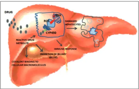
Protective effects of LA in drug-induced liver injuries
Chloroquine is commonly used as an antimalarial and antirheumatoid agent. Compared to silymarin, a reference hepatoprotective drug, orally administered LA against chloroquine-induced hepatotoxicity in Wistar rats showed significantly improved levels of plasma antioxidants, GSH, vitamin C and vitamin E, as well as decreased serum levels of AST, ALT, alka-line phosphatase, bilirubin, lipids and plasma thio-barbituric acid-reactive substances and hydroperox-ides [44]. Bromobenzene, which is primarily used as an additive to motor oil, is formed during pesticide manufacturing, chlorination of drinking water and in the rubber industry, and is resistant to biodegra-dation after outflow to the environment. It has been documented that bromobenzene hepatotoxicity can lead to hepatic necrosis. Oral administration of LA in bromobenzene-induced toxicity to albino rats signifi-cantly increased the hepatic GSH content, normalized hepatic lipid peroxidation and NO production, and preserved hepatocyte architecture [45]. Methotrexate is an effective cytotoxic drug and has been used in treatment of malignancies and inflammatory diseases, but long-term methotrexate use can cause hepatic st-eatosis, fibrosis and cirrhosis. LA treatment of Wistar albino rats reversed liver GSH levels, malondialde-hyde and (Na+/K+)-ATPase activities as well as the histopathological alterations induced by methotrex-ate [46]. The beneficial effects of LA against CCl4 and thioacetamide-induced hepatotoxicity have also been documented [47-49].
A beneficial outcome of LA supplementation was also observed in other types of liver injury, such as increased alcohol intake or intake of environmental contaminants. Additional causes of acute liver failure are neoplastic infiltration, heatstroke, mushroom in-gestion, mycotoxins, metabolic diseases such as Wil-son’s disease and viral infections (hepatitis A, B and E) [50]. Alcoholic liver disease is related to changes in the liver, including steatosis, fibrosis and cirrhosis with increased risk for the development of hepatocellular carcinoma [51]. Through cellular reduction to DHLA and the ability to normalize the NADH/NAD+ ratio,
and increase the concentration of GSH and (Na+/K+
)-ATPase activity, LA provides a valuable effect in alco-hol intoxication. Aflatoxin B1 is the most biologically active form of aflatoxins, very dangerous mycotoxins produced mainly by Aspergillus flavus and Aspergil-lus parasiticus with high toxicity in animals and hu-mans. Administration of LA prevented liver damage in broilers induced by a chronic low dose of aflatoxin B1, improved liver histopathological parameters and liver glutamic oxaloacetic transaminase and glutamic pyruvic transaminase activities [52]. Administration of lipopolysaccharide from Gram-negative pathogens contributes to the development of liver dysfunction and septic hepatic failure. Administration of LA to Wistar rats subjected to sepsis prevented the increase in pro- and antiinflammatory cytokines (IL-1β, IL-6, TNF-α, IL-10). In addition, LA treatment decreased liver antioxidant enzymes (GPx, SOD, xanthine dehy-drogenase and xanthine oxidase), lipid peroxidation and total serum NO levels [53,54].
CONCLUSION
Considering the strong antioxidant potential of LA, its use as a therapeutic agent in treatment of diseas-es whose development and progrdiseas-ession are closely linked with disturbed redox homeostasis is promis-ing. The main hepatoprotective effects of LA cover the activities that result in decreased oxidative stress, inflammation, DNA damage and fibrotic processes. Despite efforts made in unraveling the importance of antioxidant action, including the effects of LA against oxidative-stress-related diseases such as diabetes and liver diseases, as well as promising results obtained in experimental studies and clinical trials, antioxidative therapy still has to be developed. Many antioxidants
are highly effective in animal models of hepatic disor-ders, but in humans their beneficial influence in treat-ing the same liver diseases is not effective. Therefore, detailed translational research is required for the es-tablishment of antioxidant therapy in clinical practice for treating hepatic disorders triggered in diabetes as well as by drug inducers.
Acknowledgments: This work was supported by the Ministry of Education, Science and Technological Development of the Re-public of Serbia, Grant No. 173020.
Author contributions: All authors have contributed to the writing and revision of the manuscript.
Conflict of interest disclosure: The authors declare that there are no conflicts of interest relevant to this study.
REFERENCES
1. Reed LJ, DeBusk BG, Gunsalus IC, Hornberger CS Jr. Crys-talline alpha-lipoic acid; a catalytic agent associated with pyruvate dehydrogenase. Science. 1951;114:93-4.
2. Ross SM. Clinical applications of lipoic acid in type II diabe-tes mellitus. Holist Nurs Pract. 2006;20:305-6.
3. Gomes MB, Negrato CA. Alpha-lipoic acid as a pleiotropic compound with potential therapeutic use in diabetes and other chronic diseases. Diabetol Metab Syndr. 2014;6:80. 4. Singh U, Jialal I. Alpha-lipoic acid supplementation and
dia-betes. Nutr Rev. 2008;66:646-57.
5. Shay KP, Moreau RF, Smith EJ, Smith AR, Hagen TM. Alpha-lipoic acid as a dietary supplement: Molecular mecha-nisms and therapeutic potential. Biochim Biophys Acta 2009;1790(10):1149-60.
6. Jones W, Li X, Qu ZC, Perriott L, Whitesell RR, May JM. Uptake, recycling, and antioxidant actions of alpha-lipoic acid in endothelial cells. Free Radic Biol Med. 2002;33:83-93. 7. Moini H, Packer L, Saris NE. Antioxidant and prooxidant
activities of alpha-lipoic acid and dihydrolipoic acid. Toxicol Appl Pharmacol. 2002;182(1):84-90.
8. Suzuki YJ, Tsuchiya M, Packer L. Thioctic acid and dihydro-lipoic acid are novel antioxidants which interact with reactive oxygen species. Free Radic Res Commun. 1991;15:255-63. 9. Mihailović M, Arambašić J, Uskoković A, Dinić S, Grdović
N, Marković J, Poznanović G, Vidaković M. Alpha-lipoic acid preserves the structural and functional integrity of red blood cells by adjusting the redox disturbance and decreas-ing O-GlcNAc modifications of antioxidant enzymes and heat shock proteins in diabetic rats. Eur J Nutr. 2012;51(8):975-986.
10. Golbidi S, Badran M, Laher I. Diabetes and alpha lipoic Acid. Front Pharmacol. 2011;2:69.
11. Bustamante J, Lodge JK, Marcocci L, Tritschler HJ, Packer L, Rihn BH. Alpha-lipoic acid in liver metabolism and disease. Free Radic Biol Med. 1998;24(6):1023-39.
12. Singh HPP, Bowman RH. Effect of D,L-alpha lipoic acid on the citrate concentration and phosphofructokinase activity of perfused hearts from normal and diabetic rats. Biochem Biophys Res Commun. 1970;41:555-61.
13. Konrad D, Somwar R, Sweeney G, Yaworsky K, Hayashi M, Ramlal T, Klip A. The antihyperglycemic drug alpha-lipoic acid stimulates glucose uptake via both GLUT4 transloca-tion and GLUT4 activatransloca-tion: potential role of p38 mitogen-activated protein kinase in GLUT4 activation. Diabetes. 2001;50(6):1464-71.
14. Packer L, Cadenas E. Lipoic acid: energy metabolism and redox regulation of transcription and cell signaling. J Clin Biochem Nutr. 2011;48(1):26-32.
15. Gupte AA, Bomhoff GL, Morris JK, Gorres BK, Geiger PC. Lipoic acid increases heat shock protein expression and inhibits stress kinase activation to improve insulin signal-ing in skeletal muscle from high-fat-fed rats.J Appl Physiol. 2009;106(4):1425-34.
16. Jacob S, Henriksen EJ, Schiemann AL, Simon I, Clancy DE, Tritschler HJ, Jung WI, Augustin HJ, Dietze GJ. Enhance-ment of glucose disposal in patients with type 2 diabetes by alpha-lipoic acid. Arzneimittelforschung. 1995;45(8):872-4. 17. Tolman KG, Fonseca V, Dalpiaz A, Tan MH. Spectrum of liver disease in type 2 diabetes and management of patients with diabetes and liver disease. Diabetes Care 2007;30(3):734-43. 18. Wang K. Molecular mechanisms of hepatic apoptosis. Cell
Death Dis. 2014;5:e996.
19. Li S, Tan HY, Wang N, Zhang ZJ, Lao L, Wong CW, Feng Y. The Role of Oxidative Stress and Antioxidants in Liver Diseases. Int J Mol Sci. 2015;16(11):26087-124.
20. Nishikawa T, Edelstein D, Du XL, Yamagishi S, Matsumura T, Kaneda Y, Yorek MA, Beebe D, Oates PJ, Hammes HP, Giardino I, Brownlee M. Normalizing mitochondrial super-oxide production blocks three pathways of hyperglycemic damage. Nature. 2000;404(6779):787-90.
21. Brownlee M. (2001). Biochemistry and molecular cell biol-ogy of diabetic complications. Nature. 2001;414(6865):813-20. 22. Dickinson DA, Forman HJ. Cellular glutathione and thiols
metabolism. Biochem Pharmacol. 2002a;64(5-6):1019-26. 23. Dickinson DA, Forman HJ. Glutathione in defense and
signaling: lessons from a small thiol. Ann NY Acad Sci. 2002b;973:488-504.
24. Han D, Handelman G, Marcocci L, Sen CK, Roy S, Kobuchi H, Tritschler HJ, Flohé L, Packer L. Lipoic acid increases de novo synthesis of cellular glutathione by improving cystine utilization. Biofactors. 1997;6(3):321-38.
25. Suh JH, Shenvi SV, Dixon BM, Liu H, Jaiswal AK, Liu RM, Hagen TM. Decline in transcriptional activity of Nrf2 causes age-related loss of glutathione synthesis, which is reversible with lipoic acid. Proc Natl Acad Sci USA. 2004;101(10):3381-6.
27. Sadi G, Yılmaz Ö, Güray T. Effect of vitamin C and lipoic acid on streptozotocin-induced diabetes gene expression: mRNA and protein expressions of Cu-Zn SOD and catalase. Mol Cell Biochem. 2008;309(1-2):109-16.
28. Shay KP, Moreau RF, Smith EJ, Hagen TM. Is alpha-lipoic acid a scavenger of reactive oxygen species in vivo? Evi-dence for its initiation of stress signaling pathways that promote endogenous antioxidant capacity. IUBMB Life. 2008;60(6):362-7.
29. Li W, Khor TO, Xu C, Shen G, Jeong WS, Yu S, Kong AN. Activation of Nrf2-antioxidant signaling attenuates NF-κB-inflammatory response and elicits apoptosis. Biochem Phar-macol. 2008;76(11):1485-9.
30. Patel S, Santani D. Role of NF-B in the pathogenesis of dia-betes and its associated complications. Pharmacol. Rep. 2009;61(4):595-603.
31. Barnes PJ, Karin M. Nuclear factor-kappa B: a pivotal tran-scription factor in chronic inflammatory diseases. N Engl J Med. 1997;336:1066-71.
32. Ying Z, Kampfrath T, Sun Q, Parthasarathy S, Rajagopa-lan S. Evidence that α-lipoic acid inhibits NFκ-B activa-tion independent of its antioxidant funcactiva-tion. Inflamm Res. 2011;60:219-25.
33. Oksala NK, Laaksonen DE, Lappalainen J, Khanna S, Nakao C, Hanninen O, Sen CK, Atalay M. Heat shock protein 60 response to exercise in diabetes: effects of alpha-lipoic acid supplementation. J Diabetes Complicat. 2006;20:257-261. 34. Arambašić J, Mihailović M, Uskoković A, Dinić S, Grdović
N, Marković J, Poznanović G, Bajec Đ, Vidaković M. Alpha-lipoic acid upregulates antioxidant enzyme gene expression and enzymatic activity in diabetic rat kidneys through an O-GlcNAc-dependent mechanism. Eur J Nutr. 2013;52(5):1461-73.
35. Oksala NK, Lappalainen J, Laaksonen DE, Khanna S, Kaarniranta K, Sen CK, Atalay M. Alpha-lipoic Acid modulates heat shock factor-1 expression in streptozoto-cin-induced diabetic rat kidney. Antioxid Redox Signal. 2007;9:497-506.
36. Bernal W, Wendon J. Acute Liver Failure. N Engl J Med. 2013;369:2525-34.
37. Pandit A, Sachdeva T, Bafna P. Drug-Induced Hepatotoxic-ity: A Review. J App Pharm Sci. 2012;02(05):233-43. 38. Russmann S, Kullak-Ublick GA, Grattagliano I. Current
concepts of mechanism in drug-induced hepatotoxicity. Curr Med Chem. 2009;16(23):3041-53.
39. Yuan L, Kaplowitz N. Mechanisms of Drug Induced Liver Injury. Clin Liver Dis. 2013;17(4):507-18.
40. Andringa KK, Bajt ML, Jaeschke H, Bailey SM. Mitochon-drial Protein Thiol Modifications in Acetaminophen Hepa-totoxicity: effect on HMG-CoA Synthase. Toxicol Lett. 2008; 177(3):188-97.
41. Morgan RE, Trauner M, van Staden CJ, Lee PH, Ramachan-dran B, Eschenberg M, Afshari CA, Qualls CW Jr,
Lightfoot-Dunn R, Hamadeh HK. Interference with bile salt export pump function is a susceptibility factor for human liver injury in drug development. Toxicol Sci. 2010;118(2):485-500.
42. Copple IM, Goldring CE, Jenkins RE, Chia AJ, Randle LE, Hayes JD, Kitteringham NR, Park BK. The hepatotoxic metabolite of acetaminophen directly activates the Keap1-Nrf2 cell defense system. Hepatology. 2008;48(4):1292-301. 43. Niture SK, Kaspar JW, Shen J, Jaiswal AK. Nrf2 signaling and
cell survival. Toxicol Appl Pharmacol. 2010;244(1):37-42. 44. Pari L, Murugavel P. Protective effect of alpha-lipoic acid
against chloroquine-induced hepatotoxicity in rats. J Appl Toxicol. 2004;24(1):21-6.
45. Mansour DF, El-Denshary ES, Bahgat AK, Nada SA. Hepa-toprotective Effect of Alpha Lipoic Acid Against Bromo-benzene-Induced Liver Damage in Rats. Med J Cairo Univ. 2009;77(4):23-30.
46. Çakır T, Baştürk A, Polat C, Aslaner A, Durgut H, Şehirli AÖ, Gül M, Öğünç AV, Gül S, Sabuncuoglu MZ, Oruç MT. Does alfa lipoic acid prevent liver from metho-trexate induced oxidative injury in rats? Acta Cir Bras. 2015;30(4):247-52.
47. Slepneva IA, Sergeeva SV, Khramtsov VV. Reversible inhibi-tion of NADPH-cytochrome P450 reductase by alpha-lipoic acid. Biochem Biophys Res Commun. 1995;214(3):1246-53. 48. Ali SO, Darwish HA, Ismail NA. Modulatory effects of cur-cumin, silybin-phytosome and alpha-R-lipoic acid against thioacetamide-induced liver cirrhosis in rats. Chem Biol Interact. 2014;216:26-33.
49. Uskoković A, Dinić S, Arambašić Jovanović J, Poznanović G, Vidaković M, Mihailović M. Liver Diseases: Epigenetic Mecha-nisms, Oxidative stress and Use of Alpha-Lipoic Acid. In: Patel V, Preedy V, editors. The Handbook of Nutrition, Diet and Epigenetics. Springer International Publishing; 2017. p. 1-21. 50. Ichai P, Samuel D. Etiology and prognosis of fulminant
hepa-titis in adults. Liver Transpl. 2008;14:S67-S79.
51. Manzo-Avalos S, Saavedra-Molina A. Cellular and Mito-chondrial Effects of Alcohol Consumption. Int J Environ Res Public Health. 2010;7(12):4281-304.
52. Li Y, Ma QG, Zhao LH, Guo YQ, Duan GX, Zhang JY, Ji C. Protective Efficacy of Alpha-lipoic Acid against AflatoxinB1-induced Oxidative Damage in the Liver. Asian-Australas J Anim Sci. 2014; 27(6):907-15.
53. Heibashy MIA, Mazen GMA, Shahin MI. The Cura-tive Effects of some Antioxidants on Endotoxin Induced with Lipopolysaccharide in the Liver of Rats. J Am Sci. 2013;9(12):529-38.