681
© 2018 by the Serbian Biological Society How to cite this article: Kožik B, Kokanov N, Knežević-Ušaj S, Nikolić I, Davidović R, Jovanović-Ćupić S, Krajnović M. Methylation status of p16 and p14 genes in locally advanced rectal cancer: Potential clinical implication. Arch Biol Sci. 2018;70(4):681-90.
Methylation status of
p16
and
p14
genes in locally advanced rectal cancer: potential
clinical implication
Bojana Kožik1, Nikola Kokanov1, Slavica Knežević-Ušaj2, Ivan Nikolić2, Radoslav Davidović1, Snežana
Jovanović Ćupić1 and Milena Krajnović1,*
1Vinča Institute of Nuclear Sciences, University of Belgrade, 11001 Belgrade, Serbia
2Faculty of Medicine Novi Sad, Oncology Institute of Vojvodina, University of Novi Sad, 21204 Sremska Kamenica, Serbia
*Corresponding author: [email protected]
Received: March 16, 2018; Revised: June 13, 2018; Accepted: June 13, 2018; Published online: June 19, 2018
Abstract: Methylation of p16 and p14 genes is a common event in colorectal cancers; however, their exact role in the pre-diction of patients’ outcome is unclear. We conducted this retrospective study to evaluate their potential predictive and/or prognostic roles. Methylation-specific PCR was used to examine the methylation status of p16 and p14 in pretherapeutic and preoperative biopsy specimens of 60 patients with locally advanced rectal cancer. The methylation status of the examined genes did not affect the response to preoperative chemoradiotherapy (CRT), recurrence rate and overall survival. However, patients with a simultaneous presence of either p16 or p14 methylation and high vascular endothelial growth factor(VEGF) expression showed a significantly worse response to CRT (p=0.005 and p=0.038, respectively). Moreover, patients with both
p16 methylation and high VEGF expression had significantly shorter overall survival (p=0.010), while no such association was found in patients with p14 methylation and high VEGF expression. On the other hand, a subgroup of patients with p16
methylation and low VEGF and high epidermal growth factor receptor (EGFR) expression showed a significantly better response to CRT (p=0.024). The obtained results point to the importance of p16 and p14 methylation analyses in combina-tion with VEGF and EGFR expression, aimed at better predicting treatment response and patient outcome.
Key words: rectal cancer; p16;p14; DNA methylation; biomarkers; VEGF; EGFR
INTRODUCTION
Colon and rectal cancers are often considered to-gether, although these two clinical entities have many differences in etiology and treatment strategies [1]. Preoperative (neoadjuvant) CRT followed by radical surgery has become the standard treatment for pa-tients with locally advanced rectal cancer [2]. Despite reduced local recurrence rates, the responses of indi-vidual tumors to this multimodal treatment are vari-able and range from complete regression to complete resistance [3]. Better pretherapeutic patient stratifi-cation and accurate response prediction requires the development of reliable molecular biomarkers.
The predictive values of potential biomarkers have been tested, including EGFR, the marker for cellular proliferation Ki-67, and one of the key mediators of apoptosis, B-cell lymphoma 2 (Bcl-2) protein [4,5].
VEGF, a primary angiogenic factor, has been fre-quently examined since it plays a pivotal role in tu-mor angiogenesis and strongly contributes to tutu-mor malignancy [6]. However, for most marker studies, the results are conflicting and remain inconclusive.
The most investigated epigenetic modification in colorectal cancer is aberrant DNA methylation of 5’-Cytosine-phosphate-Guanine-3’ (CpG) islands within promoter regions, which is associated with gene silencing [7,8]. Specific methylation patterns of a number of tumor suppressor genes involved in color-ectal carcinogenesis have been studied with the aim of defining novel epigenetic biomarkers that could be used in clinical practice [9,10]. The INK4a/alternate reading frame (ARF) locus encodes two tumor sup-pressor proteins, P16INK4a and P14ARF, which act as key
negative regulators of the cell cycle [11]. P16INK4a is a
phosphorylation and thereby induces G1 phase arrest, while P14ARF indirectly facilitates p53-mediated
cell-cy-cle arrest and apoptosis by interaction with the mouse double minute 2 homolog (MDM2) protein [12].
In addition, there is evidence that p16 and p14 genes may have an important role in regulation of an-giogenesis through downregulation of VEGF expres-sion. This regulation takes place at the transcriptional level through inhibition of hypoxia-inducible factor 1-alpha (HIF-1α), which acts as a VEGF transcription factor [13,14]. We assumed that the methylation status of p16 and p14 genes could be related to the different levels of VEGF expression, which could have some clinical relevance.
Inactivation of p16 and p14 genes by promoter hypermethylation has been frequently reported as an early event in colorectal neoplasia [15,16]. However, correlation between the methylation status of p16 and p14 genes and clinical outcomes in patients with lo-cally advanced rectal cancers is not fully established. Hence, in the present study, the promoter methylation of these two tumor suppressor genes was examined in order to identify the relationship between their meth-ylation status and clinicopathological and immuno-histochemical parameters, and to investigate whether the methylation status of p16 and p14 genes affects the response to preoperative CRT and overall survival in 60 patients with locally advanced rectal cancer.
MATERIALS AND METHODS
Patients and tumor samples
The study was approved by the Ethical Committee of the Oncology Institute of Vojvodina, Sremska Ka-menica, and all procedures were carried out with the prior informed consent of the patients. Our retrospec-tive study included 60 patients (38 male, 22 female; median age 65 years, range 49-82 years) with locally advanced rectal cancer (clinical stage cT3b,cN0-2 with positive circumferential margin (CRM) and cT4N0-2), who were diagnosed and treated with preoperative CRT at the Oncology Institute of Vojvodina, Sremska Kamenica, Serbia, in the period 2006-2010, according to the National guidelines of the Ministry of Health of Serbia for Diagnosis, Therapy and Management
of colorectal cancer. Before the administration of pr-eoperative CRT, all patients underwent tumor biopsy for diagnostic purpose. The neoadjuvant treatment consisted of total irradiation with a dose of 50.4 Gy, that was divided into 28 fractions of 1.8 Gy, with con-comitant application of 5-fluorouracil (425 mg/m2) and leucovorin (25 mg/m2). Total mesorectal excision (TME) radical surgery was performed 8-10 weeks af-ter the end of the combined treatment. The location of the tumor was determined by MRI in the low (≤7 cm from the anal verge), high (>7 cm from the anal verge) or mid rectum (< and > than 7 cm the from anal verge). Pathological grading of primary tumor regression in posttreatment specimens was performed semiquantitatively by determining the amount of re-sidual tumor cells compared with the amount of fibro-sis. The response to neoadjuvant CRT was classified as positive when complete or partial remission (CR/PR) was detected, or as negative in the case of the presence of stable or progressive disease (SD/PD).
DNA extraction and methylation analysis of the p16 and p14 genes
The methylation status of p16 and p14 was evaluated on diagnostic tumor biopsies obtained as formalin-fixed, paraffin-embedded tumor tissues. Briefly, ge-nomic DNA was isolated from deparaffinized tumor specimens using standard proteinase K, phenol/ chloroform/isoamyl alcohol extraction and ethanol precipitation [17]. DNA methylation patterns in the promoter CpG islands of the p16 and p14 genes were determined by methylation-specific PCR (MSP). Ini-tially, sodium bisulfite conversion of genomic DNA (100-500 ng) was performed using an EZ DNA Meth-ylation-LightningTM kit (Zymo Research, Orange, CA,
cy-cler for 40 cycles (45 s at 95°C, 45 s at the annealing temperature being specific for each primer set, and 60 s at 72°, followed by final extension for 4 min at 72°C). Primer sequences and annealing temperatures used for each reaction are listed in Table 1S [18,19]. Promoter regions of both genes, including the parts used for methylation analysis are presented in Fig. 1S. DNA from peripheral blood lymphocytes from a healthy donor was used as a negative control for the methylated alleles. The same leukocyte DNA was methylated in vitro with excess SssI methyltransferase (New England Biolabs) to generate completely meth-ylated DNA at all CpG sites and used as a positive control for all genes. PCR products were separated by electrophoresis on 6% acrylamide gels, stained with silver nitrate and sodium carbonate.
Immunohistochemical analysis of VEGF, Ki-67, Bcl-2 and EGFR proteins
Immunohistochemical analysis of VEGF, Ki-67, Bcl-2 and EGFR expression was performed in the diagnostic biopsy specimens, and determined at the Oncology Institute of Vojvodina, Sremska Kamenica, as previ-ously described [20]. According to the percentage of tumor cells in the given specimens with a positive immunohistochemical (IH) reaction, tumor samples were considered to have VEGF expression as follows: absent VEGF expression (0-1% of tumor cells with a positive IH reaction), weak (1.1-10% of tumor cells with a positive IH reaction), moderate (10.1-50% of tumor cells with a positive IH reaction), high VEGF expression (50.1-100% of tumor cells with a positive IH reaction); the same criteria were applied to Ki-67 and Bcl-2 expression. In the case of EGFR, the expression of EGFR protein in 0-1% of tumor cells was consid-ered as negative, in 1.1-25% of tumor cells as weak, in 25.1-50% as moderate, in 50.1-100% of cells as strong.
Statistical analysis
Contingency tables were analyzed using Pearson’s χ2-test or Fisher’s exact two-tailed test when the
ex-pected frequencies were lower than five. Continuous variables were compared using the Student’s t-test. Overall survival distributions were estimated by the Kaplan-Meier method and differences were evaluated by the Log-rank test. In all tests, a p value ≤0.05 was
considered as statistically significant. All statistical analyses were performed using the Sigma Plot 10.0 licensed statistical analysis software package.
RESULTS
p16 and p14 methylation status
Analysis of the p16 methylation status was success-fully performed in all 60 cases, while the p14 methyla-tion status was determined in 58/60 patients. Aber-rant methylation of p16 and p14 genes was present in 43.3% (26/60) and 39.6% (23/58) of the cases, respec-tively. Concurrent methylation of p16 and p14 genes was detected in 11 out of 58 (18.9%) cases, while 38 out of 60 patients (60.3%) had at least one methyl-ated gene. Representative examples of the methylation analysis are shown in Fig. 1.
VEGF, Ki-67, Bcl-2 and EGFR expression status
Analysis of the expression statuses of VEGF, Ki-67, Bcl-2 and EGFR was successfully performed in 58/60 cases. The absence of VEGF expression was observed in 37.9% (22/58) of cases, weak VEGF expression was detected in 13.8% (8/58) of cases, moderate expres-sion in 34.5% (20/58) and high VEGF expresexpres-sion in
Fig. 1. Analysis of p16 and p14 gene methylation status by MSP. The presence of a visible PCR product in lanes U indicates the presence of unmethylated p16 (151 bp) and p14 (132 bp) genes; the presence of product in lanes M indicates the presence of meth-ylated p16 (150 bp) and p14 (122 bp) genes. Samples of rectal
cancer 49 and 60 show p16 promoter hypermethylation, while
samples 56 and 40 show p14 promoter hypermethylation. NL –
normal lymphocytes as a positive control for unmethylated
al-leles, PC – in vitro methylated DNA from normal lymphocytes
13.8% (8/58). In the case of Ki-67, weak expression was observed in 25.9% (15/58) of patients, moderate in 24.1% (14/58), while high Ki-67 expression was detected in 50% (29/58). Thirty-five out of 58 samples (60.3%) had no Bcl-2 expression, weak expression was detected in 8.6% (5/58) of patients, moderate in 15.5% (9/58), while high Bcl-2 expression was detected in 15.5% (9/58) of cases. Finally, in the case of EGFR, 41.4% (24/58) of the patients had no EGFR expres-sion, weak EGFR expression was detected in 29.3% (17/58), moderate expression in 13.8% (8/58), and high EGFR expression in 15.5% (9/58) of patients. Representative examples of IH staining for all four analyzed proteins are shown in Fig. 2. In further anal-ysis, the tumor samples were considered as having high VEGF, Ki-67 or Bcl-2 expression when at least 10% of the tumor cells expressed VEGF, Ki-67 or Bcl-2 protein, respectively. Samples were considered to have high EGFR expression if >25% of the tumor cells demonstrated membranous staining of any intensity.
Correlation of the p16 and p14 methylation statuses with clinicopathological and immunohistochemical parameters
The study of correlations between the p16 and p14 methylation statuses and clinicopathological param-eters is summarized in Table 1. No association was found between promoter methylation of either the p16 or p14 gene and the clinicopathological param-eters including age, gender, tumor location (distance to anal verge) and clinical TNM (Tumor Node Metas-tasis classification of malignant tumors) stage (p>0.05 for all variables). The data representing the number of patients according to VEGF, Ki-67, Bcl-2 and EGFR expression and the gene methylation status are given in Table 2. There was no correlation between the p16 and p14 methylation statuses and any IH parameter analyzed (p>0.05 for all variables).
p16 and p14 methylation status and response to neoadjuvant CRT
Of the 60 locally advanced rectal cancer cases, there was an equal number of patients (30/60) with positive (CR/PR) and negative (SD/PD) responses to neoadju-vant CRT. In general, a better response to neoadjuneoadju-vant CRT was observed in patients with low VEGF expres-sion (p<0.001), low VEGF and high EGFR expresexpres-sion (p=0.014) and low Ki-67 expression (p=0.004), while other analyzed parameters did not affect the response to neoadjuvant CRT (results not shown). The meth-ylation statuses of p16 and p14 genes were not related to the response to neoadjuvant CRT either (p=0.434 and p=1.000, respectively (Table 1), so we further ex-amined the response to the CRT according to the si-multaneous presence of aberrant methylation of either p16 or p14 gene, and the different levels of expression of immunohistochemically determined parameters. A significantly worse response to CRT was observed in the group of patients with a simultaneous presence of p16 methylation and high VEGF expression (p16m/ high VEGF), as compared to the other three groups of patients: unmethylated p16/low VEGF, unmethylated p16/high VEGF, methylated p16/low VEGF (p=0.005). A similar association was noted for the group of pa-tients with concurrent presence of p14 methylation and high VEGF expression (p14m/high VEGF), who showed a significantly worse response to CRT than
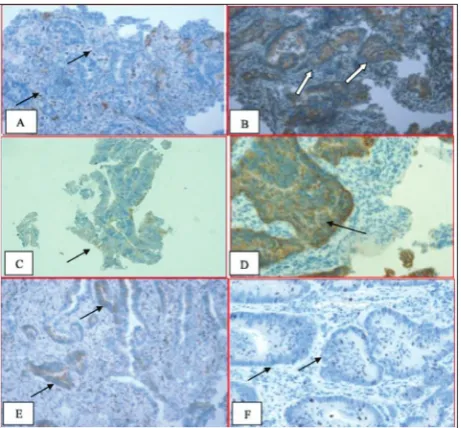
Fig. 2. Representative images for immunohistochemically
estimat-ed VEGF, EGFR, Bcl-2 and Ki-67 expression. A – Negative VEGF
the other three groups of patients according to the simultaneous examination of the p14/VEGF status (p=0.038). In addition, the tendency toward more fre-quent local recurrences and metastasis was observed among patients with the simultaneous presence of ab-errant methylation of p16 and high VEGF expression, and in the group of patients with a simultaneous pres-ence of aberrant methylation of p14 and high VEGF expression (p=0.072 and p=0.075, respectively), rather
than in the other patient groups. A better response to CRT was observed in the group of patients with si-multaneous occurrence of p16 methylation, low VEGF and high EGFR expression (p=0.024). Furthermore, the absence of local recurrences and metastasis was significantly related to cases with the simultaneous occurrence of p16 methylation and high EGFR ex-pression, as compared to the other three groups ac-cording to the simultaneous examination of the p16/
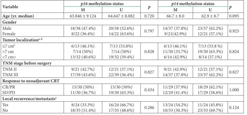
Table 1. Correlation of p16 and p14 methylation status with clinicopathological parameters.
Variable p16M methylation statusU p p14M methylation statusU p
Age (yr. median) 63.846 ± 9.124 64.647 ± 8.082 0.720 66.7 ± 8.0 62.9 ± 8.7 0.095
Gender
Male
Female 18/38 (47.4%)8/22 (36.4%) 20/38 (52.6%)14/22 (63.6%) 0.797 14/37 (37.8%)9/21(42.9%) 23/37 (62.2%)12/21 (57.1%) 0.923
Tumor localizationa, b ≤7 cmb
>7 cm <7 cm>
6/13 (46.1%) 7/14 (50%) 13/32 (40.6%)
7/13 (53.8%) 7/14 (50%)
19/32 (59.4%) 0.828
6/13 (46.1%) 11/30 (33.7%)
6/14 (42.9%)
7/13 (53.8 %) 19/30 (63.3%)
8/14 (57.1%) 0.824
TNM stage before surgery
TNM II
TNM III 17/39 (43.6%)9/21 (42.7%) 12/21 (57.1%)22/39 (56.4%) 0.827 14/37 (37.8%)9/21 (42.9%) 12/21 (57.1%)23/37 (62.2%) 0.827
Response to neoadjuvant CRT
CR/PR
SD/PD 11/30 (36.7%)15/30 (50%) 19/30 (63.3%)15/30 (50%) 0.434 11/29 (37.9%)12/29 (41.4%) 18/29 (62.1%)17/29 (58.6%) 1.000
Local recurrence/metastasisa Yes
No 18/35 (51.4%)8/24 (33.3%) 16/24 (66.7%)17/35 (48.6%) 0.286 13/24 (54.2%)10/33 (30.3%) 11/24 (45.8%)23/33 (69.7%) 0.124
M – methylated, U – unmethylated, CRT – chemoradiotherapy, CR – complete remission, PR – partial remission, SD – stable disease, PD – progressive disease. a Data are missing on one patient for given parameters. b The distance of the tumor from the anal verge.
All p values were revealed by χ2-test or Fisher’s exact two-tailed test, when expected frequencies were lower than five
Table 2. Correlation of p16 and p14 methylation status with immunohistochemical parameters.
Protein expression
levels p16M methylation statusU p p14M methylation statusU p
VEGF expressiona Low (< 10%)
High (≥ 10%) 16/30 (53.3%)10/28 (35.7%) 14/30 (46.7%)18/28 (64.3%) 0.278 11/29 (37.9%)10/27 (37%) 18/29 (62.1%)17/27 (63%) 0.836
EGFR expressiona Low (< 25%)
High (≥ 25%) 10/17 (58.8%)16/41 (39%) 7/17 (41.2%)25/41 (61%) 0.276 7/16 (43.7%)14/40 (35%) 9/16 (56.3%)26/40 (65%) 0.760
Ki67 expressiona Low (< 10%)
High (≥ 10%) 22/43 (51.2%)4/15 (26.7%) 11/15 (73.3%)21/43 (48.8%) 0.180 5/15 (33.3%)16/41 (39%) 10/15 (66.7%)25/41 (61%) 0.938
Bcl-2 expressiona Low (< 10%)
High (≥ 10%) 6/18 (33.3%)20/40 (50%) 12/18 (66.7%)20/40 (50%) 0.371 15/39 (38.5%)6/17 (35.3%) 24/39 (61.5%)11/17 (64.7%) 0.940
M – methylated, U – unmethylated, VEGF – vascular endothelial growth factor, EGFR – epidermal growth factor receptor.
a Data are missing on two patients for given parameters. All p values were revealed by χ2-test or Fisher’s exact two-tailed test, when expected
EGFR status (p=0.038). The power of the study for the observed statistically significant results was ≥0.80 at a significance level p<0.05.
Survival analysis
Follow-up data was available for 53/60 patients from our study, and the median follow-up period was 23 months (range 2-101 months). All death outcomes were cancer-related. Overall survival was significantly worse for patients who were in any of the following categories: higher VEGF expression (p=0.006), con-current presence of high VEGF and low EGFR ex-pression (p=0.007), no response to the neoadjuvant CRT (p=0.005), and occurrence of relapsed disease or distant metastasis (p<0.001). In all rectal cancer cases, we observed no significant difference in overall survival between patients with or without methylation of p16 (p=0.912, Fig. 3A) and p14 genes (p=0.911, Fig. 3B). Further, we examined the overall survival
according to the simultaneous presence of aberrant methylation of either p16 or p14 genes and differ-ent levels of expression of immunohistochemically determined parameters. The group of patients with a concurrent presence of p16 methylation and high VEGF expression showed significantly shorter overall survival when compared to the other three groups of patients (p=0.010, Fig. 3C). However, no such asso-ciation was found in the case of patients with simul-taneous p14 methylation and high VEGF expression (p=0.266, Fig. 3D). Concurrent occurrence of p16 or p14 methylation and different levels of Ki-67, Bcl-2 and EGFR expression did not affect overall survival (results not shown).
DISCUSSION
Specific gene methylation patterns can alter responses to different therapeutic agents in solid tumors, includ-ing colorectal cancer [21]. Methylation of p16 and p14 genes is a relatively frequent molecular event in the pathogenesis of colorectal cancer but its clinical rel-evance remains undetermined. Taking rectal cancer as a specific entity, only a few studies have investigated the role of p16 methylation status, while, as far as we know, our study is the first to investigate the potential predictive and prognostic value of aberrant p14 meth-ylation status solely in this type of cancer.
The reported frequency of p16 and p14 gene pro-moter hypermethylation in colorectal cancer ranges from 10% to 61% [15,22] and 28 to 50% [19,23-25], re-spectively, and our results are in agreement with this. Thirty-eight out of 60 patients (60.3%) from our study had at least one methylated gene. Thus, our findings indicate that epigenetic alterations of these two genes are common events in rectal cancer and may be im-portant to the pathogenesis of this tumor type.
We did not observe any association between the methylation statuses of p16 and p14 genes and the ex-amined clinicopathological and immunohistochemical parameters. The association with the response to CRT or patient outcomes was not observed either. Existing literature data suggest that methylation of the p16 gene is associated with a more aggressive behavior of color-ectal cancer [22,26]. Kim et al. [27] observed an asso-ciation between p16 methylation and recurrence after curative operation in rectal cancer cases specifically,
Fig. 3. Overall survival among rectal cancer patients according to the p16 or p14 methylationand VEGF expression status. A,
B – No significant difference in overall survival between patients according to the methylation status of p16 and p14 gene, respec-tively. C – The group of patients with concurrent occurrence of
p16 methylationand high VEGF expression showed significantly
shorter overall survival compared to the other three groups. D
– No significant difference in overall survival between patients
with simultaneous presence of p14 methylation and high VEGF
although preoperative CRT was not applied in these cases. According to the meta-analysis by Zhou et al. [28], no relationship was observed among aberrant p14 gene methylation and pathological features in colorec-tal cancer, except more prevalent p14 methylation in proximal colon cancers than in distal ones, while rec-tum rec-tumor location was not considered separately in the study. The situation regarding the role of p14 meth-ylation status in colorectal disease outcome is less clear. While some studies reported p14 gene methylation alone [29] or in combination with p16 [23] as a marker of worse prognosis, other reports demonstrated that p14 gene methylation in combination with aberrant methylation of another panel of genes could be related to less aggressive colorectal pathogenesis [30]. These conflicting results reveal a need for further examina-tion of the role of methylaexamina-tion of p16 and especially of p14 in rectal cancer. The fact that development of left- and right-sided colorectal cancers may involve dif-ferent molecular mechanisms should be considered in future analyses, since research focusing on the mecha-nisms behind different epigenetic profiles in colon vs rectum tumor location are rare [31].
A recent study of locally advanced rectal cancers by Kohonen-Corish et al. [32] indicated that p16 methylation itself was not associated with poor sur-vival, but the presence of both p16 methylation and Kirsten Rat Sarcoma virus (KRAS) mutation had an adverse effect on tumor recurrence and overall surviv-al. Previously, we conducted KRAS mutation analyses on the same rectal cancer samples [20]; however, we found no association between p16 or p14 methyla-tion and KRAS mutamethyla-tion status, or between the impact of these two events on therapy response and disease outcome.
In a more comprehensive analysis, we found that the simultaneous occurrence of either p16 or p14 methylation and high VEGF expression was related to a more aggressive course of the disease, which was reflected in a significantly worse response to CRT and more frequent appearance of local recurrences and distant metastasis. In colorectal cancer, VEGF is as-sociated with tumor aggressiveness and poor patient outcome [33,34], which is also demonstrated in our current study. The obtained result points to an impor-tant role of VEGF in tumor development and p16 or
p14 in tumor growth suppression. Products of p16 and p14 genes are now recognized as angiogenesis sup-pressors that achieve antiangiogenic function via the modulation of VEGF expression [13,14]. Although we did not find an association between VEGF expression and p16 or p14 methylation status, our results suggest that simultaneous VEGF overexpression and p16 and/ or p14 gene methylation may distinguish the group of rectal cancers with a more aggressive biological behavior. We speculate that this could occur through induced tumor resistance to preoperative CRT. Nev-ertheless, to clarify the direct biological relationship between p16, p14 and VEGF, additional studies of other regulatory genes included in the VEGF signal-ing pathway are necessary. It is well known that genes such as KRAS and p53 also participate in the regu-lation of VEGF expression [35,36]. However, in our recent report, a correlation between KRAS mutation status and VEGF expression was not found in locally advanced rectal cancer [20]. For future analyses, it would be useful to investigate the p53 gene status be-cause the p53 pathway seems to have a more dominant role in rectal than in colon cancer [37].
In survival analysis, we observed that the group of patients with a concurrent presence of p16 methyla-tion and high VEGF expression had a significantly shorter overall survival. Similar results were obtained in squamous cell carcinoma of the esophagus where the loss of p16 expression together with present ex-pression of VEGF protein was associated with a higher cumulative postoperative survival rate [38]. The de-termined association may be due to the more inva-sive and progresinva-sive proliferation of cancer cells with p16 alterations due to disruption of cell cycle regu-lation. Moreover, elevated VEGF expression, which was significantly related to shorter overall survival in our study, probably affects overall patient outcome indirectly through its proangiogenic effects. On the other hand, p14 methylation status in combination with VEGF expression did not influence the overall survival of patients included in our study, possibly due to the small number of preoperative biopsies analyzed and limited follow-up.
me-tastasis were significantly rare events. Moreover, in combined analysis including VEGF expression, we identified a subgroup of patients with concurrent p16 methylation, low VEGF and high EGFR expression that displayed a better response to preoperative CRT. The underlying reasons for such a finding remain unclear and our results require further elucidation. Several au-thors provided evidence that baseline EGFR expression was related to poor prognosis, poor tumor downstaging and local recurrence in rectal cancer [39-41], which was not the case in our study. Moreover, Zlobec et al. [42] found that the complete pathologic response was nearly six times more likely in EGFR-positive rectal tumors and they identified a group of VEGF-positive and EGFR-negative tumors that were highly resistant to treatment. In another study, Chakravarti et al. [43] showed that in patients with muscle-invasive bladder cancer, EGFR expression could also be a favorable prog-nostic factor since EGFR positivity was significantly associated with improved overall survival and a ten-dency toward reduced frequency of distant metasta-sis. Considering this, we assumed that increased EGFR expression could provide better sensitivity to CRT and thus indirectly allow for a better response to therapy, although a high level of EGFR itself is not good in terms of rapid proliferation of tumor cells. Several previous reports have provided evidence that VEGF and EGFR signaling pathways are interrelated [43], while Ciardiel-lo et al. [45] demonstrated that EGFR could upregulate VEGF expression. Although our results indicate that combined analysis of VEGF/EGFR expression and p16 methylation status could define a subgroup of less ag-gressive rectal cancers, further studies are needed to confirm our findings.
Our results, although limited by the small number of patients and short follow-up, suggest that methyla-tion of p16 and p14 genes, which was found as a rela-tively frequent epigenetic event among locally advanced rectal cancer patients studied, neither influences the response to CRT nor correlates with overall survival. However, after combined methylation analyses with different VEGF and EGFR expression levels in pretreat-ment and preoperative specimens, the study revealed several subgroups of patients with a more or less ag-gressive disease outcome, which could have potential predictive and/or prognostic relevance. Additional prospective studies of locally advanced rectal cancer are needed to clarify not only the clinical implications
of p16 and p14 methylation, but also the relationships between aberrant methylation of these genes and VEGF mediated angiogenesis and EGFR signaling.
Acknowledgments: This work was supported by Grant No. 173049 from the Ministry of Education, Science and Technological Development of the Republic of Serbia.
Author contributions: Bojana Kožik performed the experiments, analyzed the data and wrote the manuscript. Nikola Kokanov par-ticipated in the experiment. Slavica Knežević-Ušaj was responsible for the pathohistological and immunohistochemical analyses and assisted in the preparation of the manuscript. Ivan Nikolić was responsible for collecting the tissue samples and provided the clinicopathological data. Radoslav Davidović contributed to the design of the experiment. Snežana Jovanović Ćupić participated in the data analyses and made critical revisions of the paper. Milena Krajnović was responsible for conceiving and designing the study and critical revision of the manuscript. All authors read and ap-proved the final manuscript.
Conflict of interest disclosure: The authors declare that they have no conflict of interest.
REFERENCES
1. Schmoll HJ, Cutsem E Van, Stein A, Valentini V, Glimelius B, Haustermans K, Nordlinger B, van de Velde CJ, Bal-mana J, Regula J, Nagtegaal ID, Beets-Tan RG, Arnold D, Ciardiello F, Hoff P, Kerr D, Köhne CH, Labianca R, Price T, Scheithauer W, Sobrero A, Tabernero J, Aderka D, Bar-roso S, Bodoky G, Douillard JY, El Ghazaly H, Gallardo J, Garin A, Glynne-Jones R, Jordan K, Meshcheryakov A, Papamichail D, Pfeiffer P, Souglakos I, Turhal S, Cervantes A. ESMO Consensus Guidelines for management of patients with colon and rectal cancer. A personalized approach to clinical decision making. Ann Oncol. 2012;23:2479-516. 2. Glimelius B. Neo-adjuvant radiotherapy in rectal cancer.
World J Gastroenterol. 2013;19(46):8489-501.
3. Martin ST, Heneghan HM, Winter DC. Systematic review and meta-analysis of outcomes following pathological com-plete response to neoadjuvant chemoradiotherapy for rectal cancer. Br J Surg. 2012;99:918-28.
4. Milgrom SA, García-Aguilar J. Molecular biomarkers as pre-dictors of response to neoadjuvant chemoradiation therapy in rectal cancer. Semin Colon Rectal Surg. 2013;119-24. 5. Zeestraten EC, Kuppen PJ, van de Velde CJ, Marijnen
CA. Prediction in rectal cancer. Semin Radiat Oncol. 2012;22(2):175-83.
6. Sun W. Angiogenesis in metastatic colorectal cancer and the benefits of targeted therapy. J Hematol Oncol. 2012;5:1-9. 7. Coppedè F. Epigenetic biomarkers of colorectal cancer:
Focus on DNA methylation. Cancer Lett. 2014;342:238-47. 8. Sakai E, Nakajima A, Kaneda A. Accumulation of aberrant
9. Ng J, Yu J. Promoter Hypermethylation of Tumour Suppres-sor Genes as Potential Biomarkers in Colorectal Cancer. Int J Mol Sci. 2015;16:2472-96.
10. Draht MX, Riedl RR, Niessen H, Carvalho B, Meijer GA, Herman JG, van Engeland M, Melotte V, Smits KM. Pro-moter CpG island methylation markers in colorectal cancer: the road ahead. Epigenomics. 2012;4(2):179-94.
11. Sharpless N. INK4a/ARF: a multifunctional tumor suppres-sor locus. Mutat Res. 2005;576(1-2):22-38.
12. Sharpless N, DePinho R. The INK4A/ARF locus and its two gene products. Curr Opin Genet Dev. 1999;9:22-30. 13. Lu Y, Zhang X, Zhang J. Inhibition of Breast Tumor Cell
Growth by Ectopic Expression of p16/INK4A Via Combined Effects of Cell Cycle Arrest, Senescence and Apoptotic Induc-tion, and Angiogenesis Inhibition. J Cancer. 2012;3:333-44. 14. Kawagishi H, Nakamura H, Maruyama M, Mizutani S,
Sug-imoto K, Takagi M, SugSug-imoto M. ARF suppresses tumor angiogenesis through translational control of VEGFA mRNA. Cancer Res. 2010;70:4749-58.
15. Veganzones-de-Castro S, Rafael-Fernández S, Vidaurreta-Lázaro M, Orden V, Mediero-Valeros B, Fernández C, Mae-stro-de las Casas ML. p16 gene methylation in colorectal cancer patients with long-term follow-up. Rev Esp Enferm Dig. 2012;104:111-7.
16. Nyiraneza C, Sempoux C, Detry R, Kartheuser A, Dahan K. Hypermethylation of the 5’ CpG island of the p14ARF flanking exon 1β in human colorectal cancer displaying a restricted pattern of p53 overexpression concomitant with increased MDM2 expression. Clin Epigenetics. 2012;4:9. 17. Sambrook J, Fritsch EF, Maniatis T. Analysis and cloning of
eukariotic genomic DNA. In: Ford N, Nolan C, Ferguson M, editors. Molecular Cloning, A Laboratory Manual. New York: Cold Spring Harbor Laboratory Press; 1989. p.16-9. 18. Herman J, Graff J, Myöhänen S, Nelkin B, Baylin S.
Methyl-ation-specific PCR: a novel PCR assay for methylation status of CpG islands. Proc Natl Acad Sci U S A. 1996;93:9821-6. 19. Esteller M, Tortola S, Toyota M, Capella G, Peinado MA,
Baylin SB, Herman JG. Hypermethylation-associated inacti-vation of p14(ARF) is independent of p16(INK4a) methyla-tion and p53 mutamethyla-tional status. Cancer Res. 2000;60:129-33. 20. Krajnović M, Marković B, Knežević-Ušaj S, Nikolić I, Stanojević
M, Nikolić V, Šiljić M, Jovanović Ćupić S, Dimitrijević B. Locally advanced rectal cancers with simultaneous occurrence of KRAS mutation and high VEGF expression show invasive characteristics. Pathol Res Pract. 2016;212:598-603.
21. Azad N, Zahnow CA, Rudin CM, Baylin SB. The future of epigenetic therapy in solid tumours-lessons from the past. Nat Rev Clin Oncol. 2013;10:256-66.
22. Xing X, Cai W, Shi H, Wang Y, Li M, Jiao J, Chen M. The prognostic value of CDKN2A hypermethylation in colorec-tal cancer: a meta-analysis. Br J Cancer. 2013;108:2542-8. 23. Hibi K, Nakayama H, Koike M, Kasai Y, Ito K, Akiyama S,
Nakao A. Colorectal Cancers with both p16 and p14 Meth-ylation Show Invasive Characteristics. Jpn J Cancer Res. 2002;93:883-7.
24. Kang MY, Lee BB, Ji YI, Jung EH, Chun HK, Song SY, Park SE, Park J, Kim DH. Association of interindividual differ-ences in p14ARF promoter methylation with single
nucleo-tide polymorphism in primary colorectal cancer. Cancer. 2008;112(8):1699-707.
25. Lee M, Sup Han W, Kyoung Kim O, Hee Sung S, Sun Cho M, Lee SN, Koo H. Prognostic value of p16INK4a and p14ARF gene hypermethylation in human colon cancer. Pathol Res Pract. 2006;202:415-24.
26. Jiang W, Wang PG, Zhan Y, Zhang D. Prognostic value of p16 promoter hypermethylation in colorectal cancer: a meta-analysis. Cancer Invest. 2014;32(2):43-52.
27. Kim JC, Choi JS, Roh SA, Cho DH, Kim TW, Kim YS. Pro-moter methylation of specific genes is associated with the phenotype and progression of colorectal adenocarcinomas. Ann Surg Oncol. 2010;17:1767-76.
28. Zhou Z, Zhang H, Lai J, Diao D, Li W, Dang C, Song Y. Relationships between p14ARF gene methylation and clini-copathological features of colorectal cancer: a meta-analysis. PLoS One. 2016;11(3):e0152050.
29. Nilsson TKK, Löf-Öhlin ZM, Sun X-FF. DNA methylation of the p14ARF, RASSF1A and APC1A genes as an independent prognostic factor in colorectal cancer patients. Int J Oncol. 2013;42:127-33.
30. Hibi K, Nakao A. Lymph node metastasis is infrequent in patients with highly-methylated colorectal cancer. Antican-cer Res. 2006;26:55-8.
31. Lee MS, Menter DG, Kopetz S. Right Versus Left Colon Can-cer Biology: Integrating the Consensus Molecular Subtypes. J Natl Compr Canc Netw. 2017;15(3):411-9.
32. Kohonen-Corish MR, Tseung J, Chan C, Currey N, Dent OF, Clarke S, Bokey L, Chapuis PH. KRAS mutations and CDKN2A promoter methylation show an interactive adverse effect on survival and predict recurrence of rectal cancer. Int J Cancer. 2014;134:2820-8.
33. Giatromanolaki A, Sivridis E, Koukourakis MI. Angiogen-esis in colorectal cancer: prognostic and therapeutic implica-tions. Am J Clin Oncol. 2006;29:408-17.
34. Giralt J, Navalpotro B, Hermosilla E, de Torres I, Espin E, Reyes V, Cerezo L, de las Heras M, Ramon y Cajal S, Armen-gol M, Benavente S. Prognostic significance of vascular endothelial growth factor and cyclooxygenase-2 in patients with rectal cancer treated with preoperative radiotherapy. Oncology. 2007;71:312-9.
35. Figueras A, Arbos MA, Quiles MT, Vinals F, Germa JR, Capella G. The impact of KRAS mutations on VEGF-A production and tumour vascular network. BMC Cancer. 2013;13:125. 36. Amelio I, Melino G. The p53 family and the
hypoxia-induc-ible factors (HIFs): determinants of cancer progression. Trends Biochem Sci. 2015;40(8):425-34.
37. Kapiteijn E, Liefers GJ, Los LC, Kranenbarg EK, Hermans J, Tollenaar RA, Moriya Y, van de Velde CJ, van Krieken JH. Mechanisms of oncogenesis in colon versus rectal cancer. J Pathol. 2001;195:171-8.
38. Takeuchi H, Ozawa S, Shih C-H, Ando N, Kitagawa Y, Ueda M, Kitajima M. Loss of p16INK4a expression is associated with vascular endothelial growth factor expression in squa-mous cell carcinoma of the esophagus. Int J Cancer J Int Du Cancer. 2004;109:483-90.
predictor of tumor downstaging in locally advanced rectal cancer patients treated with preoperative chemoradiother-apy. Int J Radiat Oncol Biol Phys. 2006;66:195-200. 40. Giralt J, de las Heras M, Cerezo L, Eraso A, Hermosilla E,
Velez D, Lujan J, Espin E, Rosello J, Majó J, Benavente S, Armengol M, de Torres I; Grupo Español de Investigacion Clinica en Oncologia Radioterápica (GICOR). The expres-sion of epidermal growth factor receptor results in a worse prognosis for patients with rectal cancer treated with preop-erative radiotherapy: a multicenter, retrospective analysis. Radiother Oncol. 2005;74(2):101-8.
41. Li S, Kim JS, Kim JM, Cho MJ, Yoon WH, Song KS, Yeo SG, Kim JS. Epidermal growth factor receptor as a prognostic factor in locally advanced rectal-cancer patients treated with preoperative chemoradiation. Int J Radiat Oncol Biol Phys. 2006;65:705-12.
42. Zlobec I, Vuong T, Compton CC, Lugli A, Michel RP, Hayashi S, Jass JR. Combined analysis of VEGF and EGFR predicts complete tumour response in rectal cancer treated with preoperative radiotherapy. Br J Cancer. 2008;98:450-6. 43. Chakravarti A, Winter K, Wu CL, Kaufman D, Hammond E, Parliament M, Tester W, Hagan M, Grignon D, Heney N, Pollack A, Sandler H, Shipley W. Expression of the epi-dermal growth factor receptor and Her-2 are predictors of favorable outcome and reduced complete response rates,
respectively, in patients with muscle-invading bladder can-cers treated by concurent radiation and cisplatin-based che-motherapy: a report from the radiation therapy oncology group. Int J Radiation Oncology Biol Phys. 2005;62:309-17. 44. Roberts PJ, Der CJ. Targeting the Raf - MEK - ERK
mitogen-activated protein kinase cascade for the treatment of cancer. Oncogene. 2007;26:3291-310.
45. Ciardiello F, Troiani T, Bianco R, Orditura M, Morgillo F, Martinelli E, Morelli MP, Cascone T, Tortora G. Interaction between the epidermal growth factor receptor (EGFR) and the vascular endothelial growth factor (VEGF) pathways: a rational approach for multi-target anticancer therapy. Ann Oncol. 2006;17:109-14.
Supplementary Data
Supplementary Table S1. Primer sets used for MSP.
Available at: http://serbiosoc.org.rs/NewUploads/Uploads/Sup-plementary%20Table%201S_2771.pdf
Supplementary Fig. S1. Promoter region of p16 and p14 genes with underlined analyzed regions.