© 2019 by the Serbian Biological Society How to cite this article: Sokolović D, Drakul D, Oreščanin-Dušić Z, Tatalović N, Pecelj M, Milovanović S, Blagojević D. The role of potassium channels and calcium in the relaxation mechanism of magnesium sulfate on the isolated rat uterus. Arch Biol Sci. 2019;71(1):5-11.
The role of potassium channels and calcium in the relaxation mechanism of magnesium
sulfate on the isolated rat uterus
Dragana Sokolović1, Dragana Drakul1, Zorana Oreščanin Dušić2, Nikola Tatalović2, Milica Pecelj3,4,5,
Slobodan Milovanović1 and Duško Blagojević2,*
1Department of Pharmacology, Faculty of Medicine at Foča, University of East Sarajevo, East Sarajevo, 73301 Foča,
Republic of Srpska, Bosnia and Herzegovina
2Department for Physiology, Institute for Biological Research ‘‘Siniša Stanković’’, University of Belgrade, 11000 Belgrade, Serbia 3Faculty of Philosophy, University of East Sarajevo, Pale, Republic of Srpska, Bosnia and Herzegovina
4Geographical Institute “Jovan Cvijić” of the Serbian Academy of Sciences and Arts, 11000 Belgrade, Serbia 5Institute of Sports, Tourism and Service, South Ural State University, Chelyabinsk, Russia
*Corresponding author: [email protected]
Received: June 15, 2018; Revised: July 6, 2018; Accepted: July 6, 2018; Published online: July 16, 2018
Abstract: MgSO4 is used as a tocolytic agent. It is considered to be a calcium channel antagonist, but a different mechanism of its action might be involved. The aim of this study was to examine the contribution of calcium concentrations and potassium channels in the mechanism of MgSO4-mediated uterine relaxation. Isolated uteri from female Wister rats were treated with increasing MgSO4 concentrations (0.1-30 mM). MgSO4 induced dose-dependent inhibition of spontaneous activity. Addition of Ca2+ (6 mM and 12 mM) stimulated uterine contractile activity and attenuated the inhibitory activity of MgSO
4. In order
to analyze the role of different subtypes of potassium channels, Ca2+-stimulated uteri were pretreated with glibenclamide
(Glib), a selective ATP-sensitive potassium channel inhibitor (KATP), tetraethylammonium (TEA), a non-specific inhibitor of large conductance calcium-activated potassium channels (BKCa), and 4-aminopyridine (4-AP), a voltage-sensitive potassium channel inhibitor (Kv), at concentrations that had no effect per se. Pretreatment with 4-AP had no effect on MgSO4-mediated relaxation of Ca2+-stimulated uteri. The relaxing effect of MgSO
4 was potentiated by pretreatment with glibenclamide.
Pre-treatment with TEA attenuated the MgSO4-mediated decrease in frequency. Our results suggest that MgSO4 acts as a general calcium antagonist that influences Ca2+-mediated potassium channels. Furthermore, it seems that MgSO
4 uterine relaxation
activity is partially mediated by selective ATP-sensitive potassium channels, suggesting an ATP-dependent role.
Keywords: MgSO4; uterus; K+ channels; Ca2+ channels; tocolytic
5 INTRODUCTION
Magnesium sulfate (MgSO4, mineral salt, soluble in
water) is used as a laxative, tocolytic agent and it is known as a functional blocker of calcium channels [1,2]. Despite the long-standing experience of its
ap-plication, the use of MgSO4 in gynecology has been a
source of controversy for years. MgSO4 was first used in
1906 to prevent eclamptic attacks by Horn in Germany, when administered intrathecally [3]. Its intramuscular use was first performed in 1926 to prevent repeated attacks in women with eclampsia [4], while the first intravenous administration was in 1933 to women with eclampsia and preeclampsia [5].
The tocolytic effects of MgSO4 were originally
described by Hall et al. in 1959 [6]. Stallworth et. al. (1981) found a slight decrease in the incidence of uterine contractions, but no significant change in the
intensity of contractions during MgSO4 administration
[7]. Meta-analysis has shown that that magnesium reduces the risk of birth within 48 h by 15%, but it is considered not significant [8]. However, a
combina-tion of a betamimetic agonist and MgSO4 has been
dealing with the tocolytic effect of MgSO4, the exact mechanism of its action is still unknown.
Different types of β2-adrenergic agonists, Ca2+
channel blocker, oxytocin receptor antagonist and nonsteroidal antiinflammatory drugs are also used as tocolytics, but their insufficient effectiveness and side effects compromise their preliminary use [13,14]. Therefore, agents with potential tocolytic characteristics are still needed, and they could include calcium antago-nists, potassium channel openers and other vasodilators [15]. Potassium channels are abundant and active in the smooth muscle of the uterus [16-18]. Based on structure and function, the channels are categorized
in different groups (Kv channels, BKCa channels,
ATP-sensitive potassium channels), and each group contains many subtypes and isoforms [19]. Large conductance
calcium-activated potassium channels (BKCa) are
domi-nant and active in uterine smooth muscles, especially during gestation [20]. ATP-dependent potassium
chan-nels (KATP) in the smooth muscles of the uterus form
the connection between the metabolic state of the cell and uterine contractility [20,21,22]. Therefore, study-ing the influence of potassium channel modulators on uterine tissue has been suggested as important for finding new therapeutic concepts in the treatment of uterine contractility disturbances [15,23].
Although wide-scale examinations of drugs as modulators of contractility have been performed, a final therapeutic preference has been omitted [17,23,24].
MgSO4 is considered a general calcium antagonist,
but the potential site of MgSO4 cellular physiological
activity can also be at the level of potassium channels. Potassium channels are widespread in all living cells and very important for regulating cell membrane excitability [25]. Therefore, the aim of our study was
to explore the effect of MgSO4 on uterine contractility
with regard to the role of calcium concentrations and potassium channels.
MATERIALS AND METHODS
Experimental system
All animals were treated according to directive 2010/63/ EU of the European Parliament on the protection of animals used for scientific purposes and experiments
were approved by the Ethical Committee for the Use of Laboratory Animals of the Faculty of Medicine Foča, University of East Sarajevo, Decision No. 01-3-88. Animals were kept at 22°C, housed 3 per cage and fed
ad libitum. Uteri from intact Wistar rats (250-300 g)
in the estrus phase of the estrus cycle, determined by examination of a daily vaginal lavage [26], were used.
Reagents
MgSO4 was supplied by Galenika a.d. (Belgrade, Serbia).
Tetraethylammonium, glibenclamide and 4-aminopyri-dine were purchased from Sigma-Aldrich (St. Louis, MO, USA). Salts for De Jalon’s solution were obtained from Zorka Pharma (Šabac, Serbia), Merck (New Jersey, USA) and Centrohem d.o.o. (Stara Pazova, Serbia).
Isolated organ bath studies
All rats were killed by rapid decapitation. The uterine horns were rapidly excised, carefully cleaned of sur-rounding connective tissue and mounted vertically in a 10-mL-volume organ bath containing De Jalon’s
solution (NaCl 154 mM, KCl 5.6 mM, CaCl2 × 2H2O
0.41 mM, NaHCO3 5.9 mM and glucose 2.8 mM), under
1 g tension, aerated with 95% oxygen and 5% CO2 at
37°C. Experiments were performed after an
equilibra-tion period of about 30 min. The effect of MgSO4 was
examined on a spontaneously active uterus (incubated for 30 min in an organ bath in De Jalon’s solution at
37°C, oxygenated with 95% of O2 and 5% CO2), as well
as on calcium-stimulated (6 and 12 mM Ca2+, the latter
was referred to as double Ca) uteri. In order to analyze the possible role of different subtypes of potassium
channels, Ca2+-stimulated uteri (with 6 mM Ca2+) were
pretreated individually with Glib (10-5 M), TEA (10-3
M), or 4-aminopyridine (4-AP, 10-3 M). After 10 min,
increasing concentrations of MgSO4 (0.1-30 mM) were
added. Myometrial tension was recorded isometrically with a TSZ-04-E isolated organ bath and transducer (Experimetria, Budapest, Hungary) and an Ugo Basile isolated organ bath and a transducer (Gemonio, Italy).
Data analysis and statistical procedures
Statistical analyses (descriptive statistics, analysis of
variance (ANOVA), post hoc tests, F-test and Student’s
de-scribed by Hinkle et al. [27] and Manley [28]. Effects of the treatments on uterine contractions were calculated as the percentages of untreated control contractions. Each value is expressed as the mean±SD. Differences between groups were analyzed by two-way ANOVA on a logarithmic transformed data row, using con-centrations and pretreatments as factors (ANOVA was considered statistically significant when p<0.05), and post hoc tested by Tukey’s HSD t-test. Since con-centration-response curves were sigmoidal in shape, they were fitted according to Boltzmann functions
(the concentration axis was logarithmic) and the EC50
were calculated. Sigmoid curves were compared using
the F-test. EC50 values were compared using Student’s
t test (significance: p<0.05).
RESULTS
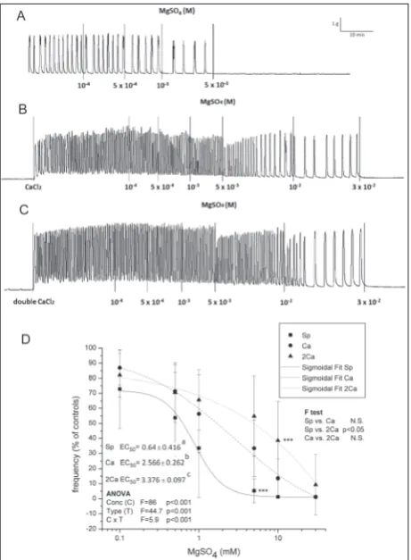
MgSO4 relaxed spontaneous uterine activity in a con-centration-dependent manner with regard to frequency (Fig. 1A). There were no significant changes in ampli-tude until complete cessation of contractions occurred
(at the highest used MgSO4 concentration of 30 mM).
The addition of Ca2+ (6 and 12 mM) caused intensive
contractile activity, and these types of uterine activity
were referred to as Ca2+- or double Ca2+-stimulated,
according to the concentration of Ca2+ used. MgSO
4
also relaxed both Ca2+- and double Ca2+-stimulated
active uteri in a concentration-dependent manner (Fig. 1B and C; ANOVA significant concentration effect,
p<0.001), but the concentration of MgSO4 necessary
for relaxation of the Ca2+- and double Ca2+-stimulated
active uteri was significantly higher (significant
dif-ference in EC50 values, ANOVA significant type and
interaction effect, p<0.001, significant post hoc Tukey
t-test; Fig. 1D). EC50 was 5 times higher for double
Ca2+-stimulated uteri than for the spontaneously
ac-tive uterus. The addition of both single and double
Ca2+ extended the MgSO
4 -induced relaxation effect
and shifted the sigmoid shape for the frequency to-ward higher concentrations (significant F-test effect, p<0.05; Fig. 1D).
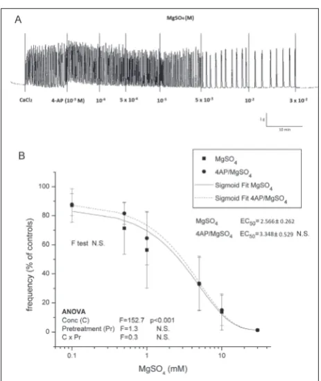
Pretreatment with 4-AP had no effect on MgSO4
-mediated relaxation of Ca2+-stimulated uteri (no
sig-nificant ANOVA pretreatment effect, no difference
between either the sigmoidal fit curve shape or EC50;
Fig. 2A and B). On the other hand, a relaxing effect
of MgSO4 was potentiated by pretreatment with
glib-enclamide (Fig. 3A and B). Pretreatment with gliben-clamide significantly deepened relaxation by decreasing frequency (significant ANOVA pretreatment effect, p<0.05). Pretreatment with glibenclamide lowered
the concentrations of MgSO4 that were needed for
relaxation (post hoc Tukey difference between degrees
of relaxation by single equivalent concentration of 5 and 10 mM). There was no difference between the
MgSO4-induced relaxing effect of Ca2+-stimulated and
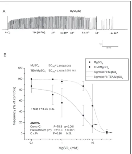
glibenclamide pretreated uteri as regards the amplitude. Pretreatment with TEA led to the elevation of frequency (significant ANOVA pretreatment effect,
Fig. 1. The effect of MgSO4 on uterine contractile activity under different Ca2+ concentrations. Graphs show original traces for A –
spontaneously active, B – Ca2+-stimulated active, and C – double
Ca2+-stimulated active. Results are presented as the means±SD
p<0.001; Fig. 4A and 4B) after application of MgSO4
concentrations above 1 mM (post hoc Tukey t test,
p<0.001). However, there was no statistically significant
difference between EC50 values for frequency since
there were large distributions of data in the middle part of the TEA curve and therefore the SD was high.
There was no difference between the MgSO4-relaxing
effect of Ca2+-stimulated and TEA pretreated uteri
regarding amplitude.
DISCUSSION
Our results showed that MgSO4 inhibited spontaneous
uterine activity in a concentration-dependent manner, and that therefore it could be considered as a uterine relaxant. This effect was in the range from 1-30 mM in the ex vivo extra uterine fluid (the EC50 for frequency
was 2.6 mM). These external MgSO4 concentrations
are not toxic, but pharmacologically they are selective with regard to the dosage, i.e. the therapeutic window is rather narrow. Magnesium ion concentrations in the plasma and extracellular fluid are approximately 1.2-1.4 mM. One-third is bound by albumin or other proteins and biochemical moieties [29]. This means that small
increases in Mg2+ in the extra uterine fluid can slow
down the spontaneous frequency, but for complete relaxation a 3-fold higher concentration is required. Literature data indicate that increasing human serum
Mg2+ concentration by 4-6 mEq/L (2-3 mmol/L)
de-creases uterine activity in preterm labor [30]. However, our results with pretreatment with glibenclamide and
TEA indicated that the effective MgSO4 concentration
range might be under the influence of applied drugs that can shift its therapeutic potential.
Fig. 2. The effect of MgSO4 on contractile activity of uteri pretreated with 4-AP. Graphs show original traces for: A – Ca2+-stimulated
active uteri pretreated with 4-AP. The results are presented as the means±SD (n=8) for frequency (B) measurements. The results were compared by two-way ANOVA for the concentration of MgSO4 (C) and the type of contractile activity (T) as factors, and F values are presented. From the curves, EC50 values were calculated, expressed as the mean±SD and compared by one-way ANOVA and post hoc Tukey’s t-test. Differences in the shape of curves were tested by the F-test.
Fig. 3. The effect of MgSO4 on contractile activity of uteri pretreated with glib. Graphs show original traces for: A – Ca2+-stimulated
Our study showed that the addition of Ca2+ to
the isolated organ bath prior toMgSO4 significantly
attenuated the relaxing effect of MgSO4. Addition of
the single Ca2+ concentration provoked an increase of
the force of spontaneous contractions by elevating both amplitude and frequency. This increase was
addition-ally elevated by the double concentration Ca2+. In both
cases, MgSO4 inhibited contractile activity, suggesting
its physiological role as a general Ca2+ antagonist. It is
known that Mg2+ inhibits the ryanodine receptor (RyR)
Ca2+-release channels by competing with Ca2+ at the
cytosolic activation sites of the channel in the mM range [31-33] and influences the fidelity of coupling between
L-type Ca2+channels and RyRs [34]. On the other hand,
cytosolic levels of H+, Ca2+, adenine nucleotides and
Mg2+ during fatigue influence the gating properties of
the SR Ca2+ channel [35], and the functional roles of
the three main intracellular ions, Na+, Ca2+ and Mg2+,
are modulated by calmodulin connected voltage-gated
Na+ channels [36].
Since increasing concentrations of Ca2+ inhibited
the relaxing effect of MgSO4 only in part, this indicated
the existence of additional cellular signaling pathways
that Mg2+ could operate through. Therefore, we partially
inhibited potassium channels by different inhibitors
prior to the addition of MgSO4. Our results showed
that the voltage-gated KV subfamilies of potassium
channels KV1-KV4were not involved in the inhibitory
action of Mg2+, since pretreatment with 4-AP had no
effect on concentration-dependent Mg2+-promoted
uterine relaxation. On the other hand, pretreatment
with TEA modified the relaxing activity of MgSO4.
TEA is a potent inhibitor of voltage-gated KV1-KV4
subfamilies of potassium channels, but it also inhibits
KV7 (KCQN) as well as BKCa potassium channel
sub-families, suggesting that KCQN as well as BKCa channels
operated during the MgSO4-induced relaxant effect.
In our experiment, pretreatment with TEA attenuated the relaxing effect as regards the frequency. Given that XE991, a KCNQ channel inhibitor, elevated the fre-quency of the murine myometrium [37], it seems that the partial blockade of the potassium channels’ pore by TEA in our experiment contributed to the elevated
frequency, and that MgSO4 operated as a mild KCQN
channel inhibitor. It is known that intracellular Mg2+
enhances the function of BKCa potassium channels [38]
through distinct binding sites and the activation is not
directly affected either by voltage or Ca2+. However,
TEA is also an efficient blocker of this type of channel, and its suppression contributed to other mechanisms
of Mg2+-induced inhibition of uterine contractility.
Moreover, BKCa channels are in neuronal cells
colocal-ized with voltage-dependent Ca2+ channels [39,40,41]
or RYR [42], and these functional couples seem to be
responsive to the Ca2+ entering into the cytosol and to
the control Ca2+ concentration [40,42,43]. Shi and Cui
[38] demonstrated that the competitive inhibition of
Ca2+-dependent activation of BK
Ca channels by Mg2+
results in a significant reduction of the Mg2+-dependent
activation at [Ca2+]i of ~0.1-100 μM. Taken together, our
results suggest that the overall interplay between Ca2+
and Mg2+ is concentration-dependent. Blocking BK
Ca
requires more Ca2+ that is prevented by the presence of
Mg2+. On the other hand, if the Ca2+ concentration is
Fig. 4. The effect of MgSO4 on contractile activity of uteri pretreated with TEA. Graphs show original traces for: A – Ca2+-stimulated
in physiological balance with Mg2+, after the addition
of Mg2+, its Ca2+ antagonist role is potentiated.
Magnesium interacts with organic phosphates such
as ATP and PIP2. Intracellular Mg2+ depresses KCNQ
currents by binding to PIP2 electrostatically, thus reduc-ing the availability of PIP2 for direct interaction with
the channels [44]. Most of cytoplasmic Mg2+ is in the
form of a complex with ATP, phosphonucleotides and
phosphometabolites and Mg2+, with ATP constituting
the largest metabolic pool capable of binding Mg2+
within the cytoplasm and the mitochondrial matrix as well [45,46]. Our results showed that a partial
block-ade of KATP channels by glibenclamide stimulated the
relaxing activity of MgSO4, which points to its
ATP-dependent role.
The results presented in our study show that MgSO4
acts only partially as a general calcium antagonist. Moreover, a part of its physiological pathway is through
BKCa channels since the blocking of BKCa channels
with TEA led to the stimulation of frequency. Since relaxing activity was predominantly achieved by the
reduction of frequency, it seems that MgSO4 is a direct
K+ channel inhibitor, but it also affects Ca2+ availability.
Furthermore, the MgSO4 uterine relaxing activity is
influenced by selective ATP-sensitive potassium chan-nels suggesting also an ATP-dependent role.
Funding: This work was funded by the Ministry of Education, Science and Technological Development, Republic of Serbia, Grant No. 173014 “Molecular Mechanisms of Redox Signaling in Homeostasis: Adaptation and Pathology”.
Author contributions: Duško Blagojević, Slobodan Milovanović conceptualized and defined the research idea and created the research design; Zorana Oreščanin-Dušić and Nikola Tatalović searched the literature data; Duško Blagojević selected the statistical tests; Dragana Jokanović, Dragana Drakul, Milica Pecelj, Zorana Oreščanin-Dušić collected and prepared the experimental data; Nikola Tatalović and Duško Blagojević performed the statistical analyses; Dragana Drakul, Milica Pecelj and Dragana Jokanović wrote the first draft of the manuscript; Duško Blagojević, Slobodan Milovanović and Zorana Oreščanin-Dušić wrote the second draft of the manuscript; Nikola Tatalović and Duško Blagojević edited the manuscript.
Conflict of interest disclosure: The authors declare that there is no conflict of interests.
REFERENCES
1. Elliott JP. Magnesium sulfate as a tocolytic agent. Am J Obstet Gynecol. 1983;147(3):277-84.
2. Gáspár R, Hajagos-Tóth J. Calcium Channel Blockers as Tocolytics: Principles of Their Actions, Adverse Effects and Therapeutic Combinations. Pharmaceuticals. 2013;6:689-99. 3. Chesley LC. History and epidemiology of preeclamp-sia-eclampsia. Clinical Obstetrics and Gynecology. 1984;27(4):801-20.
4. Dorsett L. The intramuscular injection of magnesium sul-phate for the control of convulsions in eclampsia. Am J ObstetGynecol. 1926;11:227-31.
5. Lazard EM. An analysis of 575 cases of eclamptic and pre eclamptic toxaemias treated by IV injections of magnesium sulphate. Am J Obstet Gynecol. 1933;26:647-56.
6. Hall DG, McGaughey HS, Corey EL, Thornton WN. The effects of magnesium therapy on the duration of labour. Am J Obstet Gynecol. 1959;78:27.
7. Stallworth JC, Yeh S, Petrie RH. The effect of magnesium sulphate on fetal heart rate variability and uterine activity. Am J ObstetGynecol 1981;140:702-6.
8. Crowther CA, Hiller JE, Doyle LW. Magnesium sulphate for preventing preterm birth in threatened preterm labour. Cochrane Database Syst Rev. 2002;4:CD001060.
9. Coleman FH. Safety and efficacy of combined ritodrine and magnesium sulfate for preterm labor: a method for reduc-tion of complicareduc-tions. Am J Perinatol. 1990;7(4):366-9. 10. Kosasa TS, Busse R, Wahl N, Hirata G, Nakayama RT, Hale
RW. Long-term tocolysis with combined intravenous ter-butaline and magnesium sulfate: a 10-year study of 1000 patients. Obstet Gynecol. 1994;84(3):369-73.
11. Hatjis CG, Nelson LH, Meis PJ, Swain M. Addition of magnesium sulfate improves effectiveness of ritodrine in preventing premature delivery. Am J Obstet Gynecol. 1984;150(2):142-50.
12. Ferguson JE, Hensleigh PA, Kredenster D. Adjunctive use of magnesium sulfate with ritodrine for preterm labor tocoly-sis. Am J Obstet Gynecol. 1984;148(2):166-71.
13. Kalezić I, Rodić V, Kitanović S, Milovanović G, Zgradić I, Milovanović S. The effects of ritodrine, on receptors in smooth uterine muscle and heart atria of rats. Arch Toxicol Kinet Xenobiot Metab. 1993;1:112-8.
14. van Vliet E, Dijkema GH, Schuit E, Heida KY, Roos C, van der Post J, Parry EC, McCowan L, Lyell DJ, El-Sayed YY, Carr DB, Clark AL, Mahdy ZA, Uma M, Sayin NC, Varol GF, Mol BW, Oudijk MA. Nifedipine maintenance tocolysis and perinatal outcome: an individual participant data meta-analysis. BJOG. 2016;123(11):1753-60.
15. Novaković R, Milovanović SR, Heinle H, Protić D, Gojković-Bukarica Lj. The effect of potassium channel opener pinaci-dil on non-pregnant rat uterus. Basic Clin Pharmacol Toxi-col. 2007;1742-84.
16. Oreščanin-Dušić Z, Milovanović S, Blagojevic D, Nikolic-Kokić A, Radojičić R, Spasojević I, Spasić BM. Diethyldi-thiocarbamate potentiates the effects of protamine sulfate in the isolated rat uterus. Redox Rep. 2009;14:48-54.
M, Blagojević D. Effects of protamine sulfate on spontane-ous and Ca-induced contractile activity in the rat uterus are potassium channels mediated. Gen. Physiol. Biophys. 2009;28:143-8.
18. Milovanovic S, Kordic-Bojinović J, Djordjevic S, Drakul D, Sokolovic D, Miletic N, Blagojevic D. The importance of potassium chanels in the relaxing effect of pentoxifillyne on the isolated rat uterus. Serb J Exp Clin Res. 2013;14(2):55-64. 19. Kuang Q, Purhonen P, Hebert H. Structure of potassium
channels. Cell Mol Life Sci. 2015;72:3677-93.
20. Khan RN, Matharoo BВ, Arulkumaran S, Ashford МL. Potassum channels in the human myometrium. Exp Physiol. 2001;862:255-64.
21. Morrison ЈЈ, Ashford МLЈ, Khan RN, Smith S K. The effects of potassium channel openers on isolated pregnant human myometrium before and after the onset of labor: potential for tocolysis. Аm Ј Obstet Gynecol. 1993;169:1277-85. 22. Appiah I, Nikolic-Kokic A, Orescanin-Dusic Z,
Radoji-cic R, Milovanovic S, Spasic M, Blagojevic D. Reversible Oxidation of Myometrial Voltage-Gated Potassium Chan-nels with Hydrogen Peroxide. Oxid Med Cell Longev. 2012;2012:105820.
23. Kordić-Bojinović J, Oreščanin-Dušić Z, Slavić M, Radojičić R, Spasić M, Milovanović SR, Blagojević D. Effect of indo-metacin pretreatment on protamine sulfate-mediated relax-ation of the isolated rat uterus: the role of the antioxidative defense system. Pharmacol Rep. 2011;63(4):1019-28. 24. Kordić-Bojinović J, Jokanović D, Stanković D, Janković S,
Milovanović S. Influence of modulators of relaxant effect of pentoxifylline in isolated rat uterus. Ser J Exp Clin Res. 2010;11(3):99-104.
25. Furchgott RF. Pharmacological characterization of recep-tors: its relation to radioligand-binding studies. Fed Proc. 1978;37(2):115-20.
26. Marcondes FK, Bianchi FI, Tanno AP. Determination of the estrous cycle phases of rats: some helpful considerations. Braz J Biol. 2002;62:609-14.
27. Hinkle ED, Wiersma W, Jurs GS. Applied statistics for behavioral sciences. 2nd ed. Boston: Houghton Mifflin
Com-pany; 1994.
28. Manley BFJ. Multivariate statistical methods. 2nd ed. London:
Chapman & Hall; 1986.
29. Romani AMP. Cellular magnesium homeostasis. Arch Bio-chem Biophys. 2011;512(1):1-23.
30. Guyton ST, Morey TE. Magnesium. In: Atlee JL, editor. Complications in anesthesia. Philadelphia: Saunders, Else-vier; 2007. p. 59-61.
31. Zahradnikova A, Palade P. Procaine effects on single sar-coplasmic reticulum Ca2+ release channels. Biophys J.
1993;64:991-1003.
32. Györke I, Györke S. Regulation of the cardiac ryanodine receptor channel by luminal Ca2+ involves luminal Ca2+
sens-ing sites. Biophys J. 1998;75:2801-2810.
33. Laver DR, Baynes TM, Dulhunty AF. Magnesium inhibition of ryanodine-receptor calcium channels: evidence for two independent mechanisms. J Membr Biol. 1997;156:213-229. 34. Zahradníková A, Dura M. Györke I, Escobar A.L, Zahrad-ník I, Györke S. Regulation of dynamic behavior of cardiac ryanodine receptor by Mg2+ under simulated physiological
conditions. Am J Physiol Cell Physiol 2003;285:C1059-70. 35. Coronado R, Morrissette J, Sukhareva M, Vaughan DM.
Structure and function of ryanodine receptors. Am J Physiol Cell Physiol. 1994;266:C1485-504.
36. Guo F, Zhou PD, Gao QH, Gong J, Feng R, Xu XX, Liu SY, Hu HY, Zhao MM, Adam HC, Cai JQ, Hao LY. Low-Mg2+
treatment increases sensitivity of voltage-gated Na+ channels
to Ca2+/calmodulin-mediated modulation in cultured
hippo-campal neurons. Am J Physiol Cell Physiol. 2015;308:C594-605.
37. McCallum LA, Greenwood IA, Tribe RM. Expression and function of Kv7 channels in murine myometrium through-out oestrous cycle. Pflugers Arch. 2009;457:1111-20. 38. Shi J, Cui J. Intracellular Mg2+ enhances the function
of BK-type Ca2+-activated K+ channels. J Gen Physiol.
2001;118:589-605.
39. Yazejian B, DiGregorio DA, Vergara JL, Poage RE, Meriney SD, Grinnell AD. Direct measurements of presynaptic cal-cium and calcal-cium-activated potassium currents regulating neurotransmitter release at cultured Xenopus nerve-muscle synapses. J Neurosci. 1997;17(9):2990-3001.
40. Yazejian B, Sun XP, Grinnell AD. Tracking presynaptic Ca2+
dynamics during neurotransmitter release with Ca2+
-acti-vated K+ channels. Nat Neurosci. 2000;3(6):566-71.
41. Marrion NV, Tavalin SJ. Selective activation of Ca2+-activated
K+ channels by co-localized Ca2+ channels in hippocampal
neurons. Nature. 1998;29:900-5.
42. Jaggar JH, Porter VA, Lederer WJ, Nelson MT. Calcium sparks in smooth muscle. Am J Physiol Cell Physiol. 2000;278(2):C235-56.
43. Neher E. Usefulness and limitations of linear approxima-tions to the understanding of Ca++ signals. Cell Calcium.
1998;24(5-6):345-57.
44. Suh BC, Hille B. Electrostatic interaction of internal Mg2+
with membrane PIP2 Seen with KCNQ K+ channels. J Gen
Physiol. 2007;130(3):241-56.
45. Scarpa A, Brinley FJ. In situ measurements of free cytosolic magnesium ions. Fed Proc. 1981;40(12):2646-52.
46. Lüthi D, Günzel D, McGuigan JA. Mg-ATP binding: its modification by spermine, the relevance to cytosolic Mg2+
buffering, changes in the intracellular ionized Mg2+
con-centration and the estimation of Mg2+ by 31P-NMR. Exp