Journal of Global Pharma Technology
Available Online at:
www.jgpt.co.in
RESEARCH ARTICLE
©2009-2020, JGPT. All Rights Reserved 322
Evaluation the Protective Impact of Cape Gooseberry (Physalis
peruviana L.) Extract against Benzo(a)pyrene Induced DNA
Damage and Gene Expression Change in male Rats
Lamiaa M. Salem
1*, Zeina Walid Sharawi
21Cell Biology Department, National Research Centre, 33 Bohouth St., P.O. 12622 Dokki, Giza, Egypt.
2Department of Biological Sciences, Faculty of Sciences, King Abdul Aziz University, Jeddah, Saudi
Arabia.
*Corresponding Author:Lamiaa M. Salem
Abstract
Natural bioactive compounds have been increasingly investigated against carcinogenic agents to prevent tumour progression. Thus, several active ingredients exist in different herbs and medicinal plants exhibited preventive impact against benzo(a)pyrene (BaP) induced carcinogenesis. In the present study, the efficacy of Cape gooseberry fruit (CGF) extract was investigated against BaP induced genetic toxicity in Swiss albino male rats. Male rats (n=90) were allocated in several groups exposed to BaP and/or several doses of CGF extract (100, 150 and 200 mg/kg b.w.). Assessment of DNA damage, generation of DNA adducts and expression alteration of stress (Hsp70a, CYP450) and antioxidant (GST) related genes were conducted. The results showed that treatment of BaP-exposed rats with different doses of CGF extract was able to prevent the genetic toxicity in the forms of DNA damage and DNA adducts as well as gene expression alteration induced by BaP. The results suggested that CGF extract-BaP interaction may reflect the extract efficacy against genotoxicity induced by chemotherapeutic drugs that are metabolized by antioxidant enzymes.
Keyword: Benzo(a)pyrene, Cape gooseberry, Salvia sp, DNA adducts, Gene expression, Rats.
Introduction
Human disorders development due to crushing the biological defense of the immune system against drugs, mutagens and oxidative stress is resulting mainly from oxidative damage of several biological molecules such as protein, lipid, RNA and DNA [1, 2]. Thus exposure of living organisms to mutagens and oxidative stressors affects the DNA structure, expression of gene patterns and metabolic enzymes and thus influences the immune system [3].
Benzo(a)pyrene (BaP) is one of the most powerful carcinogens. It is teratogenic and mutagenic in animal models [4, 5]. BaP is considered as a potent carcinogenic marker for contamination with hazard chemicals in an environmental sample [6, 7]. The most target organ for BaP mammals is the liver in
which the liver microsomal metabolized it and thus inducing oxidase system. Additionally, BaP is one of the most highly reactive drugs that can attach to specific DNA positions modulating the gene expression patterns inducing DNA damage, micronucleus formation and initiation of cancer in mammalian cells [8-10].
Natural compounds as chemoprevention
proffer novel tool to
©2009-2020, JGPT. All Rights Reserved
323
[14]. The mechanism of action responsible for cancer risk reduction is strangely coincided with antioxidant capacity of these active ingredients [15, 16]. Several types of dietary nutrients and flavonoids found in berry and grapes have been exhibited antioxidant capacity, cytochrome P450 suppression and anti-progression activity against several types of cancer [17].
The antioxidants existing in berry and grapes are responsible in terminating the chain radical reactions through giving atoms of hydrogen to peroxy radical building novel molecules. These new molecules are active to react with free radicals in the cell and consequently ending the propagating chain [18, 19]. Hence theses antioxidants protect cells against oxidative damage by trail off oxygen species and free radicals or increase the activity of the antioxidant enzymes [20].
Medicinal plants have been widely used for protection against oxidative damage induced by chemical exposure since ancient times in Arabian countries as exhibiting less toxic side effects. One of the very promising plants for inhibiting oxidative stress is Cape gooseberry (Physalis peruviana). The fruit of Cape gooseberry (golden berry) is containing high levels of minerals, nutrients, vitamins, β- and α-carotene [21, 22]. Several studies reported that Cape gooseberry extracts exhibited antioxidant activity [23, 24], hepatotoxicity prevention [25] and anti-inflammatory activity[25, 26].
Therefore, the present research aims to evaluate the in vivo biosafety and bioactivities of Cape gooseberry fruit extract in male rats. Additionally, the antioxidant capacity of Cape gooseberry fruit extract against genetic toxicity induced by benzo[a]pyrene in liver tissues of male rats was the main objective of current study.
Materials and Methods
ChemicalsBenzo[a]pyrene (BaP) and dimethylsulfoxide (DMSO) were purchased from Sigma (St. Louis, MO). Trizol was bought from Invitrogen (Carlsbad, CA, USA). The reverse transcription and PCR kits were obtained from Fermentas (Glen Burnie, MD, USA). SYBR Green Mix was purchased from Stratagene (La Jolla, CA, USA). All other
chemicals and reagents were of analytical grade and obtained from standard commercial suppliers.
Fruits Sampling and Preparation
Cape gooseberry fruit (CGF) were collected from private market, Giza, Egypt. The CGF were authenticated by Botany Department, National Research Center, Giza, Egypt. The Cape gooseberry were dried by means of solar energy, stored in plastic bags and kept at −20
ºC, up to use. Dry CGF powder was used to
prepare a stock solution by dissolving the powder in distilled water on a shaking plate for half hour. The CGF solution was centrifuged and the supernatant was freeze-dried by Heto DryWinner (Denmark). The extract of CGF was collected after passing through a sterilized filter (0.22 Am).
Animals
Seventy Swiss albino adult male rats (150±8.3 g) were obtained from the animal house of the National Research Centre, Giza, Egypt. They were housed in stainless steel wire meshed cages and provided free access to water and a standard rodent chow diet. All animals received human care in compliance with guidelines of the Ethical Committee of National Research Centre, Egypt, which follows the recommendations of the National Institutes of Health Guide for Care and Use of Laboratory Animals (Publication No. 85-23, revised 1985).
Rat's Treatment with BaP and CGF Extract
The rats were randomly divided into 7 groups of 10 animals and designated to receive orally either with BaP and/or CGF extract. Animals within different treatment groups were treated (daily at a 24 h interval) intragastrically for 4 weeks as follows: group 1, untreated control rats; groups 2-4, treated intragastrically with 100, 150 and 200 mg/kg b.w. of CGF extract [27]; groups 5-7, treated intragastrically with 100, 150 and 200 mg/kg b.w. of CGF extract plus intra peritoneally single dose of 50 mg/kg b.w. of BaP dissolved in 0.1% dimethylsulfoxide (DMSO) [28], group 8, treated with 50 mg/kg b.w. of BaP only; group 9, treated with DMSO.
©2009-2020, JGPT. All Rights Reserved
324
Comet Assay
The DNA damage in treated rat samples using comet assay was performed according to Blasiak et al. [29]. Homogenized liver samples of treated groups were mixed with low melting point agarose and loaded in small pieces on slides which pre-coated with normal melting agarose. The loaded samples were kept on the slides in horizontal position for half hour in dark environment at 4°C. Low melting point agarose was then pipetting above the slides including samples and the slides were left for 30 min at 4°C to harden and put afterward in lysis buffer for one hour.
Then, fresh alkaline unwinding buffer was used to submerge the slides in a dark place for one hour at 20°C. Afterwards, electrophoresis (0.8 V/cm, 300mAmps) for the slides were carried out for 30 min to assess the DNA damage in the form of tail migration in 100 cell per each animal. Specific software (TriTek corp., Comet Score, Sumerduck, VA22742) was used to determine the rate of the DNA damage per sample.
DNA Adducts (8-OHdG and 2-dG) Determination
The generation ratio of 8-OHdG/2-dG in liver tissues of supplemented animals was assessed. The total DNA was extracted from animal samples by homogenizing the tissues in lysis buffer (pH 7.4), transferring and incubating them over night (~ for 16 h) with proteinase K at 50-60oC. The incubated
samples were treated with RNase enzyme for ten min at 50oC according to Lu [30]. The
method of chloroform/isoamyl alcohol was used to isolate the genomic DNA from treated rats. The extracted DNA content was dissolved in a solution of Tris/EDTA. The generation ratio of 8-OHdG/2-dG was assessed through several electrochemical sensors using HPLC (CoulArray system, Model 5600) [31].
Expression Analysis of Stress and Antioxidant Related Genes
RNA Isolation and Reverse Transcription Reaction
TRIzol® extraction Chemical (Invitrogen) was utilized to isolate the total genomic RNA of adipose tissues of all treated animal. After completion of the isolation procedures, RNA pellet was stored in DEPC treated water.
To digest the potential DNA residues the pellet of isolated RNA was treated with RNAse-free DNAse kit (Invitrogen, Germany). RNA aliquots were stored at -20°C or utilized immediately for reverse transcription [32].
First Strand cDNA Synthesis Kit (RevertAidTM, MBI Fermentas) was used to synthesize the cDNA copy from adipose tissues via reverse transcription reaction (RT). A RT reaction program of 25°C for 10 min, then one hour at 42 °C then 5 min at 95°C was used to obtain the cDNA copy of liver genome. Finally, tubes of reaction containing cDNA copy were collected on ice up to use for cDNA amplification [33, 34].
Quantitative Real Time-PCR
SYBR® Premix Ex TaqTM kit (TaKaRa, Biotech. Co. Ltd.) was used to perform the qRT-PCR analyses using the synthesized cDNA copies from adipose tissues. For each reaction a melting curve profile was conducted. The quantitative values of the target genes were normalized on the expression of the housekeeping gene (Table 1). The 2−ΔΔCT method was used to determine
the quantitative values of the specific genes to the -actin gene [35, 36].
Statistical Analysis
General Liner Models (GLM) of Statistical Analysis System (SAS) was used to analyses the data of DNA damage, DNA adducts and gene expression assays. Afterwards, Scheffé-test was used to determine the significant differences between tested groups. The values are expressed as mean±SEM. All significance statements were based on probability of P < 0.05.
Results
Effect of Cape Gooseberry Extract on DNA Damage in BaP Exposed Rats
©2009-2020, JGPT. All Rights Reserved
325
exposed rats and were similar to those in
untreated control and DMSO rats. Moreover, treatment of Bap-exposed rats with low,
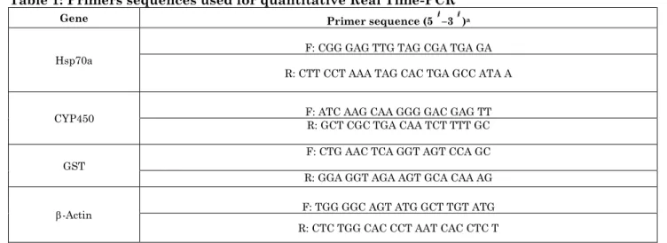
Table 1: Primers sequences used for quantitative Real Time-PCR Gene Primer sequence (5 –3 )a
Hsp70a F: CGG GAG TTG TAG CGA TGA GA
R: CTT CCT AAA TAG CAC TGA GCC ATA A
CYP450 F: ATC AAG CAA GGG GAC GAG TT R: GCT CGC TGA CAA TCT TTT GC
GST
F: CTG AAC TCA GGT AGT CCA GC
R: GGA GGT AGA AGT GCA CAA AG
-Actin F: TGG GGC AGT ATG GCT TGT ATG
R: CTC TGG CAC CCT AAT CAC CTC T
a F: forward primer; R: reverse primer. Tm: temperature
Table 2: Effect of Cape gooseberry extract on rate of DNA damage in liver tissues of male rats exposed to BaP
Treatment No. of cells Class¥ of comet DNA damaged
cells (mean ± SEM) Analyzed Total
comets 0 1 2 3
Control 5 500 37 463 33 4 7.4±0.38
d
DMSO 5 500 41 459 36 3 8.2±0.41d
CGF extract100 5 500 38 462 31 7 7.6±0.48d
CGF extract150 5 500 36 464 31 4 7.2±0.52d
CGF extract200 5 500 39 461 29 7 7.8±0.29d
BaP+CGF extract100 5 500 102 398 38 29 20.4±0.42b
BaP+CGF extract150 5 500 91 409 47 20 18.2±0.56bc
BaP+CGF extract200 5 500 77 423 39 23 15.4±0.32c
BaP 5 500 124 376 45 38 24.8±0.68a
¥: Class 0= no tail; 1= tail length < diameter of nucleus; 2= tail length between 1X and 2X the diameter of nucleus; and 3= tail length
> 2X the diameter of nucleus.(*): No of cells analyzed were 100 per an animal.
DMSO: dimethylsulfoxide; CGF: Cape gooseberry fruits; BaP: benzo(a)pyrene; Data are presented as mean ± SEM. a,b,c,d Mean values within tissue with unlike superscript letters were significantly different (P< 0.05, Scheffé-Test).
medium and high doses of CGF extracts decreased significantly the rats of DNA damage compared with the those in group of rats exposed to BaP only. Additionally, the high dose of CGF extract was the most effective dose in diminishing the rate of DNA damage induced by BaP.
Effect of Cape Gooseberry Extract on DNA Adducts in BaP Exposed Rats
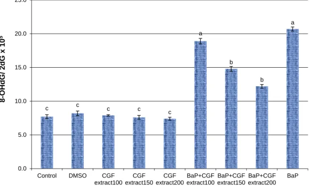
The generation of DNA adducts (8-OHdG/2-dG) ratio in liver tissues of BaP exposed rats treated with CGF extract is summarized in Figure 1. The results of this study showed that male rats exposed to BaP augmented significantly (P<0.01) the OHdG/2-dG generation rate compared with control rats. In contrary, male rats treated with low, medium and high dose of CGF extracts showed low generation ratio of DNA adducts which was similar to that in untreated control and
DMSO exposed rats. Moreover, the generation ratio of DNA adducts decreased significantly (P<0.05) in BaP-exposed rats treated with medium and high dose of CGF compared with rats exposed BaP alone. Furthermore, the high dose of CGF extract was the most efficient dose in decreasing the level of DNA adducts induced by BaP.
Effect of Cape Gooseberry Extract on Expression of Stress and Antioxidant Related Genes in BaP Exposed Rats
©2009-2020, JGPT. All Rights Reserved
326
Figure 1: Ratio of DNA adducts (8-OHdG/2dG) in liver tissues of BaP-exposed rats treated with different concentrations ssue with unlike superscript letters were significantly different (P< 0.05, Scheffé-Test)
Figure 2: Expression change of Hsp70a-mRNA levels in liver tissues of BaP-exposed rats treated with different concentrations of Cape gooseberry fruit (CGF) extract. Data are presented as mean ± SEM. a,b,c Mean values within
tissue with unlike superscript letters were significantly different (P< 0.05, Scheffé-Test)
c c c c c
a
b
b
a
0.0 5.0 10.0 15.0 20.0 25.0
Control DMSO CGF
extract100 CGF extract150
CGF extract200
BaP+CGF extract100
BaP+CGF extract150
BaP+CGF extract200
BaP
8
-O
HdG
/
2
dG
x
1
0
5
Treatment
d d d d d
b
bc
c
a
0 0.5 1 1.5 2 2.5 3 3.5
Control DMSO CGF
extract100 CGF extract150
CGF extract200
BaP+CGF extract100
BaP+CGF extract150
BaP+CGF extract200
BaP
Rel
a
tiv
e
e
x
pre
s
s
io
n
of
Hsp
7
0
a
-
m
RN
A
©2009-2020, JGPT. All Rights Reserved
327
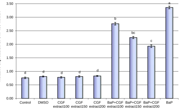
Figure 3: Expression change of CYP450-mRNA levels in liver tissues of BaP-exposed rats treated with different concentrations of Cape gooseberry fruit (CGF) extract. Data are presented as mean ± SEM. a,b,c Mean values within
tissue with unlike superscript letters were significantly different (P< 0.05, Scheffé-Test)
Figure 4: Expression change of GST-mRNA levels in liver tissues of BaP-exposed rats treated with different concentrations of Cape gooseberry fruit (CGF) extract. Data are presented as mean ± SEM. a,b,c Mean values within
tissue with unlike superscript letters were significantly different (P< 0.05, Scheffé-Test)
The results showed that the expression levels of Hsp70a and CYP450 genes in male rats exposed to BaP were up-regulated
significantly (P<0.01) compared with untreated control and DMSO groups of rats.
d d d d d
b
bc
c
a
0.00 0.50 1.00 1.50 2.00 2.50 3.00 3.50
Control DMSO CGF
extract100 CGF extract150
CGF extract200
BaP+CGF extract100
BaP+CGF extract150
BaP+CGF extract200
BaP
Rel
a
tiv
e
e
x
pre
s
s
io
n
of
CYP45
0
-m
RN
A
Treatment
a
a a a
a
d
c
b
d
0.00 0.50 1.00 1.50 2.00 2.50 3.00 3.50
Control DMSO CGF
extract100 CGF extract150
CGF extract200
BaP+CGF extract100
BaP+CGF extract150
BaP+CGF extract200
BaP
Rel
a
tiv
e
e
x
pre
s
s
io
n
of
G
ST
-
m
RN
A
©2009-2020, JGPT. All Rights Reserved
328
However, the expression levels of Hsp70a and CYP450 genes in groups of rats treated with low, medium and high doses of CGF extracts were low and reached similar levels compared to those in untreated control and DMSO groups of rats. Additionally, the expression levels of Hsp70a and CYP450 genes in BaP-exposed rats treated with low, medium and high doses of CGF extracts decreased compared to those in rats exposed to BaP only. Also, the high dose of CGF extract was the most competent dose in decreasing the levels of gene expression changes induced by BaP.
The expression levels of GST gene in male rats exposed to BaP were down-regulated significantly (P<0.01) compared with untreated control and DMSO groups of rats. In contrary, the expression levels of GST gene in groups of rats treated with low, medium and high doses of CGF extracts were high and reached similar levels compared to those in untreated control and DMSO groups of rats. Additionally, the expression levels of GST gene in BaP-exposed rats treated with low, medium
and high doses of CGF extracts increased compared to those in rats exposed to BaP only. Moreover, the high dose of CGF extract was the most effective dose in elevating the levels of GST gene expression changes induced by BaP.
Discussion
BaP is one of the most powerful carcinogens which is produced from several sources related to human activity such as charbroiled food and cigarette smoke [37]. The action mechanism of BaP inducing cancer is thought to be that it causes damage in the DNA structure through binding BaP-7,8-dihydrodiol-9,10-epoxide inducing DNA adducts (BPDE-DNA) by increasing ROS formation [38, 39].
In agreement with the above observation, the results of this study found that male rats exposed to BaP showed significant high mean rates of the DNA damage and generation ratio of DNA adducts compared to control rats. So, it could be suggested that the main reason for increase the DNA damage in the cell exposed to BaP is due to conversion of the BaP form to the BPDE metabolite [40, 41].
Araujo et al. [8] and Fanali et al. [9] reported that BaP is one of the most highly reactive
drugs that can attach to specific DNA positions modulating the gene expression patterns inducing initiation of cancer in mammalian cells. In consistent with these findings, our results revealed that expression levels of stress related genes (Hsp70a and CYP450) in male rats exposed to BaP were up-regulated significantly compared with control rats. Also, the expression levels of the antioxidant GST gene in male rats exposed to BaP were down-regulated significantly compared with control rats.
Numerous studies have been investigated to identify novel tools in prevention cancer progression through suppression the harmful effect of BaP by using promising natural products [42-46]. In the present study CGF extract was used to prevent the genetic toxicity induced by PaB mainly in liver tissues of male rats. The results found that treatment of Bap-exposed rats with all three doses, especially high dose, of CGF extracts decreased significantly the rats of DNA damage and generation ratio of DNA adducts compared with those in rats exposed to BaP only.
Several studies reported that CGF is good source for antioxidant compounds [47]. This source of compounds is thought to be the main reason for cell protection against oxidative stress. The antioxidant compounds might be have chemical structure variation which could be reacting with free radical species by donation of protons or electrons making quenching of the free radical in the cell exposed to mutgens or carcinogenic agents[48].
Natural antioxidants existing in CGF extract could inhibit the harmful impacts of BaP by free radicals scavenging or by mitigating the cell inflammatory response. Therefore, oxidative stress amelioration in BaP-exposed rats administrated with CGF extract could be attributed to its contents of antioxidants. The present study found that expression levels of antioxidant enzyme GST gene in BaP-exposed rats treated with all three doses of CGF extract, in particularly with the high dose, increased significantly compared to rats exposed to BaP only.
©2009-2020, JGPT. All Rights Reserved
329
transcription regulation is modulating through interaction between active ingredients of the plant extract and antioxidant response elements (AREs). It has been suggested that glutathione synthesis is regulated by intermediated enzyme namely, glutamylcysteine synthetase (GCS) which is activating by AREs [50].
Therefore, the main key in up-regulation of antioxidant enzymes is enhancement of GCS activity [51]. Thus, it could be possibility in the present study that the interaction between the active compounds exist in the CGF extract and AREs would result in a high expression of antioxidant enzyme GST gene explaining decrease the rate of DNA damage and DNA adducts. Several studies have been reported the interactions between Herb and drug using various phytomedicinal plants [52, 53]. Zhou et al. [54] reported that increase levels of isozymes such as Cytochrome P450 is main indication for
toxicity caused by PaB. These isozymes are vulnerable to alteration by treatment with natural active ingredients of herbs. In the same line, the present study found that expression levels of Cytochrome P450 gene in BaP-exposed rats treated with CGF extract decreased significantly compared to rats exposed to BaP only.
In conclusion, the present work showed that
treatment of male rats exposed to BaP with CGF extract was able to prevent the genetic toxicity in the forms of DNA damage and DNA adducts as well as gene expression alteration induced by BaP. Thus, CGF extract seemed to improve the antioxidant capacity of the male rats. The observed CGF extract –BaP interaction may reflect the extract efficacy against genotoxicity induced by chemotherapeutic drugs that are metabolized by antioxidant enzymes.
References
1 Borska L, Andrys C, Krejsek J, Palicka V, Vorisek V, Hamakova K, et al. Influence of dermal exposure to ultraviolet radiation and coal tar (polycyclic aromatic hydrocarbons) on the skin aging process. J Dermatol Sci 2016; 81(3):192-202.
2 Taha R, Seidman E, Mailhot G, Boudreau F, Gendron FP, Beaulieu JF, et al. Oxidative stress and mitochondrial functions in the intestinal Caco-2/15 cell line. PLoS One 2010; 5(7):e11817.
3 Havsteen BH. The biochemistry and medical significance of the flavonoids. Pharmacology and Therapeutics 2002; 96:67–202.
4 Chang J, Tao J, Xu C, Li Y, Li N, Tang Z, et al. Pollution characteristics of ambient PM(2.5)-bound benzo[a]pyrene and its cancer risks in Beijing. Sci Total Environ 2019; 654:735-741. 5 IARC. IARC Monographs on the Evaluation of
Carcinogenic Risk to Human, IARC, Lyon 1983; 32: 211
6 Shi Q, Godschalk RWL, van Schooten FJ. Inflammation and the chemical carcinogen benzo[a]pyrene: Partners in crime. Mutat Res 2017; 774:12-24.
7 WHO. Evaluation of Certain Food Additives and Contaminants; Benzo(a)pyrene. WHO Food Additives Series, WHO, Geneva, 1998; 28:27-29. 8 Araujo CS, Brito LD, Tarifa MO, Silva NJFD, Rodrigues KS, Cavalcante DGSM, et al. Protective effects of bark ethanolic extract from
Spondias dulcis Forst F. against DNA damage induced by benzo[a]pyrene and cyclophosphamide. Genet Mol Biol 2019; 42(3):643-654.
9Fanali LZ, Franco-Belussi L, Bonini-Domingos CR, de Oliveira C. Effects of benzo[a]pyrene on the blood and liver of Physalaemus cuvieri and Leptodactylus fuscus (Anura: Leptodactylidae). Environ Pollut 2018; 237:93-102.
10 Arif JM, Shappell N, Sikka HC, Kumar S, Gupta RC. 32PPostlabeling analysis of lipophilic DNA adducts resulting from interaction with (±)-3-hydroxy-trans-7,8- dihydroxy-9,10-epoxy-7,8,9,10-tetrahydro-benzo[a]pyrene. Chem Biol Interact 1999; 118:87-97.
11 Li Y, Li S, Meng X, Gan RY, Zhang JJ, Li HB. Dietary Natural Products for Prevention and Treatment of Breast Cancer. Nutrients 2017; 9(7):728.
12 Álvarez-Pérez M, Ali W, Marć MA, Handzlik J, Domínguez-Álvarez E. Selenides and Diselenides: A Review of Their Anticancer and Chemopreventive Activity. Molecules 2018; 23(3):628.
13 Edward SK, Faldo RK. Chemoprevention of lung cancer. Curr Oncol 2002; 4:341–346. 14 Nwodo JN, Ibezim A, Simoben CV, Ntie-Kang
©2009-2020, JGPT. All Rights Reserved
330 Flavonoids. Anticancer Agents Med Chem
2016;16(1):108-27.
15 Ma Q, Jiang JG, Yuan X, Qiu K, Zhu W. Comparative antitumor and anti-inflammatory effects of flavonoids, saponins, polysaccharides, essential oil, coumarin and alkaloids from Cirsium japonicum DC. Food Chem Toxicol 2019;125:422-429.
16 Block G, Patterson B, Subar A. Fruit, vegetables, and cancer prevention: a review of the epidemiological evidence. Nutr Cancer 1992;18(1):1-29.
17 Namiesnik J, Vearasilp K, Nemirovski A, Leontowicz H, Leontowicz M, Pasko P, et al. In vitro studies on the relationship between the antioxidant activities of some berry extracts and their binding properties to serum albumin. Appl Biochem Biotechnol 2014;172(6):2849-65.
18 Poprac P, Jomova K, Simunkova M, Kollar V, Rhodes CJ, Valko M. Targeting Free Radicals in Oxidative Stress-Related Human Diseases. Trends Pharmacol Sci 2017;38(7):592-607. 19 Delia D, Aiello A, Meroni L, Nicolini M, Reed
JC, Pierotti MA. Role of antioxidants and intracellular free radicals in retinamide-induced cell death. Carcinogenesis 1997;18(5):943-8.
20 Musonda CA, Helsby N, Chipman JK. Effects of quercetin on drug metabolizing enzymes and oxidation of 2',7-dichlorofluorescin in HepG2 cells. Hum Exp Toxicol 1997;16(12):700-708.
21 Ramadan MF, Moersel JT. Goldenberry: a noval fruit source of fat soluble bioactives. J Inform 2004;15:130-1.
22 Szefer P, Nriagu J. Mineral components in foods. Taylor Francis, FL: CRC Press; 2007:480.
23 Ramadan MF. Physalis peruviana pomace suppresses high-cholesterol diet-induced hypercholesterolemia in rats. J Int Fat Oil 2012;63:411-22.
24 Valdenegro M, Fuentes L, Herrera R, Moya-Leon MA. Changes in antioxidant capacity during development and ripening of goldenberry (Physalis peruviana L.) fruit and in response to 1-methylcyclopropene treatment. Postharvest Biol Technol 2012;67:110-7.
25 Chang JC, Lin CC, Wu SJ, Lin DL, Wang SS, Miaw CL, Ng LT. Antioxidative and hepatoprotective effects of Physalis peruviana extract against acetaminophen induced liver injury in rats. Pharm Biol 2008;46:724-31. 26 Arun M, Asha VV. Preliminary studies on
antihepatotoxic effect of Physalis peruviana Linn. (Solanaceae) against carbon
tetrachloride induced acute liver injury in rats. J Ethnopharmacol 2007;111:110-4. 27 Wu SJ, Ng LT, Huang YM et al. Antioxidant
activities of Physalis peruviana. Biological and Pharmaceutical Bulletin 2005; 28(6):963– 966.
28 Wang CC, Yuan JR, Wang CF, Yang N, Chen J, Liu D, et al. Anti-inflammatory Effects of Phyllanthus Emblica L on Benzopyrene-Induced Precancerous Lung Lesion by Regulating the IL-1β/miR-101/Lin28B Signaling Pathway. Integr Cancer Ther 2017; 16 (4):505-515.
29 Blasiak J, Arabski M, Krupa R, Wozniak K, Zadrozny M, Kasznikcki J, et al. DNA damage and repair in type 2 diabetes mellitus. Mutation. Mutation Res 2004; 554(1-2):297-304.
30 Lu T, Xu Y, Mericle MT, Mellgren RL. Participation of the conventional calpains in apoptosis. Biochimica et Biophysica Acta 2002; 1590:16-26.
31 Khalil WKB, Girgis E, Emam AN, Mohamed MB, Rao KV. Genotoxicity Evaluation of Nanomaterials: DNA Damage, Micronuclei, and 8-Hydroxy-2-deoxyguanosine Induced by Magnetic Doped CdSe Quantum Dots in Male Mice. Chem Res Toxicol 2011; 24(5):640-50. 32 Girgis E, Khalil WKB, Emam AN, Mohamed
MB, Rao KV. Nanotoxicity of Gold and Gold-Cobalt Nanoalloy. Chem Res Toxicol 2012; 25(5):1086-1098.
33 Salem NA, Wahba MA, Eisa WH, El-Shamarka M, Khalil W. Silver oxide nanoparticles alleviate indomethacin-induced gastric injury: a novel antiulcer agent. Inflammopharmacology. 2018; 26:1025–1035. 34 Khalil WKB, Booles HF, Hafiz NA,
El-Bassyouni GE. Ameliorative Effects of Brachidontes variabilis Calcium Carbonate Against Bone Loss in Ovariectomized Rats. International J. Pharmacology 2018; 14: 477-487.
35 Persson M, Rönnbäck L. Microglial self-defence mediated through GLT-1 and glutathione. Amino Acids 2012; 42(1):207– 219.
36 Boulland JL, Osen KK, Levy LM, Danbolt NC, Edwards RH, Storm-Mathisen J, Chaudhry FA. Cell specific expression of the glutamine transporter SN1 suggests differences in dependence on the glutamine cycle. Eur J Neurosci 2002; 15(10):1615-1631.
©2009-2020, JGPT. All Rights Reserved
331 38 Divi RL, Einem Lindeman TL, Shockley ME,
Keshava C, Weston A, Poirier MC. Correlation between CYP1A1 transcript, protein level, enzyme activity and DNA adduct formation in normal human mammary epithelial cell strains exposed to benzo[a]pyrene. Mutagenesis 2014; 29:409– 417.
39Patel B, Das SK, Patri M. Neonatal Benzo[a]pyrene Exposure Induces Oxidative Stress and DNA Damage Causing Neurobehavioural Changes during the Early Adolescence Period in Rats. Dev Neurosci 2016; 38:150–162.
40Mantey JA, Rekhadevi PV, Diggs DL, Ramesh A. Metabolism of benzo(a)pyrene by subcellular fractions of gastrointestinal (GI) tract and liver in Apc(Min) mouse model of colon cancer. Tumour Biol 2014; 35: 4929-4935.
41Wang BY, Wu SY, Tang SC, Lai CH, Ou CC, Wu MF et al. Benzo[a]pyrene-induced cell cycle progression occurs via ERK-induced Chk1 pathway activation in human lung cancer cells. Mutat Res 2015; 773:1-8.
42Kou X, Kirberger M, Yang Y, Chen N. Natural products for cancer prevention associated with Nrf2–ARE pathway. Food Sci Hum Wellness 2013; 2:22–28.
43Zhu W, Cromie MM, Cai Q, Lv T, Singh K, Gao W. Curcumin and vitamin E protect against adverse e_ects of benzo[a]pyrene in lung epithelial cells. PLoS ONE 2014; 9: e92992.
44Zhang T, Jiang S, He C, Kimura Y, Yamashita Y, Ashida H. Black soybean seed coat polyphenols prevent B(a)P-induced DNA damage through modulating drug-metabolizing enzymes in HepG2 cells and ICR mice. Mutat Res., 2013; 752:34-41.
45Sikdar S, Mukherjee A, Khuda-Bukhsh AR. Ethanolic Extract of Marsdenia condurango Ameliorates Benzo[a]pyrene-induced Lung Cancer of Rats: Condurango Ameliorates
BaP-induced Lung Cancer in Rats. J. Pharmacopunct, 2014; 17:7–17.
46 Kim KS, Kim NY, Son JY, Park JH, Lee SH, Kim HR, et al. Curcumin Ameliorates Benzo[a]pyrene-Induced DNA Damages in Stomach Tissues of Sprague-Dawley Rats. Int J Mol Sci 2019; 20:5533.
47 Sgaroba MA, Ramadan FM. Rheological behavior and physicochemical characteristics of goldenberry (Physalis peruviana) juice as affected by enzymatic treatment. J Food Proc Preserv 2011; 35:201-19.
48 Wang IK, Lin-Shiau SY, Lin JK. Induction of apoptosis by apigenin and related flavonoids through cytochrome c release and activation of caspase-9 and caspase-3 in leukaremia HL-60 cells. J Eurpean Cancer 1999; 35:1517-25. 49 Ferguson LR. Role of plant polyphenols in
genomic stability. Mutation Research 2001; 475:89-111.
50 Myhrstad MC, Carlsen H, Nordstrom O, Blomhoff R, Moskaug JO. Flavonoids increase the intracellular glutathione level by transactivation of the gamma-glutamylcysteine synthetase catalytical subunit promoter. Free Radical Biology and Medicine 2002; 32:386-393.
51 Mulcahy RT, Wartman MA, Bailey HH, Gipp JJ. Constitutive and beta-naphthoflavone-induced expression of the human gamma-glutamylcysteine synthetase heavy subunit gene is regulated by a distal antioxidant response element/TRE sequence. The Journal of Biological Chemistry 21997; 72:7445–7454. 52 Izzo AA. Herb-drug interactions: an overview
of the clinical evidence. Fundam Clin Pharmacol 2005; 19:1-16.
53 Hu ZP, Yang XX, Ho PCL, Chan SY, Heng PWS, Chan E, et al. Herb–drug interactions–a literature review. Drugs 2005; 65:1239-1282. 54 Zhou SF, Gao YH, Jiang WQ, Huang M, Xu